Desten kaybolmadan önce kaydet
Bu kartlar henüz kaydedilmedi — sayfayı terk ettiğinde silinecekler. Saklamak ve aşağıdakilerin tümünün kilidini açmak için ücretsiz bir hesap oluştur.
- Save this deck to your account
- Study with spaced repetition
- Export to Anki (.apkg) or PDF
- Process documents up to 100 pages
- Images extracted from your PDFs
- Sharper text extraction & a more advanced AI model
What are the main components found in the orbital cavity?
What structures make up the outer fibrous tunic of the eye?
List key properties of the cornea.
What are the components of the intermediate vascular tunic?
What is the function of the choroid and why is it dark?
What is the tapetum lucidum?
Describe the ciliary body's roles.
What is the ora serrata?
What structures and functions are associated with the iris?
Compare rods and cones in the retina.
What is the role of bipolar and ganglion cells in retinal signal transmission?
What role do amacrine and horizontal cells play in retinal processing?
What is unique about the optic disc?
Describe the macula lutea and its location relative to the optic disc.
What is the fovea centralis and its significance for vision?
What is the composition and primary optical function of the lens?
How does the eye achieve distance vision via lens and ciliary body changes?
Explain the accommodation process for near vision.
What is presbyopia and its cause?
How is the eye divided by the lens and what fluid fills the posterior cavity?
Describe the anterior cavity subdivisions and the role of aqueous humor.
Which extraocular muscles are labeled in the provided right eye diagram? 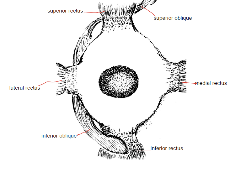
What crossing relationship between oblique and rectus muscles is noted in the diagram?
Which muscle abducts the eye?
Which muscle adducts the eye?
Which cranial nerve innervates the lateral rectus muscle?
Which muscles are antagonists to each other: medial rectus vs ?
Name an antagonist pair for vertical eye movement.
Which cranial nerve innervates the superior oblique muscle?
What are the actions of the superior oblique muscle?
Give an example of a synergist pair for elevating the eye.
What causes astigmatism and how is it corrected?
What anatomical changes produce myopia and which lens corrects it?
What anatomical changes produce hyperopia and which lens corrects it?
What phenomenon explains afterimages when bright light strikes the eye?
How does the color contrast afterimage effect arise?
Why are men more likely to be color blind?
What does the brain do for the blind spots in each eye?
During viewing of far objects, how does the lens change and what happens to the pupil?
What happens to the lens and pupil when focusing on a close object?
What is convergence in eye movements?
What is required for depth perception?
How do visual fields project across cerebral hemispheres as stated in the notes?
What is the optic chiasm?
What is the optic tract defined as in the notes?
Why is visual information processed slower than auditory information according to the notes?
Flashcards in this deck (46)
-
What are the main components found in the orbital cavity?
- Eye
- Lacrimal gland
- Extrinsic eye muscles
- Blood vessels
- Cranial nerves that innervate eye
- Orbital fat cushions
anatomy orbit -
What structures make up the outer fibrous tunic of the eye?
- Cornea (anterior, convex, refracts light, avascular, clear)
- Sclera (mostly collagen, maintains shape, protects, muscle attachments)
anatomy tunics -
List key properties of the cornea.
- Convex
- Refracts light
- Avascular
- Clear
- External part receives fluids from lacrimal glands; internal part gets nutrients from aqueous humor
cornea physiology -
What are the components of the intermediate vascular tunic?
- Choroid
- Ciliary body
- Iris
anatomy uvea -
What is the function of the choroid and why is it dark?
- Provides many capillaries that supply nutrients to the retina
- Contains melanocytes; darkness absorbs light to prevent repeated photoreceptor stimulation
choroid physiology -
What is the tapetum lucidum?
- An iridescent part of the choroid found in animals adapted to low-intensity light
choroid comparative -
Describe the ciliary body's roles.
- Contains ciliary muscles and ciliary processes
- Suspensory ligaments from ciliary body hold the lens
- Epithelial cells secrete aqueous humor
- Ciliary muscles alter lens shape by changing suspensory ligament tension
ciliary physiology -
What is the ora serrata?
- The posterior edge of the ciliary body where it meets the retina
ciliary retina -
What structures and functions are associated with the iris?
- Colored structure with central pupil that lets light in
- Contains pigment cells, blood vessels, nerves, and smooth muscles that change pupil size
iris physiology -
Compare rods and cones in the retina.
- Rods: long, narrow, mostly peripheral, detect dim light, contrast and movement, no color or fine detail
- Cones: cone-shaped, concentrated posteriorly, detect bright light and fine visual detail
retina photoreceptors -
What is the role of bipolar and ganglion cells in retinal signal transmission?
- Bipolar cells: receive graded potentials from photoreceptors and induce action potentials in ganglion cells
- Ganglion cells: axons form the optic disc and optic nerve
retina pathway -
What role do amacrine and horizontal cells play in retinal processing?
- They converge sensory information as signals from the photoreceptors.
retina photoreceptors -
What is unique about the optic disc?
- It lacks photoreceptors and creates the blind spot.
optic retina -
Describe the macula lutea and its location relative to the optic disc.
- The Macula Lutea is a yellow spot lateral to the optic disc.
macula retina -
What is the fovea centralis and its significance for vision?
- Center of macula with densely packed cones and almost no rods; provides sharpest vision.
fovea vision -
What is the composition and primary optical function of the lens?
- Transparent, made of organelle-free cells filled with crystallin; focuses light onto the retina.
lens optics -
How does the eye achieve distance vision via lens and ciliary body changes?
- Ciliary body moves posteriorly, increasing tension on suspensory ligaments and flattening the lens for distance vision.
accommodation lens -
Explain the accommodation process for near vision.
- Parasympathetic signals cause ciliary muscles to contract and move closer to lens; suspensory ligaments relax and lens rounds to focus near images.
accommodation parasympathetic -
What is presbyopia and its cause?
- Age-related inflexibility of the lens causing inability to stay rounded and difficulty seeing close objects; reading glasses are needed.
presbyopia lens -
How is the eye divided by the lens and what fluid fills the posterior cavity?
- Lens divides the eye into anterior and posterior cavities; posterior cavity is filled with vitreous humor, a gelatinous fluid that maintains eye shape, transmits light, and supports the retina.
cavities vitreous -
Describe the anterior cavity subdivisions and the role of aqueous humor.
- Anterior cavity is anterior to lens; iris divides it into anterior chamber (between iris and cornea) and posterior chamber (between lens and iris). Aqueous humor is a filtrate of blood plasma produced by ciliary epithelium, secreted into posterior chamber, flows through the pupil to anterior chamber to provide nutrients and remove waste.
aqueous cavities -
Which extraocular muscles are labeled in the provided right eye diagram?
- Superior rectus
- Superior oblique
- Lateral rectus
- Medial rectus
- Inferior oblique
- Inferior rectus
muscles extraocular -
What crossing relationship between oblique and rectus muscles is noted in the diagram?
- Superior oblique crosses under superior rectus, and inferior oblique crosses over inferior rectus.
muscles anatomy -
Which muscle abducts the eye?
- Lateral rectus
muscles eye -
Which muscle adducts the eye?
- Medial rectus
muscles eye -
Which cranial nerve innervates the lateral rectus muscle?
- Abducens
cranialnerve eye -
Which muscles are antagonists to each other: medial rectus vs ?
- Lateral rectus
antagonist muscles -
Name an antagonist pair for vertical eye movement.
- Superior rectus and inferior rectus
antagonist eye -
Which cranial nerve innervates the superior oblique muscle?
- Trochlear
cranialnerve eye -
What are the actions of the superior oblique muscle?
- Medial rotation, depression, abduction
muscles actions -
Give an example of a synergist pair for elevating the eye.
- Superior rectus and inferior oblique
synergist eye -
What causes astigmatism and how is it corrected?
- Blurry lines because lens or cornea do not refract light well; LASIK reshapes the cornea
optics clinical -
What anatomical changes produce myopia and which lens corrects it?
- Elongated eyeball or too curved cornea; corrected with a concave lens
refractive myopia -
What anatomical changes produce hyperopia and which lens corrects it?
- Eyeball too short or cornea too flat; corrected with a convex lens
refractive hyperopia -
What phenomenon explains afterimages when bright light strikes the eye?
- Light bleaches photopigment in rods and cones; pigment resynthesis is slow, causing afterimages
vision photoreceptors -
How does the color contrast afterimage effect arise?
- Bleached cones for a color become unavailable, so opposing cone types respond and produce the opposite color
color vision -
Why are men more likely to be color blind?
- Color vision genes are on the X chromosome and the defect is recessive, so men are more affected
genetics color -
What does the brain do for the blind spots in each eye?
- The brain fills in information in blind spots
vision perception -
During viewing of far objects, how does the lens change and what happens to the pupil?
- Lens flattens to focus on fovea; the pupil enlarges to admit more light
accommodation reflex -
What happens to the lens and pupil when focusing on a close object?
- Lens becomes more rounded
- More light refraction
- Pupil decreases in size
 accommodation lens eye
 -
What is convergence in eye movements?
Medial rotation of the eyes for close-up objects
convergence eye movement -
What is required for depth perception?
Depth perception requires both eyes/retinas
depth perception vision -
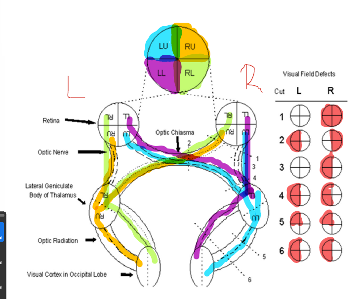
How do visual fields project across cerebral hemispheres as stated in the notes?
Everything in the right field crosses to the left field, and everything in the left field goes to the right field
visualfield crossing vision -
What is the optic chiasm?
The location where the optic nerves merge
opticchiasm anatomy vision -
What is the optic tract defined as in the notes?
Neuron axons from the thalamus to the occipital lobe
 optictract thalamus occipital
 -
Why is visual information processed slower than auditory information according to the notes?
Because the visual pathway is more complex
processing vision auditory
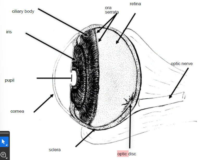
Eye anatomy — overview
- Orbital cavity components: eye, lacrimal gland, extrinsic eye muscles, blood vessels, cranial nerves, orbital fat.
- Main functions: protect the eye, produce/tear fluids, move the eye, transmit visual signals to brain.
Wall of the eye (three tunics)
1. Outer fibrous tunic
- Cornea: anterior, transparent, avascular, convex — refracts light; external surface nourished by tears, internal surface by aqueous humor.
- Sclera: posterior, collagenous, provides shape/protection and attachment for extrinsic muscles.
2. Intermediate vascular tunic (uvea)
- Choroid: posterior, vascular, pigmented (absorbs stray light), supplies retina.
- Tapetum lucidum: reflective, found in some animals for low-light vision.
- Ciliary body: contains ciliary muscle and processes; suspensory ligaments (zonules) attach the lens; epithelial cells secrete aqueous humor.
- Ora serrata: junction between ciliary body and retina.
- Iris: pigmented diaphragm with central pupil; smooth muscles change pupil diameter to control light.
3. Inner tunic (retina)
- Photoreceptors:
- Rods: sensitive in dim light, peripheral vision, detect contrast and motion, no color.
- Cones: require brighter light, concentrated at posterior retina, mediate color and high-acuity vision.
- Bipolar cells: transmit graded potentials from photoreceptors to ganglion cells.
- Ganglion cells: produce action potentials; their axons form the optic nerve at the optic disc.
- Horizontal & amacrine cells: integrate and modulate signals between photoreceptors/bipolars/ganglion cells.
- Optic disc: blind spot (no photoreceptors).
- Macula lutea & fovea centralis: macula is lateral to optic disc; fovea is center of macula with densely packed cones for sharpest vision.
Alt text: Diagram of the human eye labeling major structures.
Lens and focusing
- Structure: transparent, elastic, made of denucleated fiber cells filled with crystallin; focuses light onto retina.
- Accommodation (near vision): parasympathetic activation contracts the ciliary muscle, reduces tension on suspensory ligaments, lens becomes more rounded, increasing refractive power.
- Distance vision: ciliary muscle relaxes (or moves posteriorly), suspensory ligaments tense, lens flattens to focus distant objects.
- Presbyopia: age-related loss of lens elasticity → reduced accommodation; reading glasses correct this.
Cavities and humors
- Anterior cavity (in front of lens): contains aqueous humor; subdivided into anterior chamber (between cornea and iris) and posterior chamber (between iris and lens).
- Aqueous humor is produced by ciliary epithelium, flows from posterior chamber through the pupil into anterior chamber, supplies nutrients and maintains pressure.
- Posterior cavity (vitreous chamber): between lens and retina; filled with gelatinous vitreous humor that maintains globe shape and supports the retina.
Extraocular muscles — actions and innervation
| Muscle | Primary action | Cranial nerve |
|---|---|---|
| Lateral rectus | Abducts eye | Abducens (VI) |
| Medial rectus | Adducts eye | Oculomotor (III) |
| Superior rectus | Elevates eye (and intorsion) | Oculomotor (III) |
| Inferior rectus | Depresses eye (and extorsion) | Oculomotor (III) |
| Superior oblique | Intorsion, depression, abduction | Trochlear (IV) |
| Inferior oblique | Elevates eye when adducted | Oculomotor (III) |
Alt text: Diagram showing the muscles of the eye and their positions.
Antagonists and synergists
- Antagonists: medial vs lateral rectus; superior vs inferior rectus; superior oblique vs inferior oblique.
- Synergists: superior rectus with inferior oblique; inferior rectus with superior oblique.
Visual tests and common refractive errors
- Visual acuity test: uses eye chart; reduced acuity suggests refractive error or pathology.
- Myopia (nearsighted): eyeball too long or cornea too curved; image focuses in front of retina; corrected with concave (diverging) lenses.
- Hyperopia (farsighted): eyeball too short or cornea too flat; image focuses behind retina; corrected with convex (converging) lenses.
- Astigmatism: irregular corneal/lens curvature causes blurred lines; LASIK reshapes cornea.
- Afterimages & color contrast: intense light bleaches photopigments; temporary afterimages occur while pigments regenerate; color afterimages reflect which cone types were fatigued.
- Color blindness: often X-linked recessive; caused by absent/nonfunctional cone pigments or cones.
Reflexes: accommodation, pupil, convergence
- Near response: accommodation (lens rounds), pupillary constriction, and convergence (medial rotation) act together to focus on close objects.
- Far response: lens flattens, pupils may dilate to admit more light, eyes diverge for distant viewing.
Visual pathway and field organization
- Retina → optic nerve → optic chiasm → optic tract → lateral geniculate nucleus (LGN) of thalamus → optic radiations → visual cortex (occipital lobe).
- Decussation pattern: fibers from the nasal (medial) half of each retina cross at the optic chiasm; temporal retinal fibers remain ipsilateral. This creates contralateral visual field representation in each cerebral hemisphere (right visual field → left cortex).
- Optic tract lesions produce predictable visual field deficits depending on where fibers are interrupted.
Alt text: Visual pathway from retina through chiasm to visual cortex.
Key clinical correlations / quick facts
- Optic disc = physiological blind spot; brain "fills in" missing information.
- Macula/fovea = highest acuity; central vision loss (e.g., macular degeneration) severely impairs detail vision.
- Aqueous humor drainage blockage raises intraocular pressure → glaucoma risk.
- Damage to cranial nerves III, IV, or VI causes characteristic eye movement deficits and diplopia.
Study tips
- Memorize muscle–action–nerve table and antagonistic/synergistic pairs.
- Relate lens shape changes to ciliary muscle tension: "contract → lens rounds; relax → lens flattens."
- Use diagrams (retina cross-section and visual pathway) to visualize signal flow and lesion effects.