在牌组消失前保存
这些记忆卡还没保存——离开页面后会消失。创建一个免费账户来保留它们,并解锁下面的所有功能。
- Save this deck to your account
- Study with spaced repetition
- Export to Anki (.apkg) or PDF
- Process documents up to 100 pages
- Images extracted from your PDFs
- Sharper text extraction & a more advanced AI model
What is the general chemical process by which carbohydrates, fats, and proteins are broken down for absorption?
How does hydrolysis reverse the condensation that formed disaccharides?
What are the dietary forms of carbohydrates named in the text?
Why is cellulose not considered a food for humans?
What is the structure of most dietary fats and how are they hydrolyzed?
How are proteins digested chemically?
What is true about digestive enzymes themselves?
What enzyme in saliva begins starch digestion and what type of enzyme is it?
Approximately what percentage of ingested starch is hydrolyzed by salivary amylase before swallowing?
How long can starch digestion by salivary amylase continue in the stomach before gastric secretions inactivate it?
At what pH does salivary amylase become essentially inactive in the stomach?
What is the role and relative strength of pancreatic amylase in intestinal carbohydrate digestion?
How quickly does pancreatic amylase digest carbohydrates after chyme enters the duodenum?
Which intestinal epithelial enzymes split disaccharides and small glucose polymers on the brush border?
What are the monosaccharide products of lactose, sucrose, and maltose digestion?
What are the final products of carbohydrate digestion and where are they absorbed?
In an ordinary diet, what proportion of final carbohydrate digestion products is glucose?
How are dietary proteins chemically described in the text?
At what pH range is pepsin most active, and above what pH is it inactive?
What condition must stomach juices meet for pepsin to digest protein effectively?
What do the gastric glands secrete in large quantity?
Which cells in the gastric glands secrete hydrochloric acid?
At approximately what pH is hydrochloric acid secreted by parietal cells?
What pH range is highly favorable for pepsin activity in the stomach?
What unique protein can pepsin digest effectively that other digestive enzymes affect little?
Approximately what percentage of total protein digestion is initiated by pepsin in the stomach?
Which pancreatic proteolytic enzymes attack protein breakdown products in the duodenum and jejunum?
What is the primary action of trypsin and chymotrypsin on proteins?
What specific cleavage does carboxypolypeptidase perform on polypeptides?
How is proelastase activated and what does elastase digest?
After pancreatic digestion, in what forms do most proteins remain before enterocyte processing?
Which two peptidases on enterocyte microvilli are especially important for final peptide digestion?
What happens to dipeptides and tripeptides inside enterocytes before entering the blood?
What proportion of final absorbed protein products are individual amino acids?
What rare consequence can result from absorption of a few intact whole protein molecules?
What are the main products when tristearin is hydrolyzed by lipase as shown in the figure?
What is the chemical composition of a neutral fat (triglyceride)?
Which dietary source contains more neutral fats: animal or plant foods?
Name three other lipid types present in the usual diet besides neutral fats.
Why are phospholipids and cholesterol esters considered fats?
Does cholesterol contain fatty acids, and is it considered a dietary fat?
What is another name for neutral fats?
Where does most dietary fat digestion occur?
Which enzyme in the mouth/stomach digests a small amount of triglycerides?
What is the first step in fat digestion and what causes it?
Does bile contain digestive enzymes?
How do bile salts and lecithin make fat globules easier to fragment?
Why does lowering interfacial tension help fat emulsification?
By how much can emulsification increase the total surface area of intestinal fat particles?
What is the primary enzyme that digests dietary triglycerides in the small intestine?
Into which products are most dietary triglycerides split by pancreatic lipase?
How do bile salts accelerate fat digestion beyond emulsification?
What concentration-dependent structure do bile salts form in water that aids fat digestion?
What is the typical size and molecular composition of bile salt micelles?
How is each bile salt molecule structurally arranged to solubilize fat?
Where is the fat located within a bile salt micelle and how are polar groups oriented?
Why do bile salt micelles remain dissolved and stable in digestive fluids?
What role do bile salt micelles play for monoglycerides and free fatty acids?
What happens to bile salts after they deliver monoglycerides and free fatty acids to the intestinal brush border?
Which pancreatic enzyme hydrolyzes dietary cholesterol esters to free fatty acids?
Which pancreatic enzyme hydrolyzes phospholipids to free fatty acids?
What role do bile salt micelles play in intestinal absorption of cholesterol?
Besides cholesterol, what other lipid digestates do bile salt micelles transport?
What is the total daily fluid volume that intestines must absorb?
How much fluid passes from the small intestine into the colon each day?
Why is the stomach a poor absorptive area of the GI tract?
Which types of substances can be absorbed in small quantities from the stomach?
By approximately how much do folds of Kerckring, villi, and microvilli increase small intestinal mucosal absorptive area?
How much do the valvulae conniventes (folds of Kerckring) increase absorptive surface area and how far can they protrude?
How long do villi project from the mucosal surface and how does their distribution change along the small intestine?
How much does the combination of folds of Kerckring, villi, and microvilli increase small intestinal absorptive area?
What are the typical dimensions of individual microvilli on intestinal epithelial cells?
What structural feature on intestinal epithelial cells further increases absorptive surface area by about 20-fold?
What movements occur within each microvillus and what structures cause them?
Approximately how much carbohydrate, fat, protein, ions, and water are absorbed daily by the small intestine under normal conditions?
What is the absorptive capacity of the small intestine compared to normal daily absorption?
How is water transported across the intestinal mucosa and what determines its direction?
How much sodium must the intestines absorb daily to prevent net sodium loss, and why is this important?
What small-scale physical process allows some substances to be absorbed by enterocytes without transporter proteins?
View the microscopic brush border image and recall its relevance to intestinal absorption.
What role does sodium play in intestinal absorption of sugars and amino acids?
What is the basic mechanism that powers sodium absorption from the intestine?
Which enzyme class in the cell membrane catalyzes the energy process for sodium active transport?
How does chloride become absorbed along with sodium in the intestine?
What is the approximate intracellular sodium concentration after active basolateral transport?
How does intestinal sodium concentration in chyme compare to intracellular sodium after absorption?
Are the principles of intestinal sodium absorption unique to the intestine?
What visual structure can illustrate intestinal epithelial cells with apical hair-like projections?
How does sodium move from intestinal chyme into epithelial cells?
What is the role of the Na+–K+ ATPase in intestinal epithelial cells?
Which specific sodium co-transporter mediates sodium-linked glucose uptake in the intestine?
Through which pathways does water move during intestinal absorption?
Why does water undergo osmosis into the paracellular spaces during absorption?
How does aldosterone affect intestinal sodium absorption?
How are chloride ions absorbed in the upper small intestine?
What exchanger absorbs chloride across the brush border in parts of the ileum and large intestine?
How is bicarbonate (HCO3-) reabsorbed in the duodenum and jejunum?
What happens to the CO2 produced when HCO3- combines with H+ in the intestines?
What happens to the water produced when HCO3- combines with H+ in the intestines?
What is meant by the 'active absorption of HCO3-' described in intestinal epithelium?
What ion exchange occurs in the epithelial cells of the ileum and large intestine involving bicarbonate?
Why do the ileum and large intestine secrete alkaline HCO3-?
Where are immature intestinal epithelial cells that secrete sodium chloride and water located?
What happens to the sodium chloride and water secreted by immature epithelial cells?
How can cholera toxin cause extreme diarrheal fluid loss?
How much fluid loss can cholera-like secretion cause per day?
What is the usual life-saving treatment for severe cholera fluid loss?
Where is calcium actively absorbed most, and what controls its absorption?
What percentage of absorbed carbohydrate calories is usually glucose?
Which monosaccharides make up most of the remaining absorbed carbohydrates besides glucose?
What transporters are named in the intestinal monosaccharide absorption figure?
How do monovalent and bivalent ions differ in intestinal absorption?
How does the maximum absorption of calcium compare to sodium according to the text?
Why is glucose absorption negligible without sodium transport?
What are the two stages of sodium and glucose transport across the intestinal epithelium?
What triggers sodium from the lumen to move into intestinal epithelial cells via secondary active transport?
What is SGLT1's role in intestinal glucose absorption?
How is glucose transported from the intestinal lumen into epithelial cells?
What transporter facilitates glucose exit from the intestinal epithelial cell into the blood?
What provides the driving force for glucose uptake across intestinal membranes?
How is galactose absorbed across the intestinal epithelium?
How is fructose transported across intestinal epithelium?
Why is fructose transport slower than glucose or galactose?
In what forms are most digested proteins absorbed by intestinal epithelial cells?
What mechanism supplies energy for most peptide and amino acid uptake into enterocytes?
How are monoglycerides and free fatty acids delivered to intestinal epithelial microvilli?
What are two properties of bile micelles that allow them to transport lipid digestion products in chyme?
How many different transport proteins for amino acids and peptides have been found in intestinal epithelial cells?
What is the primary function of bile micelles in fat digestion?
How much dietary fat is absorbed in the presence of abundant bile micelles?
What happens to fatty acids and monoglycerides after entering intestinal epithelial cells?
How do chylomicrons enter the circulation after formation in epithelial cells?
Why are short- and medium-chain fatty acids absorbed directly into the portal blood?
How much chyme typically passes into the large intestine daily and how much fluid remains to be excreted as feces?
What proportion of ions are lost in feces after colonic absorption?
What are the primary functions of the proximal and distal colon?
What is a primary mechanism driving chloride absorption in the large intestine?
How do the tight junctions of large intestinal epithelium compare with those of the small intestine?
How does aldosterone affect sodium transport in the large intestine?
What exchange process involving HCO3- occurs in the large intestine?
What role does HCO3- secretion play in the large intestine?
How does absorption of sodium and chloride affect water movement in the large intestine?
What is the maximum daily capacity of the large intestine to absorb fluid and electrolytes?
What can cause severe diarrhea by increasing intestinal secretion to 10 or more liters per day?
What minor nutritional contribution do colon bacteria provide to humans?
Which vitamins are formed by bacterial activity in the colon?
Which gases produced by colonic bacteria contribute to flatus?
What is the approximate water and solid composition of normal feces?
What are the main components of the solid fraction of feces by approximate percentages?
What pigments cause the brown color of feces?
Which bacterial products principally cause the odor of feces?
Flashcards in this deck (151)
-
What is the general chemical process by which carbohydrates, fats, and proteins are broken down for absorption?
Hydrolysis: digestive enzymes add H+ and OH- from H₂O to split macromolecules into absorbable units.
digestion -
How does hydrolysis reverse the condensation that formed disaccharides?
Hydrolysis returns H+ and OH- from water to the disaccharide (R"-R') to yield R'OH and RH, separating monosaccharides.
carbohydrates -
What are the dietary forms of carbohydrates named in the text?
- Sucrose
- Lactose
- Starches
carbohydrates -
Why is cellulose not considered a food for humans?
Humans do not secrete enzymes capable of hydrolyzing cellulose, so it cannot be digested or absorbed.
carbohydrates -
What is the structure of most dietary fats and how are they hydrolyzed?
Most dietary fats are triglycerides (three fatty acids + glycerol); enzymes add three H₂O molecules to split fatty acids from glycerol.
fats -
How are proteins digested chemically?
Proteins are chains of amino acids linked by peptide bonds; proteolytic enzymes hydrolyze these bonds by adding H+ and OH- from water to yield amino acids.
proteins -
What is true about digestive enzymes themselves?
All digestive enzymes are proteins secreted by gastrointestinal glands.
enzymes -
What enzyme in saliva begins starch digestion and what type of enzyme is it?
- Ptyalin (saliva)
- An α-amylase that hydrolyzes starch into maltose and small glucose polymers
carbohydrates enzymes -
Approximately what percentage of ingested starch is hydrolyzed by salivary amylase before swallowing?
- About 5% of all starches are hydrolyzed by salivary amylase before food is swallowed
carbohydrates digestion -
How long can starch digestion by salivary amylase continue in the stomach before gastric secretions inactivate it?
- Up to 1 hour in the body and fundus of the stomach before mixing with gastric secretions inactivates the enzyme
carbohydrates stomach -
At what pH does salivary amylase become essentially inactive in the stomach?
- Below about pH 4.0 salivary amylase is essentially inactive
enzymes ph -
What is the role and relative strength of pancreatic amylase in intestinal carbohydrate digestion?
- Pancreatic α-amylase functions like salivary amylase but is several times more powerful, digesting most carbohydrates after chyme reaches the duodenum
carbohydrates pancreas -
How quickly does pancreatic amylase digest carbohydrates after chyme enters the duodenum?
- Within 15 to 30 minutes after chyme mixes with pancreatic juice, virtually all carbohydrates are digested
digestion timing -
Which intestinal epithelial enzymes split disaccharides and small glucose polymers on the brush border?
- Lactase, sucrase, maltase, and α-dextrinase located on enterocyte microvilli (brush border)
intestinal enzymes -
What are the monosaccharide products of lactose, sucrose, and maltose digestion?
- Lactose → galactose + glucose
- Sucrose → fructose + glucose
- Maltose/small polymers → glucose
carbohydrates products -
What are the final products of carbohydrate digestion and where are they absorbed?
- Monosaccharides (glucose, galactose, fructose) that are water soluble and absorbed immediately into the portal blood
absorption carbohydrates -
In an ordinary diet, what proportion of final carbohydrate digestion products is glucose?
- Glucose represents more than 80% of the final products; galactose and fructose each seldom exceed 10%
carbohydrates composition -
How are dietary proteins chemically described in the text?
- Long chains of amino acids bound together by peptide linkages
proteins definition -
At what pH range is pepsin most active, and above what pH is it inactive?
- Most active at pH 2.0–3.0; inactive at pH above about 5.0
proteins pepsin -
What condition must stomach juices meet for pepsin to digest protein effectively?
- Stomach juices must be acidic so pepsin can be active
proteins stomach -
What do the gastric glands secrete in large quantity?
- Hydrochloric acid (HCl)
physiology stomach -
Which cells in the gastric glands secrete hydrochloric acid?
- Parietal (oxyntic) cells
cells stomach -
At approximately what pH is hydrochloric acid secreted by parietal cells?
- About pH 0.8
physiology acid -
What pH range is highly favorable for pepsin activity in the stomach?
pH 2.0 to 3.0
pepsin digestion -
What unique protein can pepsin digest effectively that other digestive enzymes affect little?
Collagen
pepsin collagen -
Approximately what percentage of total protein digestion is initiated by pepsin in the stomach?
10% to 20%
pepsin protein -
Which pancreatic proteolytic enzymes attack protein breakdown products in the duodenum and jejunum?
- Trypsin
- Chymotrypsin
- Carboxypolypeptidase
- Elastase
pancreas proteases -
What is the primary action of trypsin and chymotrypsin on proteins?
They split proteins into small polypeptides
trypsin chymotrypsin -
What specific cleavage does carboxypolypeptidase perform on polypeptides?
It cleaves individual amino acids from the carboxyl end of polypeptides
carboxypolypeptidase enzymology -
How is proelastase activated and what does elastase digest?
Proelastase is converted to elastase, which digests elastin fibers
elastase activation -
After pancreatic digestion, in what forms do most proteins remain before enterocyte processing?
Mostly as dipeptides and tripeptides, with only small percentages as free amino acids
peptides absorption -
Which two peptidases on enterocyte microvilli are especially important for final peptide digestion?
- Aminopolypeptidase
- Dipeptidases
enterocyte peptidases -
What happens to dipeptides and tripeptides inside enterocytes before entering the blood?
Cytosolic peptidases digest them to single amino acids, which then pass into the blood
enterocyte aminoacids -
What proportion of final absorbed protein products are individual amino acids?
More than 99%
absorption aminoacids -
What rare consequence can result from absorption of a few intact whole protein molecules?
They can sometimes cause serious allergic or immunologic reactions
allergy immunology -
What are the main products when tristearin is hydrolyzed by lipase as shown in the figure?
- 2-Monoglyceride
- Two stearic acid molecules
lipase fat -
What is the chemical composition of a neutral fat (triglyceride)?
- A glycerol nucleus bound to three fatty acid side chains
fats triglyceride -
Which dietary source contains more neutral fats: animal or plant foods?
- Animal foods contain much more neutral fat than plant foods
fats diet -
Name three other lipid types present in the usual diet besides neutral fats.
- Phospholipids
- Cholesterol
- Cholesterol esters
lipids diet -
Why are phospholipids and cholesterol esters considered fats?
- Because they contain fatty acids, so they can be considered fats
phospholipids fats -
Does cholesterol contain fatty acids, and is it considered a dietary fat?
- Cholesterol contains no fatty acid but is considered a dietary fat because it is derived from fats and is metabolized similarly
cholesterol fats -
What is another name for neutral fats?
- Triglycerides
terminology fats -
Where does most dietary fat digestion occur?
- Small intestine
fat digestion anatomy -
Which enzyme in the mouth/stomach digests a small amount of triglycerides?
- Lingual lipase
enzymes lipase -
What is the first step in fat digestion and what causes it?
- Emulsification by bile acids and lecithin
fat emulsification bile -
Does bile contain digestive enzymes?
- No; bile contains bile salts and lecithin but no digestive enzymes
bile physiology -
How do bile salts and lecithin make fat globules easier to fragment?
- Their fat-soluble portions dissolve in the fat surface while polar portions project into water, lowering interfacial tension
bile mechanism emulsification -
Why does lowering interfacial tension help fat emulsification?
- Lower interfacial tension lets agitation break nonmiscible fat into many tiny particles more easily
physics emulsification -
By how much can emulsification increase the total surface area of intestinal fat particles?
- Up to 1000-fold (average particle diameter falls to <1 micrometer)
quantitative emulsification -
What is the primary enzyme that digests dietary triglycerides in the small intestine?
- Pancreatic lipase
enzymes pancreas lipase -
Into which products are most dietary triglycerides split by pancreatic lipase?
- Free fatty acids and 2-monoglycerides
products lipid -
How do bile salts accelerate fat digestion beyond emulsification?
- They form micelles that remove monoglycerides and free fatty acids from the digestion site, preventing reversal and allowing continued hydrolysis
micelles bile digestion -
What concentration-dependent structure do bile salts form in water that aids fat digestion?
Bile salts form micelles when their concentration is high enough in water.
bile micelle -
What is the typical size and molecular composition of bile salt micelles?
- Diameter: 3 to 6 nanometers
- Composed of 20 to 40 molecules of bile salt
micelle size -
How is each bile salt molecule structurally arranged to solubilize fat?
Each bile salt has a fat-soluble sterol nucleus and a water-soluble polar group.
structure bile -
Where is the fat located within a bile salt micelle and how are polar groups oriented?
The sterol nucleus surrounds the fat digestate in the micelle center, while polar groups project outward to cover the micelle surface.
micelle orientation -
Why do bile salt micelles remain dissolved and stable in digestive fluids?
The polar groups are negatively charged, allowing the entire micelle to dissolve in water and remain in stable solution.
stability bile -
What role do bile salt micelles play for monoglycerides and free fatty acids?
Micelles transport monoglycerides and free fatty acids to the brush border of intestinal epithelial cells for absorption.
transport fat -
What happens to bile salts after they deliver monoglycerides and free fatty acids to the intestinal brush border?
Bile salts are released back into the chyme to be reused repeatedly for ferrying lipids.
recycling bile -
Which pancreatic enzyme hydrolyzes dietary cholesterol esters to free fatty acids?
The enzyme cholesterol ester hydrolase hydrolyzes cholesterol esters.
enzymes cholesterol -
Which pancreatic enzyme hydrolyzes phospholipids to free fatty acids?
The enzyme phospholipase A2 hydrolyzes phospholipids.
enzymes phospholipid -
What role do bile salt micelles play in intestinal absorption of cholesterol?
Bile salt micelles 'ferry' free cholesterol and phospholipid digestates into absorptive sites; essentially no cholesterol is absorbed without this micellar function.
lipids bile absorption -
Besides cholesterol, what other lipid digestates do bile salt micelles transport?
They ferry monoglycerides and free fatty acids as well as phospholipid digestates.
lipids micelles -
What is the total daily fluid volume that intestines must absorb?
Approximately 8 to 9 liters per day (≈1.5 L ingested fluid plus ≈7 L secreted).
fluid physiology -
How much fluid passes from the small intestine into the colon each day?
About 1.5 liters pass through the ileocecal valve into the colon each day.
intestine fluid -
Why is the stomach a poor absorptive area of the GI tract?
Because it lacks villus absorptive membrane and has tight junctions between epithelial cells, limiting absorption.
stomach absorption anatomy -
Which types of substances can be absorbed in small quantities from the stomach?
A few highly lipid-soluble substances such as alcohol and some drugs (e.g., aspirin).
stomach drugs absorption -
By approximately how much do folds of Kerckring, villi, and microvilli increase small intestinal mucosal absorptive area?
They increase the mucosal absorptive area by nearly 1000-fold.
surface-area intestine anatomy -
How much do the valvulae conniventes (folds of Kerckring) increase absorptive surface area and how far can they protrude?
They increase surface area about threefold and can protrude up to 8 millimeters into the lumen, especially in the duodenum and jejunum.
kerckring intestine anatomy -
How long do villi project from the mucosal surface and how does their distribution change along the small intestine?
Villi project about 1 millimeter from the mucosa; they lie close together and often touch in the upper small intestine but are less profuse in the distal small intestine.
villi intestine anatomy -
How much does the combination of folds of Kerckring, villi, and microvilli increase small intestinal absorptive area?
- Increases total absorptive area approximately 1000-fold, yielding about 250 m²
anatomy surface-area -
What are the typical dimensions of individual microvilli on intestinal epithelial cells?
- ~1 micrometer in length
- ~0.1 micrometer in diameter
microvilli anatomy -
What structural feature on intestinal epithelial cells further increases absorptive surface area by about 20-fold?
- The brush border of microvilli increases exposed surface area by at least 20-fold
brush-border surface-area -
What movements occur within each microvillus and what structures cause them?
- Multiple actin filaments extend into microvilli and contract rhythmically, causing continual movement
microvilli cytoskeleton -
Approximately how much carbohydrate, fat, protein, ions, and water are absorbed daily by the small intestine under normal conditions?
- Several hundred grams carbohydrates
- ≥100 g fat
- 50–100 g amino acids
- 50–100 g ions
- 7–8 L water
absorption quantities -
What is the absorptive capacity of the small intestine compared to normal daily absorption?
- Capacity is much greater: up to several kg carbohydrates, 500 g fat, 500–700 g proteins, and ≥20 L water can be absorbed per day
absorption capacity -
How is water transported across the intestinal mucosa and what determines its direction?
- Water crosses entirely by diffusion/osmosis; direction depends on osmotic gradient (water moves to make chyme isosmotic with plasma)
water osmosis -
How much sodium must the intestines absorb daily to prevent net sodium loss, and why is this important?
- Intestines must absorb 25–35 g Na+ per day to offset secretions and intake, preventing depletion of body sodium
sodium electrolytes -
What small-scale physical process allows some substances to be absorbed by enterocytes without transporter proteins?
- Pinocytosis: infolding of enterocyte membrane forms vesicles that trap and absorb small amounts of fluid and solutes
pinocytosis absorption -
View the microscopic brush border image and recall its relevance to intestinal absorption.
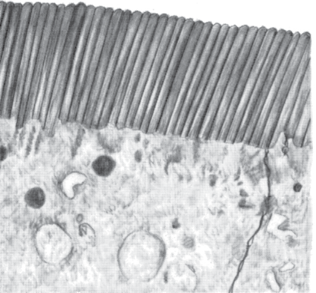
- Image shows hair-like microvilli that form the brush border and increase absorptive surface
image brush-border -
What role does sodium play in intestinal absorption of sugars and amino acids?
- Sodium helps absorb sugars and amino acids by enabling their transport across the intestinal mucosa.
sodium absorption -
What is the basic mechanism that powers sodium absorption from the intestine?
- Active transport of Na+ from epithelial cells through basolateral membranes into paracellular spaces, driven by energy-dependent transporters.
mechanism sodium -
Which enzyme class in the cell membrane catalyzes the energy process for sodium active transport?
- Adenosine triphosphatase (ATPase) enzymes in the cell membrane.
atpase physiology -
How does chloride become absorbed along with sodium in the intestine?
- Chloride is mainly passively 'dragged' by the positive electrical charges of sodium ions.
chloride electrolytes -
What is the approximate intracellular sodium concentration after active basolateral transport?
- About 50 mEq/L intracellular sodium concentration.
concentration sodium -
How does intestinal sodium concentration in chyme compare to intracellular sodium after absorption?
- Chyme sodium is normally about 142 mEq/L, which is much higher than the intracellular value of ≈50 mEq/L.
chyme sodium -
Are the principles of intestinal sodium absorption unique to the intestine?
- No. The principles are essentially the same for sodium absorption from the gallbladder and renal tubules.
comparative physiology -
What visual structure can illustrate intestinal epithelial cells with apical hair-like projections?
- Microscopic view of intestinal epithelial cells with apical microvilli as illustration:
image microvilli - Microscopic view of intestinal epithelial cells with apical microvilli as illustration:
-
How does sodium move from intestinal chyme into epithelial cells?
- Down its electrochemical gradient through the brush border
- Co-transport via SGLT1, sodium–amino-acid transporters, and Na+–H+ exchanger
physiology sodium -
What is the role of the Na+–K+ ATPase in intestinal epithelial cells?
- Pumps Na+ out across the basolateral membrane, powering secondary active absorption of glucose and amino acids
physiology transport -
Which specific sodium co-transporter mediates sodium-linked glucose uptake in the intestine?
- SGLT1 (sodium-glucose co-transporter 1)
carbohydrate transport -
Through which pathways does water move during intestinal absorption?
- Paracellular pathway (through tight junctions)
- Transcellular pathway (through epithelial cells)
water absorption -
Why does water undergo osmosis into the paracellular spaces during absorption?
- Because elevated ion concentration in paracellular spaces creates a large osmotic gradient that draws water
water osmotic -
How does aldosterone affect intestinal sodium absorption?
- Increases activation of enzymes and transport mechanisms for sodium absorption, enhancing Na+, chloride, and water uptake (notably in the colon)
hormone aldosterone -
How are chloride ions absorbed in the upper small intestine?
- Mainly by diffusion: chloride follows sodium due to electrical gradient created by Na+ absorption
chloride absorption -
What exchanger absorbs chloride across the brush border in parts of the ileum and large intestine?
- A brush border chloride–bicarbonate exchanger
chloride bicarbonate -
How is bicarbonate (HCO3-) reabsorbed in the duodenum and jejunum?
- Na+ absorption is coupled to H+ secretion into lumen; H+ combines with HCO3- to form H2CO3, which then dissociates for reabsorption
bicarbonate duodenum -
What happens to the CO2 produced when HCO3- combines with H+ in the intestines?
The CO2 is readily absorbed into the blood and subsequently expired through the lungs.
physiology respiration -
What happens to the water produced when HCO3- combines with H+ in the intestines?
The water remains as part of the chyme in the intestines.
digestion fluid -
What is meant by the 'active absorption of HCO3-' described in intestinal epithelium?
It refers to the mechanism where HCO3- is converted to CO2 and H2O, with CO2 absorbed into blood and expired, similar to a mechanism in kidney tubules.
physiology kidney -
What ion exchange occurs in the epithelial cells of the ileum and large intestine involving bicarbonate?
Epithelial cells secrete HCO3- in exchange for absorption of chloride ions.
electrolytes intestine -
Why do the ileum and large intestine secrete alkaline HCO3-?
To neutralize acidic end products formed by bacteria in the large intestine.
microbiota homeostasis -
Where are immature intestinal epithelial cells that secrete sodium chloride and water located?
Immature epithelial cells are located deep in the spaces between the intestinal epithelial folds and secrete sodium chloride and water into the lumen.
intestinal epithelium -
What happens to the sodium chloride and water secreted by immature epithelial cells?
The sodium chloride and water secreted into the lumen are reabsorbed by older epithelial cells outside the folds, providing flow of water for absorption of intestinal digestates.
absorption epithelium -
How can cholera toxin cause extreme diarrheal fluid loss?
A cholera toxin subunit enters epithelial cells, stimulates excess cyclic AMP, opens many chloride channels causing Cl− secretion into crypts, which draws Na+ and then water by osmosis, producing large fluid loss.
cholera diarrhea -
How much fluid loss can cholera-like secretion cause per day?
This secretion can cause a loss of about 5 to 10 liters of water and sodium chloride per day in severe cases.
cholera fluid -
What is the usual life-saving treatment for severe cholera fluid loss?
Administration of large amounts of sodium chloride solution to replace the lost fluid and electrolytes.
treatment cholera -
Where is calcium actively absorbed most, and what controls its absorption?
Calcium is actively absorbed especially from the duodenum, and its absorption is exactly controlled by parathyroid hormone and activated vitamin D.
calcium absorption -
What percentage of absorbed carbohydrate calories is usually glucose?
Glucose usually accounts for more than 80% of the carbohydrate calories absorbed.
carbohydrates glucose -
Which monosaccharides make up most of the remaining absorbed carbohydrates besides glucose?
The remaining roughly 20% of absorbed monosaccharides is composed almost entirely of galactose and fructose.
carbohydrates monosaccharides -
What transporters are named in the intestinal monosaccharide absorption figure?
The figure names SGLT1, GLUT2, and GLUT5 as intestinal monosaccharide transporters.
transporters carbohydrates -
How do monovalent and bivalent ions differ in intestinal absorption?
- Monovalent ions: absorbed with ease and in great quantities
- Bivalent ions: normally absorbed only in small amounts
physiology ions absorption -
How does the maximum absorption of calcium compare to sodium according to the text?
- Calcium maximum absorption: about 1/50 of the normal absorption of sodium
physiology calcium ions -
Why is glucose absorption negligible without sodium transport?
- Because glucose absorption occurs by co-transport with active sodium transport; without sodium transport, virtually no glucose is absorbed
physiology glucose sodium -
What are the two stages of sodium and glucose transport across the intestinal epithelium?
- Stage 1: Active transport of Na+ through basolateral membranes into interstitial fluid, depleting intracellular Na+
- Stage 2: Secondary active transport of Na+ from lumen into cells via SGLT1 coupled with glucose
physiology transport glucose -
What triggers sodium from the lumen to move into intestinal epithelial cells via secondary active transport?
- A decrease of sodium inside epithelial cells (caused by basolateral active Na+ transport) triggers lumenal Na+ to enter via secondary active transport
physiology sodium transport -
What is SGLT1's role in intestinal glucose absorption?
- SGLT1 is a transport protein that binds sodium and will transport sodium into the cell only when it also combines with glucose
physiology sglt1 glucose -
How is glucose transported from the intestinal lumen into epithelial cells?
Glucose binds SGLT1 and is co-transported with Na+ into the cell using the sodium electrochemical gradient.
physiology carbohydrates transport -
What transporter facilitates glucose exit from the intestinal epithelial cell into the blood?
GLUT2 facilitates diffusion of glucose through the basolateral membrane into the paracellular space and then into blood.
physiology carbohydrates glut2 -
What provides the driving force for glucose uptake across intestinal membranes?
Active transport of Na+ through the basolateral membrane of epithelial cells creates the force that drives Na+-glucose cotransport.
physiology transport sodium -
How is galactose absorbed across the intestinal epithelium?
Galactose is absorbed by the same mechanism as glucose using SGLT1 for luminal uptake and GLUT2 for basolateral exit.
physiology carbohydrates galactose -
How is fructose transported across intestinal epithelium?
Fructose is transported by facilitated diffusion: GLUT5 mediates uptake from lumen and GLUT2 mediates exit to the paracellular space.
physiology carbohydrates fructose -
Why is fructose transport slower than glucose or galactose?
Because fructose is not co-transported with Na+, its overall transport rate is about one-half that of glucose or galactose.
physiology carbohydrates kinetics -
In what forms are most digested proteins absorbed by intestinal epithelial cells?
Most digested proteins are absorbed as dipeptides, tripeptides, and a few free amino acids.
physiology proteins absorption -
What mechanism supplies energy for most peptide and amino acid uptake into enterocytes?
A sodium co-transport (secondary active transport) supplies the energy: peptides/amino acids bind transporters that require Na+ binding before transport.
physiology proteins sodium -
How are monoglycerides and free fatty acids delivered to intestinal epithelial microvilli?
They dissolve in the central lipid portions of bile micelles, which carry them to microvilli where they diffuse out of micelles into epithelial cells.
physiology fats micelles -
What are two properties of bile micelles that allow them to transport lipid digestion products in chyme?
Micelles are 3–6 nm in diameter and have a highly charged exterior, making them soluble in chyme.
physiology fats micelles -
How many different transport proteins for amino acids and peptides have been found in intestinal epithelial cells?
At least 10 different types of transport proteins for amino acids and peptides have been found.
physiology proteins transporters -
What is the primary function of bile micelles in fat digestion?
Bile micelles ferry monoglycerides and fatty acids in the chyme to facilitate their absorption.
fat bile micelles -
How much dietary fat is absorbed in the presence of abundant bile micelles?
About 97% of the fat is absorbed when bile micelles are abundant.
fat absorption bile -
What happens to fatty acids and monoglycerides after entering intestinal epithelial cells?
They are taken up by the smooth endoplasmic reticulum and mainly reconverted into triglycerides, then released as chylomicrons.
fat chylomicrons endoplasmicreticulum -
How do chylomicrons enter the circulation after formation in epithelial cells?
Chylomicrons are released at the base of epithelial cells, flow upward through the thoracic lymph duct, and empty into the blood.
chylomicrons lymph fat -
Why are short- and medium-chain fatty acids absorbed directly into the portal blood?
Because they are more water soluble and are mostly not reconverted into triglycerides, allowing diffusion into capillary blood.
fat portal absorption -
How much chyme typically passes into the large intestine daily and how much fluid remains to be excreted as feces?
About 1500 ml of chyme enters the large intestine daily, leaving less than 100 ml of fluid to be excreted in the feces.
colon fluid absorption -
What proportion of ions are lost in feces after colonic absorption?
Only about 1 to 5 mEq each of sodium and chloride ions are lost in the feces.
electrolytes colon absorption -
What are the primary functions of the proximal and distal colon?
- Proximal colon: absorbs most water and electrolytes (absorbing colon)
- Distal colon: stores feces until excretion (storage colon)
colon function absorption -
What is a primary mechanism driving chloride absorption in the large intestine?
Chloride absorption is driven by the electrical potential gradient created by active sodium absorption.
electrolytes colon -
How do the tight junctions of large intestinal epithelium compare with those of the small intestine?
The tight junctions in the large intestine are much tighter than those in the small intestine, preventing back-diffusion of ions.
epithelium colon -
How does aldosterone affect sodium transport in the large intestine?
Aldosterone greatly enhances sodium transport capability, allowing more complete absorption against higher concentration gradients.
aldosterone sodium -
What exchange process involving HCO3- occurs in the large intestine?
The large intestinal mucosa secretes HCO3- while simultaneously absorbing an equal number of chloride ions in an exchange transport process.
bicarbonate exchange -
What role does HCO3- secretion play in the large intestine?
HCO3- helps neutralize the acidic end products of bacterial action in the large intestine.
bicarbonate ph -
How does absorption of sodium and chloride affect water movement in the large intestine?
Absorption of sodium and chloride creates an osmotic gradient across the mucosa that causes absorption of water.
water osmosis -
What is the maximum daily capacity of the large intestine to absorb fluid and electrolytes?
The large intestine can absorb a maximum of 5 to 8 liters of fluid and electrolytes each day.
capacity colon -
What can cause severe diarrhea by increasing intestinal secretion to 10 or more liters per day?
Toxins from cholera or certain other bacterial infections causing crypt secretion in the terminal ileum and large intestine.
diarrhea cholera -
What minor nutritional contribution do colon bacteria provide to humans?
Colon bacteria can digest small amounts of cellulose, providing a few calories of extra nutrition.
microbiota cellulose -
Which vitamins are formed by bacterial activity in the colon?
- Vitamin K
- Vitamin B12
- Thiamine
- Riboflavin
vitamins microbiota -
Which gases produced by colonic bacteria contribute to flatus?
- Carbon dioxide (CO2)
- Hydrogen gas (H2)
- Methane (CH4)
gases flatus -
What is the approximate water and solid composition of normal feces?
Feces are about three-fourths water and one-fourth solid matter.
feces composition -
What are the main components of the solid fraction of feces by approximate percentages?
- ~30% dead bacteria
- 10–20% fat
- 10–20% inorganic matter
- 2–3% protein
- ~30% undigested roughage and dried digestive constituents
feces components -
What pigments cause the brown color of feces?
Stercobilin and urobilin, derivatives of bilirubin, cause the brown color of feces.
feces pigments -
Which bacterial products principally cause the odor of feces?
Indole, skatole, mercaptans, and hydrogen sulfide are principal odoriferous products.
feces odour
Overview
- Digestion breaks carbohydrates, fats, and proteins into absorbable units by hydrolysis (enzymes add H+ and OH- from water to split bonds).
- Absorption occurs mainly in the small intestine via specialized epithelium (folds, villi, microvilli) using active, secondary active, and passive transport.
Chemical basis of digestion (general)
- Hydrolysis reverses condensation: food polymers + H2O --enzymes--> monomers.
- All digestive enzymes are proteins and are secreted by salivary, gastric, pancreatic, biliary, and intestinal glands.
Carbohydrate digestion and absorption
- Major dietary carbs: starch, sucrose, lactose; humans cannot digest cellulose.
- Mouth and stomach:
- Salivary a-amylase (ptyalin) starts starch digestion; only a small fraction of total starch is hydrolyzed in the mouth (food residence is short).
- Salivary amylase activity diminishes when gastric pH falls below \(4.0\).
- Small intestine:
- Pancreatic a-amylase completes starch → maltose and short glucose polymers (within minutes after duodenal entry).
- Brush-border enzymes (enterocyte microvilli) split disaccharides and small polymers:
- Maltase / a-dextrinase → glucose
- Lactase → glucose + galactose
- Sucrase → glucose + fructose
- Final absorbable forms: monosaccharides (mainly glucose: \(>80\%\), plus galactose and fructose).
- Absorption mechanisms:
- Glucose & galactose: secondary active transport via SGLT1 (Na+ co-transport across apical membrane) then exit via GLUT2 across basolateral membrane.
- Fructose: facilitated diffusion via GLUT5 (apical) and GLUT2 (basolateral); transport rate ~ half that of glucose.
Protein digestion and absorption
- Proteins → peptides → amino acids via staged proteolysis:
- Stomach: pepsin active at \(pH\;2.0\text{–}3.0\) begins cleavage (important for collagen).
- Pancreas: trypsin, chymotrypsin, elastase, carboxypolypeptidase break proteins into small polypeptides and many di-/tripeptides.
- Brush border & enterocyte cytosol: aminopeptidases and dipeptidases finish digestion to mostly single amino acids.
- Absorbed forms: mainly amino acids (>\(99\%\)); di- and tripeptides are taken up too and rapidly hydrolyzed intracellularly.
- Transport: most amino acids and peptides use Na+ co-transport (multiple specific carriers); a few use facilitated diffusion.
Fat digestion and absorption
- Dietary fats are predominantly triglycerides (neutral fats); also present: cholesterol, cholesterol esters, phospholipids.
- Initial step: emulsification
- Bile salts and lecithin (phospholipid) coat fat droplets, reducing interfacial tension and allowing fragmentation by intestinal motility.
- Emulsification hugely increases surface area (particles ≲1 µm), enabling enzyme action.
- Hydrolysis: pancreatic lipase converts triglycerides → 2-monoglycerides + free fatty acids (major pathway).
- Cholesterol esters → free cholesterol + fatty acid by cholesterol ester hydrolase.
- Phospholipids → lysophospholipids + fatty acid by phospholipase A2.
- Bile salt micelles (3–6 nm) ferry monoglycerides, free fatty acids, cholesterol, and lysophospholipids to the brush border for uptake.
- With bile micelles: fat absorption ≈ \(97\%\); without them absorption falls to \(40\%-50\%\).
- Enterocyte processing:
- Lipid uptake into enterocyte → re-esterification to triglycerides in smooth ER → assembly into chylomicrons → exocytosis into lymph (central lacteal) → thoracic duct → systemic blood.
- Short/medium-chain fatty acids are relatively water-soluble and can enter portal blood directly without chylomicron packaging.
Intestinal anatomy for absorption
- Surface amplification: folds of Kerckring (valvulae conniventes) × villi × microvilli ≈ total absorptive area ≈ tennis-court scale (≈250 m²).
- Villi structure: enterocyte epithelium, central lacteal (lymph), dense capillary network for portal blood transport.
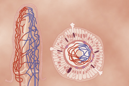 Alt: Longitudinal and cross-sectional view of intestinal villus with capillaries.
- Microvilli (brush border) contain digestive enzymes and increase apical surface; they move continually to mix luminal contents.
Alt: Electron micrograph showing enterocyte brush border microvilli.
Fluid and electrolyte balance
- Daily fluid handled by GI tract: ingested \(\,\approx 1.5\ \text{L}\) + secretions \(\approx 7\ \text{L}\) → total \(\approx 8\text{–}9\ \text{L}\); most absorbed in small intestine, leaving ≈ \(1.5\ \text{L}\) to colon.
- Water moves by osmosis; when chyme is hypo- or hyperosmotic, water shifts rapidly to achieve near-isosmotic conditions.
Sodium, chloride, bicarbonate
- Sodium: absorbed actively via basolateral Na+/K+ ATPase that keeps intracellular [Na+] low; apical entry occurs passively and via co-transporters (SGLT1, amino-acid co-transporters, Na+/H+ exchanger).
- Water follows Na+ by transcellular and paracellular routes (tight junctions and cells).
- Aldosterone increases intestinal Na+ absorption (especially colon) and thus conserves NaCl and water.
- Chloride: follows Na+ passively in much of small intestine; in ileum and colon it is absorbed by Cl-/HCO3- exchangers.
- Bicarbonate: large loads from pancreas/bile are reclaimed indirectly via H+ secretion and CO2 diffusion (carbonic acid → CO2 + H2O), similar to renal tubule handling.
Other ions
- Calcium: active, regulated absorption (duodenum); controlled by vitamin D and parathyroid hormone.
- Iron: actively absorbed and regulated to match body needs.
- K+, Mg2+, PO4^3-: absorbed variably; monovalent ion absorption is easier than divalent.
Large intestine: absorption and fecal formation
- About \(1.5\ \text{L}\) enters colon daily; colon absorbs most water and electrolytes, leaving < \(100\ \text{mL}\) fecal water.
- Proximal colon = absorbing; distal colon = storage.
- Colon has tighter junctions than small intestine → can absorb Na+ against higher gradients (augmented by aldosterone).
- Maximal colonic absorption capacity ≈ \(5\text{–}8\ \text{L/day}\); overwhelm → diarrhea.
- Bacterial flora:
- Ferment some fiber → small caloric contribution and short-chain fatty acids.
- Synthesize vitamin K and some B vitamins.
- Produce gases (CO2, H2, CH4) and odorous compounds (indole, skatole, mercaptans).
- Feces composition (typical): ~ \(75\%\) water, \(25\%\) solids (dead bacteria, undigested fiber, fat \(10\%-20\%\), inorganic matter, sloughed cells). Brown color from stercobilin/urobilin.
Pathophysiologic highlight: secretory diarrhea (e.g., cholera)
- Cholera toxin enters enterocytes and raises intracellular cAMP, opening numerous Cl- channels.
- Massive Cl- secretion into lumen drags Na+ and water, producing up to \(5\text{–}10\ \text{L/day}\) diarrheal fluid loss.
- Treatment principle: aggressive replacement of NaCl and water (oral or IV rehydration).
Key formulas / numbers (MathJax)
- Salivary amylase contribution: \(\approx 5\%\) (mouth), but up to \(30\%-40\%\) before mixing with gastric juice.
- Pancreatic amylase completes digestion: \(\approx 50\%-80\%\) (stated in figure context).
- Pepsin optimum: \(pH\;2.0\text{–}3.0\), inactive if \(pH>5.0\).
- Typical daily intestinal fluid handling: \(1.5\ \text{L}\) (ingested) \(+7\ \text{L}\) (secretions) \(\approx 8\text{–}9\ \text{L}\).
- Fat absorption with bile micelles: \(\approx 97\%\); without micelles: \(\approx 40\%-50\%\).
High-yield takeaways
- Hydrolysis is the common chemical theme: carbs → monosaccharides, proteins → amino acids, fats → monoglycerides + fatty acids.
- Emulsification (bile salts, lecithin) + pancreatic lipase + micelle transport = efficient fat absorption.
- Na+ active transport underpins absorption of glucose, amino acids, and water (SGLT1 is central).
- Brush-border enzymes and transporters (SGLT1, GLUT2, GLUT5, multiple amino-acid carriers) do the final steps at the apical membrane.
- Colon primarily recovers water and electrolytes; bacterial flora contribute vitamins and gases.