Save your deck before it's gone
These flashcards aren't stored yet — they'll disappear when you leave. Create a free account to keep them, and unlock everything below.
- Save this deck to your account
- Study with spaced repetition
- Export to Anki (.apkg) or PDF
- Process documents up to 100 pages
- Images extracted from your PDFs
- Sharper text extraction & a more advanced AI model


What are the main components of the oral cavity?
- Lips
- Palate
- Teeth and associated structures
- Tongue
- Major salivary glands
- Waldeyer's tonsillar ring
How is the oral cavity epithelium generally described?
- Stratified squamous epithelium, keratinized or nonkeratinized depending on location
What forms the mucosa of the oral cavity?
- Epithelium and subjacent connective tissue (epithelial ridges interdigitate with connective tissue papillae)
What types of connective tissue compose the oral mucosa?
- Dense connective tissue in maxilla and mandible
- Loose connective tissue in soft tissues
Name the three oral mucosa subtypes and a key feature of each.
- Masticatory mucosa: keratinized; dorsal tongue, hard palate, gingivae
- Specialized mucosa: nonkeratinized; contains taste buds
- Lining mucosa: nonkeratinized; remainder of oral cavity
Into which regions are the lips divided?
- External (skin)
- Vermilion zone
- Internal (mucosa)
Describe the epithelial covering of the lip regions.
- External and vermilion: stratified squamous keratinized epithelium
- Internal: wet stratified squamous nonkeratinized epithelium
What structures are present in the external and internal regions of the lips?
- External: sebaceous glands, sweat glands, hair follicles
- Internal: minor salivary glands
- Occasional: Fordyce's granules (nonfunctional sebaceous glands) in internal and vermilion
What composes the core of the lips beneath the mucosa?
- Dense irregular collagenous connective tissue enveloping skeletal muscle
What is Waldeyer's tonsillar ring's role in immunity?
- Waldeyer's tonsillar ring: 2nd-3rd line of immune defense
How can a lip histology micrograph be used as an illustration?
- The image shows oral mucosa, vermilion zone, skin, hair follicles, glands, muscle, and minor salivary glands as histologic features
What is the primary function of the palate?
Separates the nasal cavity from the oral cavity (anterior portion).
What embryologic failure leads to cleft lip or cleft palate?
Failure of separation between the nasal and oral cavities during embryology, causing cleft lip or cleft palate.
What epithelium lines the nasal aspect of the palate (except the uvula)?
Pseudostratified ciliated columnar epithelium with glands (respiratory epithelium).
How is the palate grossly divided?
- Hard palate (anterior, bony shelf core)
- Soft palate (posterior, skeletal muscle core)
- Uvula (posterior terminus of soft palate)
What type of epithelium lines the oral aspect of the hard palate?
Stratified squamous parakeratinized to stratified squamous keratinized epithelium.
What connective tissue features are present in the oral aspect of the hard palate?
Contains anterior adipose tissue and posterior minor mucous salivary glands in the mucoperiosteum.
What epithelium and glands are found in the oral aspect of the soft palate?
Stratified squamous nonkeratinized epithelium and minor mucous salivary glands in the connective tissue.
What are the tissues composing a tooth?
- Enamel and cementum (surface layers)
- Dentin (between surface and pulp)
- Pulp (internal soft tissue)
What are the four types of teeth and how do they differ?
- Molars
- Premolars
- Incisors
- Canine They differ only by shape.
Which structure separates the nasal from the oral cavity and may be illustrated below?
The palate separates the nasal and oral cavities.
What is the composition of dental enamel?
- 96% inorganic material (hydroxyapatite)
- 4% organic material (keratin, enamelin, other proteins)
Which cells elaborate enamel and when are they active?
- Ameloblasts elaborate enamel during crown formation and are no longer active after tooth eruption
What are the main features of dentin?
- Dentin surrounds the pulp chamber and root canal
- Manufactured by odontoblasts that persist and produce dentin lifelong
- Calcified matrix contains type I collagen
How is cementum characterized and produced?
- Cementum has a type I collagen-containing calcified matrix
- Produced by cementoblasts that continue making cementum after eruption
What are the key components and properties of dental pulp?
- Gelatinous, richly vascularized connective tissue
- Single peripheral layer of odontoblasts, fibroblasts, mesenchymal cells, thin type I and III collagen
- Contains afferent nerve fibers; sensations are interpreted as pain
Describe the process and timing of predentin mineralization.
- Predentin is secreted by odontoblasts and mineralizes to dentin in approximately 1 day as hydroxyapatite crystals form
What structural extension forms from odontoblasts during dentin formation?
- An apical extension called the odontoblast process forms and becomes surrounded by new matrix, lengthening as dentin-predentin thickens
Which cell types are commonly origins of tumors related to teeth?
- Tumors commonly arise from ameloblasts or odontoblasts
Give the eruption ages for the upper permanent central incisor and upper 1st molar.
- Upper central incisor: 7–8 years
- Upper 1st molar: 6–7 years
What happens to the calcified portion of human teeth during root canal procedures?
- During root canal procedures the calcified portion of the tooth is maintained
What are dentinal tubules?
- Dentinal tubules are canals within the dentin.
How far do dentinal tubules extend within the dentin?
- They run through the full thickness of the dentin.
What is the composition and fiber arrangement of the periodontal ligament (PDL)?
Composed of dense irregular collagenous connective tissue with type I collagen fibers arranged to extend from the cementum to bone.
Name two functions or features of the periodontal ligament.
- Suspends the tooth in its alveolus
- More prominent during inflammation & disease
What epithelium covers the gingivae?
Stratified squamous keratinized or parakeratinized epithelium.
List the five principal gingival fiber bundles.
- Alveologingival
- Dentogingival
- Circular
- Dentoperiosteal
- Transseptal
What is the gingival sulcus and what clinical problem can occur there?
A shallow groove where epithelium is reflected onto enamel surrounding the tooth neck; improper oral care can lead to inflammation & infection entering the sulcus.
What is the junctional epithelium and its role?
A narrow band of epithelial cells at the base of the sulcus that acts as a barrier between the oral cavity and the connective tissue elements of the gingiva.
Compare dorsal and ventral epithelial keratinization of the tongue.
- Dorsal: stratified squamous, parakeratinized to keratinized
- Ventral: stratified squamous, nonkeratinized and highly vascularized
What tissues underlie both epithelial surfaces of the tongue?
A lamina propria and submucosa of dense irregular collagenous connective tissue; core of skeletal muscle forms the bulk of the tongue.
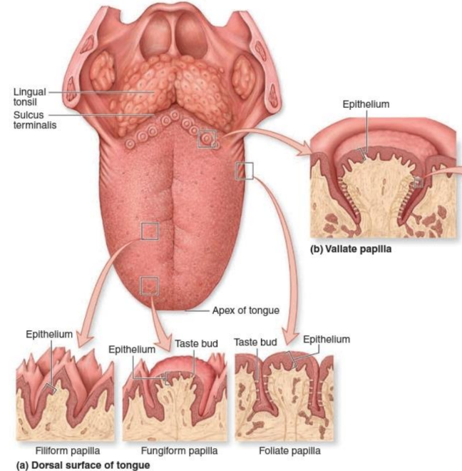
Where are lingual papillae located on the tongue?
On the dorsal surface of the anterior two-thirds of the tongue.
What are glands of von Ebner and their function?
Minor serous salivary glands that deliver serous secretion into papillary furrows, assisting taste buds in perceiving stimuli and into foliate papillae furrows.
How do serous and mucinous glands appear in H&E staining?
Serous glands appear basic/blueish & granular/grainy; mucinous glands appear lighter, resembling vesicles.
Describe filiform papillae.
Short, narrow, highly keratinized structures lacking taste buds; purpose is to make the tongue rough; located on the anterior portion.
Describe fungiform papillae.
Mushroom-shaped structures interspersed among filiform papillae with occasional taste buds on their superior aspect; located anteriorly.
Describe foliate papillae and the fate of their taste buds.
Shallow longitudinal furrows on the lateral posterior region of the anterior two-thirds; taste buds here usually degenerate shortly after the second year of life.
Give key features of circumvallate papillae.
10 to 15 large circular papillae each surrounded by a moat-like furrow; lie just anterior to the sulcus terminalis and possess taste buds.
What anatomical landmark distinguishes anterior from posterior tongue?
The circumvallate papillae: the portion in front is the anterior tongue; behind them is the posterior tongue.
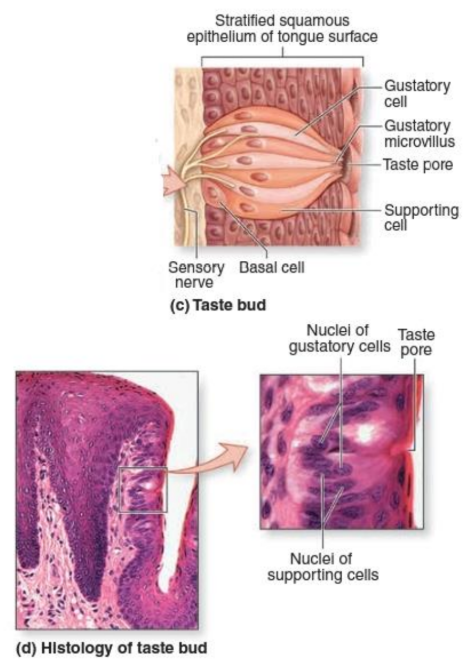
How many taste buds are typically found on the tongue?
- 2,000 to 8,000 taste buds
What is the cellular composition of a taste bud?
- 60 to 80 spindle-shaped cells forming a barrel-shaped intraepithelial structure
Where on the tongue are taste buds located?
- Superior surface of fungiform papillae
- Lateral surfaces of circumvallate (vallate) papillae
- Walls of moat-like furrows around circumvallate papillae
What is the taste pore and what projects from it?
- Taste pore: small opening of each taste bud
- Microvilli (taste hairs) project from the pore into the oral cavity or surrounding moat
Name the four cell types recognized in taste buds.
- Dark cells (Type I)
- Light cells (Type II)
- Intermediate cells (Type III)
- Short regenerative basal cells
Which taste bud cell types form synapses with afferent nerve fibers?
- Type I, Type II, and Type III cells form synapses with afferent nerve fibers
What is notable about the lifespan of intermediate (Type III) taste bud cells?
- Intermediate (Type III) cells have short life spans
When basal cells divide in a taste bud, what daughter cells do they produce?
- A basal cell and a dark cell
What are the three morphological cell types in a taste bud?
- Dark cell
- Light cell
- Intermediate cell
How long does the maturation-to-degeneration cycle of a taste bud cell take?
About 10 days from newly formed dark cell to dying intermediate cell
What type of cells are the taste receptor cells described in the notes?
They are neuroepithelial cells whose microvilli (taste hairs) recognize specific tastants
How are most taste bud receptors kept functional for chemical detection?
Receptors for taste and smell are moist so chemicals dissolve in saliva or mucus to be perceived
Which tastants stimulate sodium and hydrogen ion channels?
- Salt stimulates sodium channels
- Sour stimulates hydrogen ion channels
Which tastants stimulate G protein-linked receptors?
- Sweet
- Bitter
- Umami
How do fatty tastants stimulate taste cells?
Fatty tastants stimulate fatty acid transporters
How is the muscle core of the tongue organized?
Bundles of skeletal muscle fibers arranged in three planes with minor salivary glands interspersed
Where are lingual tonsils located on the tongue?
On the dorsal surface of the posterior one-third of the tongue
What immune roles do lingual tonsils perform?
- Part of the 2nd line of defense via macrophages (APCs)
- Part of the 3rd line of defense when developing antibodies
How are salivary glands classified anatomically?
They are compound tubulo-acinar glands
Name the three paired major salivary glands.
- Parotid
- Submandibular
- Sublingual
What proportion of daily saliva is produced by the major salivary glands?
The three major paired glands produce about 90% of saliva
What is the typical daily saliva volume produced by major and minor glands together?
About 0.75–1.50 L of saliva daily
What products do salivary glands synthesize and secrete?
Products include secretory component, proteins, lysozyme, lactoferrin, thiocyanate, and salivary amylase
How are salivary glands classified by acinar type?
Classified as serous, mucous, or mixed depending on the secretory acini present
What are serous demilunes in salivary gland acini?
Crescent-shaped collections of serous cells capping mucous acini
What cells assist acinar secretion by sharing the basal lamina?
Myoepithelial cells that share the basal lamina of acinar cells
Describe the secretion process of acinar cells in salivary glands.
Acinar cells release a primary secretion resembling extracellular fluid that is modified in ducts to form the final saliva
What type of connective tissue capsule surrounds salivary glands?
A capsule of dense irregular collagenous connective tissue with septa subdividing into lobes and lobules
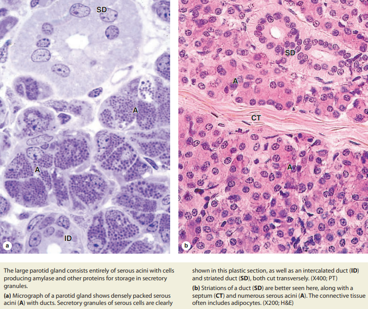
What is a defining feature of parotid gland acini?
Parotid glands consist of serous acini
What distinctive feature is noted for sublingual glands?
Sublingual glands consist mostly of mucous tubules capped with serous demilunes (mixed)
How are neurovascular elements conveyed to salivary gland acinar cells?
Neurovascular elements are conveyed to acinar cells within the connective tissue septa
What autonomic fibers control saliva release from salivary gland acini and ducts?
Cholinergic parasympathetic fibers (acetylcholine-releasing) control saliva release from the acini and ducts.
What effect do cholinergic parasympathetic fibers have on acinar cells?
They induce the acinar cells to release their primary saliva.
What are the two main secretory cell types in the submandibular gland lobule?
- Serous cells (protein-secreting, rounded nuclei)
- Mucous cells (flattened basal nuclei with condensed chromatin)
Describe key ultrastructural features of serous acinar cells.
Serous acini have rounded nuclei, basal accumulation of RER, and apical ends filled with secretory granules.
How are mucous tubule cell nuclei described?
Mucous tubule cells have flattened, basal nuclei with condensed chromatin.
What is a 'serous demilune' in mixed salivary secretory units?
A serous demilune is a distal cluster of serous cells capping short mucous tubules in mixed tubuloacinar units.
What epithelium lines the short intercalated ducts in salivary glands?
Short intercalated ducts are lined with low cuboidal epithelium.
Which duct cells show adaptations for ion transport, and what are those features?
Striated duct cells are columnar with basal membrane invaginations and mitochondrial accumulations for ion transport.
Where are myoepithelial cells located in relation to serous acini?
Myoepithelial cells are situated around the serous acini.
What type of acini does the parotid gland consist of and what do its cells produce?
- Entirely serous acini
- Produce amylase and other proteins stored in secretory granules
What is the composition of the submandibular gland?
- Mixed serous and mucous acini (serous cells predominate)
- Tubuloacinar arrangement
How is the sublingual gland described in terms of acini type?
- Mixed but largely mucous gland
- Tubuloacinar arrangement with poorly stained mucous cells
What causes the crescent-shaped serous demilunes seen in mixed glands?
- They arise at least in part artifactually due to disproportionate swelling of adjacent mucous cells during slide preparation
What is the typical epithelium of salivary ducts in the secretory portion and how does it change in the meatus?
- Mostly simple cuboidal in the secretory portion
- Transitions to squamous in the meatus
Where do intercalated ducts originate and what is one function they may have?
- Originate in the acini and join to form striated ducts
- May deliver bicarbonate ions into the primary secretion
What ion-transport roles do striated (intralobular) ducts perform in saliva modification?
- Remove Na+ and Cl- from luminal fluid
- Actively pump K+ and HCO3- into the fluid, transforming primary to secondary saliva
How are interlobular (excretory) ducts formed and where do they drain?
- Formed by convergence of striated ducts and travel in connective tissue septa
- Drain into the main duct of each gland, which empties into the oral cavity
What type of epithelium covers the gingiva?
- Stratified squamous epithelium over connective tissue of the lamina propria (LP).
With which structures is the gingival connective tissue continuous?
- Periosteum covering alveolar bone (P/B)
- Periodontal ligament (PL)
What features characterize the periodontal ligament (PDL)?
- Many blood vessels (V)
- Insertions into alveolar bone (B)
- Serves as periosteum of the alveolar socket
What material covers dentin at the tooth root and which cells secrete it?
- Cementum covers dentin and is secreted by cementoblasts.
How is cementum described microscopically?
- A thin layer of bone-like material secreted by large, elongated cementoblasts.
What continuity is shown between alveolar bone and periodontal ligament collagen?
- Collagen fiber continuity between alveolar bone bundles and periodontal ligament bundles.
Describe the main characteristics of ameloblasts during enamel formation.
- Tall polarized cells whose apical ends contact dentin; form a cell layer with basal connective tissue.
What matrix do ameloblasts secrete and what protein is emphasized?
- A matrix lacking collagens but rich in proteins such as amelogenin that initiate calcium hydroxyapatite formation.
State two key facts about enamel based on histology.
- Enamel is the hardest material in the body.
- Enamel consists of enamel rods (prisms); each rod is the product of one ameloblast.
What happens to the layer of ameloblasts and enamel during tooth eruption or decalcification?
- Ameloblast layer is completely lost during eruption.
- Teeth decalcified for histology typically lose their enamel layer completely.
What microscopic structures are visible in dentin and enamel in ground tooth preparations?
- Dentin shows fine, long tubules.
- Enamel may show faint rods and prominent incremental growth lines from cyclic secretion.
Flashcards in this deck (112)
-
-
-
-
-
What are the main components of the oral cavity?
- Lips
- Palate
- Teeth and associated structures
- Tongue
- Major salivary glands
- Waldeyer's tonsillar ring
oral anatomy -
How is the oral cavity epithelium generally described?
- Stratified squamous epithelium, keratinized or nonkeratinized depending on location
mucosa epithelium -
What forms the mucosa of the oral cavity?
- Epithelium and subjacent connective tissue (epithelial ridges interdigitate with connective tissue papillae)
mucosa histology -
What types of connective tissue compose the oral mucosa?
- Dense connective tissue in maxilla and mandible
- Loose connective tissue in soft tissues
connective mucosa -
Name the three oral mucosa subtypes and a key feature of each.
- Masticatory mucosa: keratinized; dorsal tongue, hard palate, gingivae
- Specialized mucosa: nonkeratinized; contains taste buds
- Lining mucosa: nonkeratinized; remainder of oral cavity
mucosa classification -
Into which regions are the lips divided?
- External (skin)
- Vermilion zone
- Internal (mucosa)
lips anatomy -
Describe the epithelial covering of the lip regions.
- External and vermilion: stratified squamous keratinized epithelium
- Internal: wet stratified squamous nonkeratinized epithelium
lips epithelium -
What structures are present in the external and internal regions of the lips?
- External: sebaceous glands, sweat glands, hair follicles
- Internal: minor salivary glands
- Occasional: Fordyce's granules (nonfunctional sebaceous glands) in internal and vermilion
lips glands -
What composes the core of the lips beneath the mucosa?
- Dense irregular collagenous connective tissue enveloping skeletal muscle
lips connective -
What is Waldeyer's tonsillar ring's role in immunity?
- Waldeyer's tonsillar ring: 2nd-3rd line of immune defense
tonsils immunity -
How can a lip histology micrograph be used as an illustration?
- The image shows oral mucosa, vermilion zone, skin, hair follicles, glands, muscle, and minor salivary glands as histologic features
image lips -
What is the primary function of the palate?
Separates the nasal cavity from the oral cavity (anterior portion).
palate anatomy -
What embryologic failure leads to cleft lip or cleft palate?
Failure of separation between the nasal and oral cavities during embryology, causing cleft lip or cleft palate.
embryology clinical -
What epithelium lines the nasal aspect of the palate (except the uvula)?
Pseudostratified ciliated columnar epithelium with glands (respiratory epithelium).
epithelium palate -
How is the palate grossly divided?
- Hard palate (anterior, bony shelf core)
- Soft palate (posterior, skeletal muscle core)
- Uvula (posterior terminus of soft palate)
palate anatomy -
What type of epithelium lines the oral aspect of the hard palate?
Stratified squamous parakeratinized to stratified squamous keratinized epithelium.
hardpalate epithelium -
What connective tissue features are present in the oral aspect of the hard palate?
Contains anterior adipose tissue and posterior minor mucous salivary glands in the mucoperiosteum.
hardpalate connective -
What epithelium and glands are found in the oral aspect of the soft palate?
Stratified squamous nonkeratinized epithelium and minor mucous salivary glands in the connective tissue.
softpalate epithelium -
What are the tissues composing a tooth?
- Enamel and cementum (surface layers)
- Dentin (between surface and pulp)
- Pulp (internal soft tissue)
teeth histology -
What are the four types of teeth and how do they differ?
- Molars
- Premolars
- Incisors
- Canine They differ only by shape.
teeth dentition -
Which structure separates the nasal from the oral cavity and may be illustrated below?
The palate separates the nasal and oral cavities.
palate illustration -
What is the composition of dental enamel?
- 96% inorganic material (hydroxyapatite)
- 4% organic material (keratin, enamelin, other proteins)
enamel teeth histology -
Which cells elaborate enamel and when are they active?
- Ameloblasts elaborate enamel during crown formation and are no longer active after tooth eruption
ameloblasts enamel development -
What are the main features of dentin?
- Dentin surrounds the pulp chamber and root canal
- Manufactured by odontoblasts that persist and produce dentin lifelong
- Calcified matrix contains type I collagen
dentin odontoblasts teeth -
How is cementum characterized and produced?
- Cementum has a type I collagen-containing calcified matrix
- Produced by cementoblasts that continue making cementum after eruption
cementum cementoblasts periodontium -
What are the key components and properties of dental pulp?
- Gelatinous, richly vascularized connective tissue
- Single peripheral layer of odontoblasts, fibroblasts, mesenchymal cells, thin type I and III collagen
- Contains afferent nerve fibers; sensations are interpreted as pain
pulp teeth histology -
Describe the process and timing of predentin mineralization.
- Predentin is secreted by odontoblasts and mineralizes to dentin in approximately 1 day as hydroxyapatite crystals form
predentin odontoblasts dentin -
What structural extension forms from odontoblasts during dentin formation?
- An apical extension called the odontoblast process forms and becomes surrounded by new matrix, lengthening as dentin-predentin thickens
odontoblasts odontoblast_process dentin -
Which cell types are commonly origins of tumors related to teeth?
- Tumors commonly arise from ameloblasts or odontoblasts
pathology teeth tumors -
Give the eruption ages for the upper permanent central incisor and upper 1st molar.
- Upper central incisor: 7–8 years
- Upper 1st molar: 6–7 years
eruption teeth dentition -
What happens to the calcified portion of human teeth during root canal procedures?
- During root canal procedures the calcified portion of the tooth is maintained
rootcanal clinical teeth -
What are dentinal tubules?
- Dentinal tubules are canals within the dentin.
teeth dentin -
How far do dentinal tubules extend within the dentin?
- They run through the full thickness of the dentin.
dentin structure -
What is the composition and fiber arrangement of the periodontal ligament (PDL)?
Composed of dense irregular collagenous connective tissue with type I collagen fibers arranged to extend from the cementum to bone.
periodontium pdl -
Name two functions or features of the periodontal ligament.
- Suspends the tooth in its alveolus
- More prominent during inflammation & disease
periodontium pdl function -
What epithelium covers the gingivae?
Stratified squamous keratinized or parakeratinized epithelium.
gingiva epithelium -
List the five principal gingival fiber bundles.
- Alveologingival
- Dentogingival
- Circular
- Dentoperiosteal
- Transseptal
gingiva fibers -
What is the gingival sulcus and what clinical problem can occur there?
A shallow groove where epithelium is reflected onto enamel surrounding the tooth neck; improper oral care can lead to inflammation & infection entering the sulcus.
gingiva sulcus clinical -
What is the junctional epithelium and its role?
A narrow band of epithelial cells at the base of the sulcus that acts as a barrier between the oral cavity and the connective tissue elements of the gingiva.
gingiva junctional -
Compare dorsal and ventral epithelial keratinization of the tongue.
- Dorsal: stratified squamous, parakeratinized to keratinized
- Ventral: stratified squamous, nonkeratinized and highly vascularized
tongue epithelium -
What tissues underlie both epithelial surfaces of the tongue?
A lamina propria and submucosa of dense irregular collagenous connective tissue; core of skeletal muscle forms the bulk of the tongue.
tongue histology -
Where are lingual papillae located on the tongue?
On the dorsal surface of the anterior two-thirds of the tongue.
tongue papillae -
What are glands of von Ebner and their function?
Minor serous salivary glands that deliver serous secretion into papillary furrows, assisting taste buds in perceiving stimuli and into foliate papillae furrows.
tongue salivary taste -
How do serous and mucinous glands appear in H&E staining?
Serous glands appear basic/blueish & granular/grainy; mucinous glands appear lighter, resembling vesicles.
histology staining -
Describe filiform papillae.
Short, narrow, highly keratinized structures lacking taste buds; purpose is to make the tongue rough; located on the anterior portion.
papillae filiform -
Describe fungiform papillae.
Mushroom-shaped structures interspersed among filiform papillae with occasional taste buds on their superior aspect; located anteriorly.
papillae fungiform -
Describe foliate papillae and the fate of their taste buds.
Shallow longitudinal furrows on the lateral posterior region of the anterior two-thirds; taste buds here usually degenerate shortly after the second year of life.
papillae foliate -
Give key features of circumvallate papillae.
10 to 15 large circular papillae each surrounded by a moat-like furrow; lie just anterior to the sulcus terminalis and possess taste buds.
papillae circumvallate -
What anatomical landmark distinguishes anterior from posterior tongue?
The circumvallate papillae: the portion in front is the anterior tongue; behind them is the posterior tongue.
tongue landmark -
How many taste buds are typically found on the tongue?
- 2,000 to 8,000 taste buds
taste histology tongue -
What is the cellular composition of a taste bud?
- 60 to 80 spindle-shaped cells forming a barrel-shaped intraepithelial structure
taste cells histology -
Where on the tongue are taste buds located?
- Superior surface of fungiform papillae
- Lateral surfaces of circumvallate (vallate) papillae
- Walls of moat-like furrows around circumvallate papillae
taste papillae anatomy -
What is the taste pore and what projects from it?
- Taste pore: small opening of each taste bud
- Microvilli (taste hairs) project from the pore into the oral cavity or surrounding moat
taste microvilli structure -
Name the four cell types recognized in taste buds.
- Dark cells (Type I)
- Light cells (Type II)
- Intermediate cells (Type III)
- Short regenerative basal cells
taste celltypes histology -
Which taste bud cell types form synapses with afferent nerve fibers?
- Type I, Type II, and Type III cells form synapses with afferent nerve fibers
taste synapse neuroscience -
What is notable about the lifespan of intermediate (Type III) taste bud cells?
- Intermediate (Type III) cells have short life spans
taste celltypes lifespan -
When basal cells divide in a taste bud, what daughter cells do they produce?
- A basal cell and a dark cell
taste regeneration basal -
What are the three morphological cell types in a taste bud?
- Dark cell
- Light cell
- Intermediate cell
taste histology -
How long does the maturation-to-degeneration cycle of a taste bud cell take?
About 10 days from newly formed dark cell to dying intermediate cell
taste cellbiology -
What type of cells are the taste receptor cells described in the notes?
They are neuroepithelial cells whose microvilli (taste hairs) recognize specific tastants
taste celltypes -
How are most taste bud receptors kept functional for chemical detection?
Receptors for taste and smell are moist so chemicals dissolve in saliva or mucus to be perceived
taste physiology -
Which tastants stimulate sodium and hydrogen ion channels?
- Salt stimulates sodium channels
- Sour stimulates hydrogen ion channels
taste mechanism -
Which tastants stimulate G protein-linked receptors?
- Sweet
- Bitter
- Umami
taste receptors -
How do fatty tastants stimulate taste cells?
Fatty tastants stimulate fatty acid transporters
taste lipids -
How is the muscle core of the tongue organized?
Bundles of skeletal muscle fibers arranged in three planes with minor salivary glands interspersed
tongue anatomy -
Where are lingual tonsils located on the tongue?
On the dorsal surface of the posterior one-third of the tongue
tonsils anatomy -
What immune roles do lingual tonsils perform?
- Part of the 2nd line of defense via macrophages (APCs)
- Part of the 3rd line of defense when developing antibodies
tonsils immunology -
How are salivary glands classified anatomically?
They are compound tubulo-acinar glands
saliva histology -
Name the three paired major salivary glands.
- Parotid
- Submandibular
- Sublingual
saliva glands -
What proportion of daily saliva is produced by the major salivary glands?
The three major paired glands produce about 90% of saliva
saliva physiology -
What is the typical daily saliva volume produced by major and minor glands together?
About 0.75–1.50 L of saliva daily
saliva physiology -
What products do salivary glands synthesize and secrete?
Products include secretory component, proteins, lysozyme, lactoferrin, thiocyanate, and salivary amylase
saliva secretions -
How are salivary glands classified by acinar type?
Classified as serous, mucous, or mixed depending on the secretory acini present
saliva classification -
What are serous demilunes in salivary gland acini?
Crescent-shaped collections of serous cells capping mucous acini
acini histology -
What cells assist acinar secretion by sharing the basal lamina?
Myoepithelial cells that share the basal lamina of acinar cells
acini cells -
Describe the secretion process of acinar cells in salivary glands.
Acinar cells release a primary secretion resembling extracellular fluid that is modified in ducts to form the final saliva
acini physiology -
What type of connective tissue capsule surrounds salivary glands?
A capsule of dense irregular collagenous connective tissue with septa subdividing into lobes and lobules
saliva structure -
What is a defining feature of parotid gland acini?
Parotid glands consist of serous acini
parotid acini -
What distinctive feature is noted for sublingual glands?
Sublingual glands consist mostly of mucous tubules capped with serous demilunes (mixed)
sublingual acini -
How are neurovascular elements conveyed to salivary gland acinar cells?
Neurovascular elements are conveyed to acinar cells within the connective tissue septa
saliva neurovascular -
What autonomic fibers control saliva release from salivary gland acini and ducts?
Cholinergic parasympathetic fibers (acetylcholine-releasing) control saliva release from the acini and ducts.
salivary innervation -
What effect do cholinergic parasympathetic fibers have on acinar cells?
They induce the acinar cells to release their primary saliva.
salivary physiology -
What are the two main secretory cell types in the submandibular gland lobule?
- Serous cells (protein-secreting, rounded nuclei)
- Mucous cells (flattened basal nuclei with condensed chromatin)
salivary histology -
Describe key ultrastructural features of serous acinar cells.
Serous acini have rounded nuclei, basal accumulation of RER, and apical ends filled with secretory granules.
serous acini -
How are mucous tubule cell nuclei described?
Mucous tubule cells have flattened, basal nuclei with condensed chromatin.
mucous cells -
What is a 'serous demilune' in mixed salivary secretory units?
A serous demilune is a distal cluster of serous cells capping short mucous tubules in mixed tubuloacinar units.
serous demilune -
What epithelium lines the short intercalated ducts in salivary glands?
Short intercalated ducts are lined with low cuboidal epithelium.
ducts intercalated -
Which duct cells show adaptations for ion transport, and what are those features?
Striated duct cells are columnar with basal membrane invaginations and mitochondrial accumulations for ion transport.
striated ducts -
Where are myoepithelial cells located in relation to serous acini?
Myoepithelial cells are situated around the serous acini.
myoepithelial salivary -
What type of acini does the parotid gland consist of and what do its cells produce?
- Entirely serous acini
- Produce amylase and other proteins stored in secretory granules
salivary parotid histology -
What is the composition of the submandibular gland?
- Mixed serous and mucous acini (serous cells predominate)
- Tubuloacinar arrangement
salivary submandibular histology -
How is the sublingual gland described in terms of acini type?
- Mixed but largely mucous gland
- Tubuloacinar arrangement with poorly stained mucous cells
salivary sublingual histology -
What causes the crescent-shaped serous demilunes seen in mixed glands?
- They arise at least in part artifactually due to disproportionate swelling of adjacent mucous cells during slide preparation
histology artifact salivary -
What is the typical epithelium of salivary ducts in the secretory portion and how does it change in the meatus?
- Mostly simple cuboidal in the secretory portion
- Transitions to squamous in the meatus
ducts epithelium histology -
Where do intercalated ducts originate and what is one function they may have?
- Originate in the acini and join to form striated ducts
- May deliver bicarbonate ions into the primary secretion
ducts intercalated salivary -
What ion-transport roles do striated (intralobular) ducts perform in saliva modification?
- Remove Na+ and Cl- from luminal fluid
- Actively pump K+ and HCO3- into the fluid, transforming primary to secondary saliva
ducts striated physiology -
How are interlobular (excretory) ducts formed and where do they drain?
- Formed by convergence of striated ducts and travel in connective tissue septa
- Drain into the main duct of each gland, which empties into the oral cavity
ducts interlobular salivary -
What type of epithelium covers the gingiva?
- Stratified squamous epithelium over connective tissue of the lamina propria (LP).
oral gingiva -
With which structures is the gingival connective tissue continuous?
- Periosteum covering alveolar bone (P/B)
- Periodontal ligament (PL)
gingiva connective -
What features characterize the periodontal ligament (PDL)?
- Many blood vessels (V)
- Insertions into alveolar bone (B)
- Serves as periosteum of the alveolar socket
periodontium pdl -
What material covers dentin at the tooth root and which cells secrete it?
- Cementum covers dentin and is secreted by cementoblasts.
tooth cementum -
How is cementum described microscopically?
- A thin layer of bone-like material secreted by large, elongated cementoblasts.
cementum histology -
What continuity is shown between alveolar bone and periodontal ligament collagen?
- Collagen fiber continuity between alveolar bone bundles and periodontal ligament bundles.
collagen periodontium -
Describe the main characteristics of ameloblasts during enamel formation.
- Tall polarized cells whose apical ends contact dentin; form a cell layer with basal connective tissue.
ameloblasts enamel -
What matrix do ameloblasts secrete and what protein is emphasized?
- A matrix lacking collagens but rich in proteins such as amelogenin that initiate calcium hydroxyapatite formation.
ameloblasts amelogenin -
State two key facts about enamel based on histology.
- Enamel is the hardest material in the body.
- Enamel consists of enamel rods (prisms); each rod is the product of one ameloblast.
enamel tooth -
What happens to the layer of ameloblasts and enamel during tooth eruption or decalcification?
- Ameloblast layer is completely lost during eruption.
- Teeth decalcified for histology typically lose their enamel layer completely.
ameloblasts enamel clinical -
What microscopic structures are visible in dentin and enamel in ground tooth preparations?
- Dentin shows fine, long tubules.
- Enamel may show faint rods and prominent incremental growth lines from cyclic secretion.
dentin enamel histology
Digestive System — Oral Cavity (Histology)
Concise notes for the oral cavity, teeth, tongue, tonsils, periodontium, and salivary glands.
Overview
- The oral cavity includes lips, palate, teeth, tongue, major salivary glands, and Waldeyer's ring (part of mucosal immunity).
- Most surfaces are lined by stratified squamous epithelium (keratinized or nonkeratinized depending on function).
- Mucosa = epithelium + lamina propria (connective tissue); submucosa present in some regions.
Mucosae (functional types)
- Masticatory mucosa: keratinized (hard palate, gingiva, dorsal tongue regions under mechanical stress).
- Specialized mucosa: contains taste buds (dorsum of tongue, soft palate); often nonkeratinized.
- Lining mucosa: nonkeratinized regions (most of oral cavity).
Lips
- Three zones: external skin (hair, sebaceous/sweat glands), vermilion (thin, lightly keratinized epidermis), internal mucosa (wet nonkeratinized epithelium).
- Core contains skeletal muscle and dense irregular connective tissue.
- Minor salivary glands in internal mucosa; Fordyce spots = ectopic sebaceous glands in vermilion/internal mucosa.
Palate
- Separates nasal and oral cavities; failure of fusion causes cleft lip/palate.
- Hard palate: oral surface = parakeratinized/keratinized stratified squamous epithelium; mucoperiosteum with minor glands posteriorly.
- Soft palate: oral surface = nonkeratinized stratified squamous epithelium; muscle core and mucous glands.
- Nasal (superior) side mostly respiratory epithelium (pseudostratified ciliated columnar) except uvula.
Teeth — overall structure and development
- Tooth tissues: enamel (surface crown), dentin (bulk, collagen-containing), cementum (covers root), and dental pulp (vascular, innervated connective tissue).
- Humans have 2 sets; permanent tooth eruption ages vary by tooth (molars, premolars, canines, incisors).
Enamel
- Formed by ameloblasts during crown development; after eruption ameloblasts are lost so enamel cannot regenerate.
- ~96% inorganic (hydroxyapatite), ~4% organic; hardest body material.
Dentin
- Secreted by odontoblasts (derived from dental papilla); odontoblasts persist and add secondary dentin lifelong.
- Contains dentinal tubules with odontoblastic processes; matrix rich in type I collagen.
Cementum
- Bone-like tissue on the root surface produced by cementoblasts; anchors periodontal ligament fibers.
Dental pulp
- Loose, vascularized connective tissue with peripheral odontoblasts, fibroblasts, mesenchymal cells and sensory nerve fibers; infections cause pain.
 Alt text: Dentin and odontoblasts micrograph.
Periodontium
- Components: periodontal ligament (PDL), cementum, alveolar bone, and gingiva.
- PDL: dense irregular collagenous tissue (type I collagen) connecting cementum to alveolar bone; acts as shock absorber and periosteum for alveolus.
- Gingiva: keratinized/parakeratinized stratified squamous epithelium; collagen fiber groups (alveologingival, dentogingival, circular, dentoperiosteal, transseptal).
- Junctional epithelium at sulcus base forms a barrier; poor oral care allows bacterial invasion leading to periodontitis.
 Alt text: Gingiva and periodontal ligament micrograph.
Tongue
- Surface: dorsum = parakeratinized-to-keratinized stratified squamous epithelium with papillae; ventral = nonkeratinized, highly vascular (sublingual absorption).
- Core: skeletal muscle in three planes + minor salivary glands.
Lingual papillae (anterior two-thirds)
- Filiform: most numerous, highly keratinized, no taste buds, roughen surface.
- Fungiform: mushroom-shaped, contain occasional taste buds on top.
- Foliate: lateral folds with taste buds (often regress in childhood).
- Circumvallate (vallate): 10–15 large papillae in a row anterior to sulcus terminalis; each surrounded by a moat and rich in taste buds.
- Von Ebner glands (serous) empty into circumvallate moats to flush tastants.
 Alt text: Submandibular gland lobule micrograph.
Taste buds
- Each taste bud = 60–80 spindle-shaped neuroepithelial cells forming a barrel; has a taste pore with microvilli (taste hairs).
- Cell types: Type I (support/"dark"), Type II (light), Type III (intermediate) — all form synapses with afferent nerves; plus basal regenerative cells.
- Turnover ~10 days (basal → dark → light → intermediate → die).
- Tastant detection: salt/sour via ion channels; sweet/bitter/umami via G-protein coupled receptors; fatty via fatty acid transporters.
- Taste receptors require dissolution of chemicals in saliva/mucus to be detected.
Tonsils (lingual)
- Located on dorsal posterior one-third of tongue; part of Waldeyer's ring.
- Provide antigen sampling (macrophages/APCs) and adaptive responses (antibody production).
Salivary glands
- Compound tubuloacinar exocrine glands: three major paired glands — parotid (serous), submandibular (mixed, serous > mucous), sublingual (mostly mucous).
- Minor salivary glands throughout mucosa produce lubrication.
Acini and cells
- Serous acini: pyramidal protein-secreting cells (amylase, lysozyme).
- Mucous acini: mucin-secreting with pale-staining cytoplasm; may have serous demilunes in mixed glands.
- Myoepithelial cells: contractile cells between acinar/ductal cells and basal lamina aiding secretion.
Duct system and ion transport
- Intercalated ducts: short, cuboidal; may add bicarbonate.
- Striated (intralobular) ducts: columnar cells with basal striations (mitochondria and membrane infoldings) that reabsorb Na+/Cl- and secrete K+/HCO3-, converting primary to final saliva.
- Interlobular (excretory) ducts: larger ducts in septa that drain to oral cavity.
- Parasympathetic (cholinergic) stimulation increases watery saliva secretion; sympathetic input modifies protein content.
Clinical correlations / key facts to remember
- Enamel cannot regenerate after ameloblast loss; dental caries affect enamel and can progress to pulp infections.
- Odontoblasts persist and produce secondary dentin throughout life.
- Junctional epithelium integrity is critical to prevent periodontal disease.
- Sublingual mucosa is used for rapid systemic drug absorption (sublingual route avoids first-pass metabolism).
Quick checkpoint (answers)
- Highest taste bud concentration: e. Vallate (circumvallate) papillae.
- Oral cavity epithelium type: e. Stratified squamous epithelium.
- T/F: Mucosa is connective tissue only — False (mucosa = epithelium + lamina propria).
- Mucosa involved in mechanical stress: Masticatory mucosa.
- T/F: Mucosa of soft palate is same as gingivae — False.
- Fordyce granules: d. Found in the internal region of the lips (ectopic, nonfunctional sebaceous glands).
- Cells that make up dental pulp: Fibroblasts, mesenchymal cells, and odontoblasts (peripheral layer).
- Structure that suspends tooth in alveolus: Periodontal ligament.
- Highly vascularized surface of tongue: Ventral surface.
- Papillae lacking taste buds: Filiform papillae.
References / further reading
- Junqueira's Basic Histology (Mescher) — chapters on oral cavity and salivary glands.
- Lecture slides, Dr. Jon Alex Mosuela.