Guarda tu mazo antes de que desaparezca
Estas tarjetas aún no se han guardado — desaparecerán cuando salgas. Crea una cuenta gratuita para conservarlas y desbloquear todo lo de abajo.
- Save this deck to your account
- Study with spaced repetition
- Export to Anki (.apkg) or PDF
- Process documents up to 100 pages
- Images extracted from your PDFs
- Sharper text extraction & a more advanced AI model
What are the main components of the descending motor pathways overview?
Which structures are included in the pyramidal system?
Name the two corticospinal tracts mentioned.
What core concept pertains to extrapyramidal pathways?
What is the role of the corticoponto‑cerebellar pathway in the core concepts?
Which clinical localization topics are included among the core concepts?
What are the three descending motor systems?
What does the pyramidal system directly link?
Which two tracts compose the pyramidal system and their targets?
What is the corticoponto-cerebellar pathway?
Name the two pyramidal (corticospinal) spinal tracts and their crossing status.
How is the extrapyramidal system linked to the spinal cord?
Which extrapyramidal tracts originate from the midbrain and what is their crossing status?
Which extrapyramidal tracts originate from the vestibular nuclei?
Which extrapyramidal tracts originate from the reticular formation?
What other extrapyramidal descending fibers are listed besides midbrain, vestibular, and reticular tracts?
Show an illustrative image of the motor system pathways (supplementary).
What are the two main divisions of the pyramidal motor system?
What is the primary function of the corticospinal tract?
What body regions does the corticobulbar tract control?
Name the two subdivisions of the corticospinal tract listed.
What is the main listed function of the rubrospinal tract?
According to the notes, what are the two opposing functions of the reticulospinal tract subdivisions?
What is the primary function of the vestibulospinal tract?
What function is attributed to the tectospinal (colliculospinal) tract?
Which major motor systems are compared in the diagram provided with the lecture?
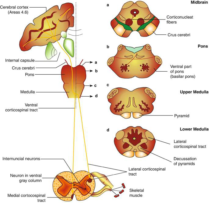
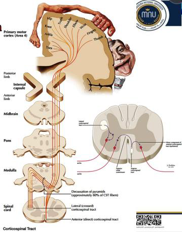
Where does the corticospinal tract travel from and to, as shown in the pathway diagram?
What are the cortical origins and their percentage contributions to the corticospinal tract?
What proportion of corticospinal fibers are large and arise from Betz (giant pyramidal) cells?
Name the main structures/levels the corticospinal fibers descend through from cortex to medulla.
What happens to 85% of corticospinal fibers in the lower medulla?
What is the fate of the 15% of corticospinal fibers that remain uncrossed?
Briefly describe the corticospinal tract pathway from motor cortex to spinal cord.
Flashcards in this deck (34)
-
What are the main components of the descending motor pathways overview?
- Pyramidal (corticospinal/corticobulbar) system
- Extrapyramidal tracts
- Corticoponto‑cerebellar pathway
- Clinical localization (decussation, UMN vs LMN signs)
motor overview -
Which structures are included in the pyramidal system?
- Corticospinal tract
- Corticobulbar tract
pyramidal tracts -
Name the two corticospinal tracts mentioned.
- Lateral corticospinal tract
- Ventral (anterior) corticospinal tract
corticospinal tracts -
What core concept pertains to extrapyramidal pathways?
- Extrapyramidal tracts and their functions
extrapyramidal functions -
What is the role of the corticoponto‑cerebellar pathway in the core concepts?
- Corticoponto‑cerebellar pathway for coordination
Answer illustration:
 corticopontocerebellar coordination
 -
Which clinical localization topics are included among the core concepts?
- Decussation and lesion localization
- Upper motor neuron (UMN) vs lower motor neuron (LMN) clinical signs
clinical localization -
What are the three descending motor systems?
- Pyramidal system
- Extrapyramidal system
- Corticoponto-cerebellar pathway
motor pathways -
What does the pyramidal system directly link?
- Cerebral cortex to spinal cord and brainstem (direct link)
pyramidal anatomy -
Which two tracts compose the pyramidal system and their targets?
- Corticospinal tract: to anterior horn cells (AHCs) of spinal cord
- Corticobulbar tract: to motor nuclei in the brainstem
pyramidal tracts -
What is the corticoponto-cerebellar pathway?
- Pathway to the cerebellum
cerebellum pathway -
Name the two pyramidal (corticospinal) spinal tracts and their crossing status.
- Lateral corticospinal tract: crossed
- Ventral corticospinal tract: uncrossed
corticospinal decussation -
How is the extrapyramidal system linked to the spinal cord?
- Indirect link between cerebral cortex and spinal cord through the brainstem; tracts reach the spinal cord outside the medullary pyramid
extrapyramidal anatomy -
Which extrapyramidal tracts originate from the midbrain and what is their crossing status?
- Rubrospinal tract (from midbrain, crossed)
- Tectospinal tract (from midbrain, crossed)
extrapyramidal midbrain -
Which extrapyramidal tracts originate from the vestibular nuclei?
- Lateral vestibulospinal tract
- Medial vestibulospinal tract
vestibular extrapyramidal -
Which extrapyramidal tracts originate from the reticular formation?
- Lateral reticulospinal tract
- Medial reticulospinal tract
reticular extrapyramidal -
What other extrapyramidal descending fibers are listed besides midbrain, vestibular, and reticular tracts?
- Raphe-spinal tract
- Descending autonomic fibers (hypothalamospinal tract)
extrapyramidal others -
Show an illustrative image of the motor system pathways (supplementary).
- Illustration of motor system pathways:
(Use as supplementary illustration; facts are testable without the image.)
illustration motor -
What are the two main divisions of the pyramidal motor system?
- Corticospinal tract
- Corticobulbar tract
motor pyramidal -
What is the primary function of the corticospinal tract?
- Voluntary control of the body
corticospinal pyramidal -
What body regions does the corticobulbar tract control?
- Face
- Head
- Neck
corticobulbar pyramidal -
Name the two subdivisions of the corticospinal tract listed.
- Lateral corticospinal
- Anterior (ventral) corticospinal
corticospinal anatomy -
What is the main listed function of the rubrospinal tract?
- Fine motor control
rubrospinal extrapyramidal -
According to the notes, what are the two opposing functions of the reticulospinal tract subdivisions?
- Medial reticulospinal: contraction, increases tone
- Lateral reticulospinal: relaxation, decreases tone
reticulospinal extrapyramidal -
What is the primary function of the vestibulospinal tract?
- Balance and posture
vestibulospinal extrapyramidal -
What function is attributed to the tectospinal (colliculospinal) tract?
- Head co-ordination
tectospinal extrapyramidal -
Which major motor systems are compared in the diagram provided with the lecture?
- Pyramidal system (corticospinal, corticobulbar)
- Extrapyramidal system (rubrospinal, reticulospinal, vestibulospinal, tectospinal)
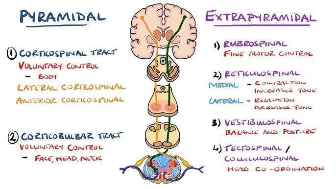 diagram overview
 -
Where does the corticospinal tract travel from and to, as shown in the pathway diagram?
- From cerebral cortex to skeletal muscle, passing through midbrain, pons, medulla; includes decussation of pyramids and lateral/ventral corticospinal paths
 pathway corticospinal
 -
What are the cortical origins and their percentage contributions to the corticospinal tract?
- 40% from upper 2/3 of primary motor area (area 4)
- 40% from premotor area (area 6)
- 20% from general sensory areas (areas 3, 1 & 2)
anatomy corticospinal origin -
What proportion of corticospinal fibers are large and arise from Betz (giant pyramidal) cells?
- 3% of the fibers are large and arise from giant pyramidal (Betz) cells
anatomy betz corticospinal -
Name the main structures/levels the corticospinal fibers descend through from cortex to medulla.
- Medullary center of hemisphere (corona radiata)
- Posterior limb of internal capsule (anterior 1/2 of lenticulothalamic part)
- Midbrain (middle 3/5 of crus cerebri)
- Pons (basis pontis)
- Medulla (pyramid)
anatomy pathway descent -
What happens to 85% of corticospinal fibers in the lower medulla?
- 85% cross to the opposite side at the pyramidal (motor) decussation and form the lateral corticospinal tract
anatomy decussation lateral -
What is the fate of the 15% of corticospinal fibers that remain uncrossed?
- 15% remain uncrossed to form the ventral (anterior) corticospinal tract; they cross in the cervical and upper thoracic spinal cord
anatomy ventral corticospinal -
Briefly describe the corticospinal tract pathway from motor cortex to spinal cord.
- Axons from motor/sensory cortices converge as corona radiata → posterior limb of internal capsule → middle 3/5 crus cerebri → basis pontis → medullary pyramid → 85% decussate to lateral corticospinal, 15% form ventral corticospinal
 anatomy overview pathway
 -
Motor System — concise summary
- The descending motor system comprises three main pathways: pyramidal, extrapyramidal, and corticoponto‑cerebellar; these control voluntary movement, posture, tone, and coordination.
Overview of descending systems
- Pyramidal system: direct cortical control of motor nuclei in brainstem and anterior horn cells of the spinal cord (voluntary movement).
- Extrapyramidal system: indirect cortical influence via brainstem centers; modulates tone, posture, balance, and automatic movement.
- Corticoponto‑cerebellar pathway: cortex → pontine nuclei → cerebellum; essential for motor planning and coordination.
Alt text: Diagram comparing pyramidal and extrapyramidal motor systems.
Pyramidal system
Components
- Corticospinal tract — major pathway to spinal anterior horn cells; controls limb and trunk voluntary movement.
- Corticobulbar tract — projects to brainstem motor nuclei to control face, head and neck muscles.
Corticospinal tract — origin
- Axons arise from pyramidal (Betz and other) cells in cortex: roughly \(40\%\) from primary motor cortex (area 4), \(40\%\) from premotor cortex (area 6), and \(20\%\) from primary somatosensory areas (areas 3, 1, 2).
- About \(3\%\) of fibers are large Betz cell axons.
Corticospinal tract — course
- Corona radiata → posterior limb of internal capsule (anteriormost lenticulothalamic part).
- Crus cerebri (middle \(3/5\) of cerebral peduncle).
- Basis pontis → medullary pyramid.
-
At the caudal medulla most fibers decussate (pyramidal decussation) forming the lateral corticospinal tract; remaining fibers continue as the ventral (anterior) corticospinal tract.
-
About \(85\%\) of fibers cross in the pyramidal decussation → form lateral corticospinal tract controlling distal limb muscles.
- About \(15\%\) remain uncrossed in the ventral corticospinal tract and cross segmentally to influence axial and proximal musculature.
Alt text: Corticospinal tract pathway from motor cortex to spinal cord.
Corticobulbar tract — key points
- Descends with corticospinal fibers to brainstem motor nuclei (cranial nerve motor nuclei).
- Innervation is largely bilateral for most cranial nuclei, providing redundancy.
- Important exceptions (clinical relevance):
- Lower facial nucleus (lower half of CN VII) receives predominantly contralateral input → UMN lesion produces contralateral lower facial weakness.
- Hypoglossal nucleus (CN XII) — mainly contralateral to protrude tongue toward side of lesion.
- Other nuclei (jaw, eye movement) have predominantly bilateral input; deficits vary.
Clinical correlates — pyramidal (UMN) lesion signs
- Weakness (more of distal than proximal in lateral corticospinal lesions), increased tone (spasticity), hyperreflexia, positive Babinski sign, minimal wasting early but disuse atrophy later.
- Lesion location rule: above pyramidal decussation → contralateral weakness; below decussation → ipsilateral weakness.
Extrapyramidal tracts — major tracts and functions
-
Originate in brainstem nuclei and modulate spinal motor neurons indirectly.
-
Rubrospinal tract (from red nucleus, midbrain)
- Facilitates distal flexor tone and fine motor control, especially for upper limbs.
- Vestibulospinal tracts
- Lateral vestibulospinal (ipsilateral): facilitates extensor tone for posture and balance.
- Medial vestibulospinal: coordinates head and neck position (bilateral control via medial longitudinal fasciculus).
- Reticulospinal tracts
- Medial (pontine) reticulospinal: facilitates extensor tone; increases muscle tone and aids posture.
- Lateral (medullary) reticulospinal: generally inhibitory to extensor tone; promotes relaxation and modulation.
- Tectospinal (colliculospinal)
- From superior colliculus; orients head/neck to visual/auditory stimuli.
- Raphespinal
- Modulates pain transmission and influences tone (serotonergic influence).
- Descending autonomic fibers (hypothalamospinal)
-
Carry sympathetic control to spinal intermediolateral cell column.
-
Extrapyramidal pathways are important for automatic aspects of movement (posture, gait, tone) rather than direct voluntary activation.
Corticoponto‑cerebellar pathway
- Originates in widespread cortical areas → synapses in pontine nuclei → crosses and enters cerebellum via the middle cerebellar peduncle.
- Function: conveys motor planning and cortical intent to cerebellum for coordination and timing.
Clinical application & localization tips
- Distal limb weakness and loss of fine skilled movements → lesion of lateral corticospinal.
- Predominant axial weakness / posture problems → lesion of ventral corticospinal or extrapyramidal (vestibulo/reticulospinal) pathways.
- Facial weakness pattern helps localize corticobulbar involvement (upper vs lower motor neuron):
- UMN lesion of corticobulbar → contralateral lower facial paralysis with spared forehead.
- LMN lesion of facial nerve → ipsilateral entire half of face weak.
- Changes in tone: increased tone (spasticity) suggests UMN/pyramidal or pontine reticulospinal involvement; decreased tone suggests LMN or medullary reticulospinal dysfunction.
High‑yield facts / mnemonics
- Lateral corticospinal = crossed, controls limbs; ventral corticospinal = uncrossed then segmental cross, controls axial muscles.
- "Pyramidal" = direct cortical → motor neurons; "extrapyramidal" = indirect modulation via brainstem.
- Corticoponto‑cerebellar = cortex → pons → cerebellum (middle peduncle) for coordination.
Quick reference table
| Tract | Origin | Main function | Crossed? |
|---|---|---|---|
| Lateral corticospinal | Motor cortex | Distal limb voluntary movement | Yes (\(85\%\) at medulla) |
| Ventral corticospinal | Motor cortex | Axial/proximal control | Cross segmentally (uncrossed initially) |
| Corticobulbar | Motor cortex | Motor cranial nuclei | Mostly bilateral (some exceptions) |
| Rubrospinal | Red nucleus | Distal flexor facilitation | Crossed (midbrain) |
| Lateral vestibulospinal | Vestibular nuclei | Tone/posture (extensors) | Ipsilateral |
| Medial vestibulospinal | Vestibular nuclei | Head/neck coordination | Bilateral |
| Reticulospinal (medial/lateral) | Reticular formation | Tone and automatic gait | Mixed |