덱이 사라지기 전에 저장해 둬
이 플래시카드는 아직 저장되지 않았어 — 페이지를 나가면 사라져. 무료 계정을 만들면 저장되고 아래 기능들도 모두 이용할 수 있어.
- Save this deck to your account
- Study with spaced repetition
- Export to Anki (.apkg) or PDF
- Process documents up to 100 pages
- Images extracted from your PDFs
- Sharper text extraction & a more advanced AI model
What is the definition of sensation?
What does decussation mean in neural anatomy?
What do the terms contralateral and ipsilateral mean?
How are ascending sensory tracts organized from receptor to cortex?
Which cortical area receives ascending somatosensory information?
Where is the 1st order neuron cell body located in sensory pathways?
Do the axons of 1st order sensory neurons cross (decussate)?
Where are 2nd order sensory neurons located?
Do the axons of 2nd order sensory neurons cross (decussate)?
Where is the 3rd order sensory neuron located?
Do the axons of 3rd order sensory neurons cross (decussate)?
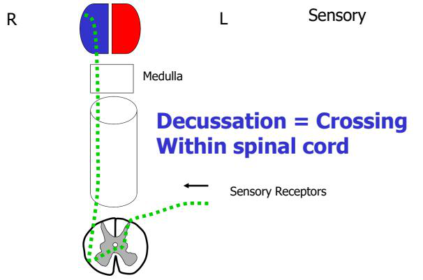
What is the definition of 'decussation' in neuroanatomy?
What defines a white matter tract (bundle or fasciculus)?
What are 'long tracts' and their general directions?
What general role do long tracts serve between brain and spinal cord?
Name major ascending tracts listed in the spinal cord cross-section.
Which anatomical feature in the spinal cord is explicitly named where crossing occurs?
Where does the text state decussation occurs apart from the spinal cord?
Provide a visual diagram showing decussation within the spinal cord pathways.
What types of sensory impulses do ascending tracts carry?
What are the two main destinations of ascending sensory pathways?
Describe the three-neuron sequence of conscious ascending pathways.
Which thalamic nucleus contains the 3rd order neuron for conscious sensory pathways?
Name spinocerebellar tracts or related pathways mentioned for subconscious proprioception.
Which spinocerebellar inputs serve the upper body (above T6)?
Which spinocerebellar inputs serve the lower body (below T6)?
Which primary afferent fiber types are routed to spinocerebellar tracts in the diagram?
What structure is mentioned as part of the subconscious pathway entry to the cerebellum?
Flashcards in this deck (28)
-
What is the definition of sensation?
Conscious or unconscious awareness of external or internal stimuli.
neuroscience sensation -
What does decussation mean in neural anatomy?
Crossing (nerve fibres crossing to the opposite side).
neuroanatomy decussation -
What do the terms contralateral and ipsilateral mean?
- Contralateral: opposite side
- Ipsilateral: same side
neuroanatomy terms -
How are ascending sensory tracts organized from receptor to cortex?
They carry sensory information from peripheral receptor → dorsal root ganglion (DRG) and travel in a three‑order neuron chain to the primary somatosensory cortex in the post‑central gyrus.
sensory ascending -
Which cortical area receives ascending somatosensory information?
Primary somatosensory cortex in the post‑central gyrus.
cortex sensory -
Where is the 1st order neuron cell body located in sensory pathways?
- Dorsal root ganglion
neuroanatomy sensory -
Do the axons of 1st order sensory neurons cross (decussate)?
- No — its axons never cross
neuroanatomy decussation -
Where are 2nd order sensory neurons located?
- Spinal cord or brain stem
neuroanatomy sensory -
Do the axons of 2nd order sensory neurons cross (decussate)?
- Yes — its axons always cross
neuroanatomy decussation -
Where is the 3rd order sensory neuron located?
- Ventral posterior lateral nucleus (VPL) of thalamus
neuroanatomy thalamus -
Do the axons of 3rd order sensory neurons cross (decussate)?
- No — its axons never cross
neuroanatomy decussation -
What is the definition of 'decussation' in neuroanatomy?
- Decussation = Crossing
terminology decussation -
What defines a white matter tract (bundle or fasciculus)?
- Fibers with the same origin, termination and function
neuroanatomy white_matter -
What are 'long tracts' and their general directions?
- Ascending (sensory/afferent)
- Descending (motor/efferent)
neuroanatomy tracts -
What general role do long tracts serve between brain and spinal cord?
- Serve to join the brain to the spinal cord
neuroanatomy function -
Name major ascending tracts listed in the spinal cord cross-section.
- Fasciculus gracilis
- Fasciculus cuneatus
- Posterior spinocerebellar tract
- Anterior spinocerebellar tract
- Lateral spinothalamic tract
- Anterior spinothalamic tract
 neuroanatomy ascending
 -
Which anatomical feature in the spinal cord is explicitly named where crossing occurs?
- Anterior white commissure
neuroanatomy decussation -
Where does the text state decussation occurs apart from the spinal cord?
- Within medulla oblongata
neuroanatomy decussation -
Provide a visual diagram showing decussation within the spinal cord pathways.
- Diagram of spinal-cord decussation
 visual decussation
 -
What types of sensory impulses do ascending tracts carry?
- Pain
- Thermal
- Tactile
- Muscle and joint receptors
sensory ascending -
What are the two main destinations of ascending sensory pathways?
- Conscious: cerebral cortex
- Subconscious: cerebellum
sensory pathways -
Describe the three-neuron sequence of conscious ascending pathways.
- 1st order: dorsal root ganglion
- 2nd order: spinal grey matter or medulla oblongata
- 3rd order: thalamus (ventral posterior nucleus)
pathways three-neuron -
Which thalamic nucleus contains the 3rd order neuron for conscious sensory pathways?
- Ventral posterior (VP) thalamic nucleus
thalamus sensory -
Name spinocerebellar tracts or related pathways mentioned for subconscious proprioception.
- Dorsal spinocerebellar tract (DSCT)
- Ventral spinocerebellar tract (VSCT)
- Rostral spinocerebellar tract (RSCT)
- Cuneocerebellar tract
spinocerebellar cerebellum -
Which spinocerebellar inputs serve the upper body (above T6)?
- Cuneocerebellar tract, RSCT, VSCT (upper body above T6)
upper spinocerebellar -
Which spinocerebellar inputs serve the lower body (below T6)?
- Dorsal spinocerebellar tract (DSCT) (lower body below T6)
lower spinocerebellar -
Which primary afferent fiber types are routed to spinocerebellar tracts in the diagram?
- Ia fibers (to cuneocerebellar or DSCT)
- Ib fibers (to RSCT or VSCT)
afferent fibers -
What structure is mentioned as part of the subconscious pathway entry to the cerebellum?
- Inferior cerebellar peduncle
cerebellum peduncle
Overview
- Sensory system & ascending tracts: pathways that transmit sensory information from peripheral receptors to the brain. They include conscious pathways (reach cerebral cortex) and subconscious pathways (reach cerebellum).
Key definitions
- Receptor: peripheral sensory ending that detects stimuli.
- Dorsal root ganglion (DRG): location of 1st order neuron cell bodies.
- Decussation: anatomical crossing of fibres from one side to the other.
- Contralateral: opposite side; Ipsilateral: same side.
Organization of conscious ascending pathways (general rules)
- Most conscious sensory pathways use a three-neuron chain: receptor → spinal cord/brainstem → thalamus → cortex.
- Typical sequence:
- 1st order neuron — cell body in DRG; peripheral receptor to spinal cord or medulla; axons do not cross.
- 2nd order neuron — in spinal cord gray matter or brainstem nuclei; axons do cross (decussate) and ascend to thalamus.
- 3rd order neuron — in ventral posterior lateral nucleus (VPL) of thalamus; projects to primary somatosensory cortex (postcentral gyrus); axons do not cross.
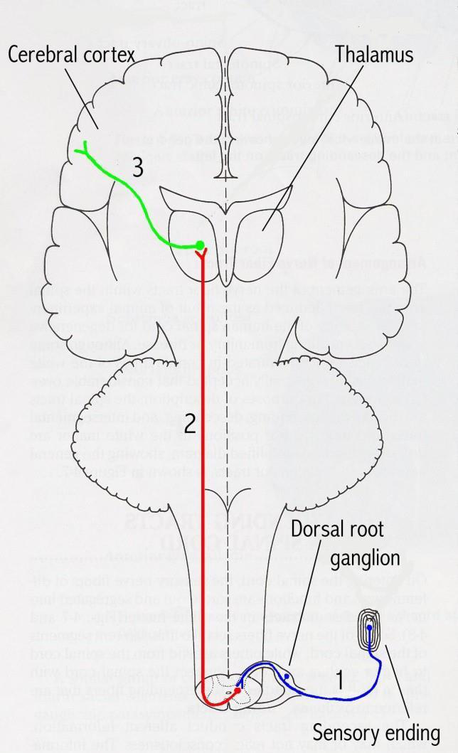 Alt text: Sensory pathway from receptor to cortex
Major ascending tracts — what they carry and key features
- Dorsal column–medial lemniscus (DCML)
- Tracts: Fasciculus gracilis (lower body) and Fasciculus cuneatus (upper body).
- Function: fine touch, vibration, conscious proprioception.
-
Path: 1st order in DRG → ascend ipsilaterally in dorsal columns → synapse in nucleus gracilis/cuneatus (medulla) → 2nd order decussate as internal arcuate fibers → ascend as medial lemniscus → VPL thalamus → cortex.
-
Spinothalamic tracts (anterolateral system)
- Lateral spinothalamic: pain and temperature.
- Anterior spinothalamic: crude touch and pressure.
-
Path: 1st order in DRG → synapse in dorsal horn → 2nd order decussate within 1–2 spinal segments via anterior white commissure → ascend contralaterally to VPL → cortex.
-
Spinocerebellar tracts (subconscious proprioception)
- Dorsal spinocerebellar tract (DSCT): lower limb proprioception to ipsilateral cerebellum.
- Ventral spinocerebellar tract (VSCT) and rostral/cuneocerebellar tracts: integrate spinal interneuron signals, upper body, and coordinate movement.
- These pathways largely project to the cerebellum (subconscious) and may be ipsilateral or double-crossed.
Alt text: Spinal cord cross-section with ascending and descending tracts
Decussation and clinical localization
- Level of lesion determines deficits:
- Lesion below decussation of a pathway produces ipsilateral deficits for that pathway (e.g., dorsal columns below medulla).
- Lesion above decussation produces contralateral deficits.
- Example: DCML decussates in the medulla → spinal cord lesions cause ipsilateral loss of vibration/proprioception; brainstem/cortical lesions cause contralateral loss.
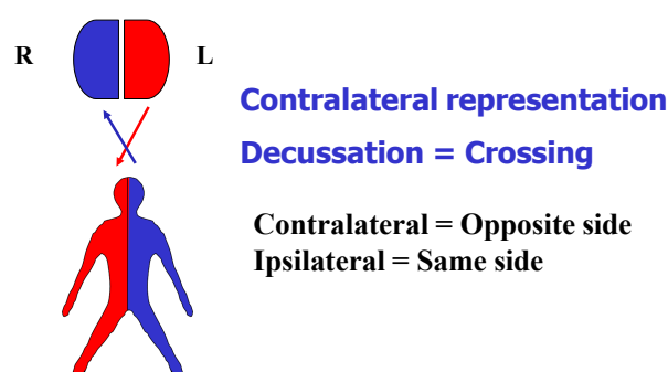 Alt text: Diagram showing decussation and contralateral representation
Practical study tips
- Memorize the three-neuron rule and where each neuron synapses (DRG → spinal gray/brainstem nuclei → VPL thalamus → cortex).
- Use the tract location in the spinal cord cross-section to predict which sensations are lost with focal lesions.
- Distinguish conscious (cortex via VPL) vs subconscious (cerebellum) pathways — this guides clinical signs (sensory loss vs ataxia).
Quick reference table
| Order neuron | Location of cell body | Crosses? | Typical relay |
|---|---|---|---|
| 1st order | Dorsal root ganglion | No | Peripheral receptor → spinal cord/medulla |
| 2nd order | Spinal cord gray or brainstem nucleus | Yes (usually) | Ascends to VPL thalamus |
| 3rd order | VPL nucleus of thalamus | No | VPL → primary somatosensory cortex |
One-sentence summary
- Ascending tracts transmit sensory modalities via organized bundles (DCML, spinothalamic, spinocerebellar) using a 3-neuron chain and characteristic decussation points for clinical localization.