덱이 사라지기 전에 저장해 둬
이 플래시카드는 아직 저장되지 않았어 — 페이지를 나가면 사라져. 무료 계정을 만들면 저장되고 아래 기능들도 모두 이용할 수 있어.
- Save this deck to your account
- Study with spaced repetition
- Export to Anki (.apkg) or PDF
- Process documents up to 100 pages
- Images extracted from your PDFs
- Sharper text extraction & a more advanced AI model
What is the primary definition of periodontal instrumentation?
What is the primary goal of conserving dental cementum during periodontal instrumentation?
What occurs as biofilm matures?
The biofilm stops requiring an inflammatory response from the immune system.
The pH level of the biofilm becomes more alkaline.
The quality of the biofilm changes, with more gram-negative and anaerobic bacteria appearing.
The biofilm becomes exclusively composed of gram-positive aerobic bacteria.
How does an increase in mature biofilm affect the local environment in the periodontal pocket?
Why does the immune system initiate an inflammatory response to bacterial biofilm?
What is the first step in maintaining periodontal health regarding bacterial composition?
What are the primary benefits of mechanically disrupting and removing biofilm?
Why does biofilm exhibit resistance to antibiotics?
How does professional debridement influence the subgingival environment?
Why must calculus be removed to effectively manage periodontal health?
What is a key difference between supragingival and subgingival calculus in terms of appearance and composition?
What are the two main types of cementum involved in PDL attachment to the root surface?
What is the consequence for periodontal fibers if cementum is absent?
What are the three main components that compose a dental hand instrument?
What is the mechanical action performed by hand instruments during debridement?
Which instruments are mentioned as being available for supragingival scaling?
Endo scaler, sickle scaler, McCall scaler, power scalers, and gracey curettes
Power scalers and explorers only
Only sickle scalers and endo scalers
Only gracey curettes
What are the cross-sectional characteristics of a dental scaler?
Round in cross-section with 1 cutting edge
Oval in cross-section with no cutting edges
Triangular in cross-section with 2 cutting edges
Square in cross-section with 4 cutting edges
Which specific instrument is identified for use on anterior teeth during supragingival scaling?
Which instruments are indicated for subgingival scaling?
Endo scalers and power scalers with any tips
McCall scalers and sickle scalers
Only sickle scalers
Powered scalers with specific tips and gracey curettes
Why are scalers with sharp pointed ends restricted to supra-gingival use?
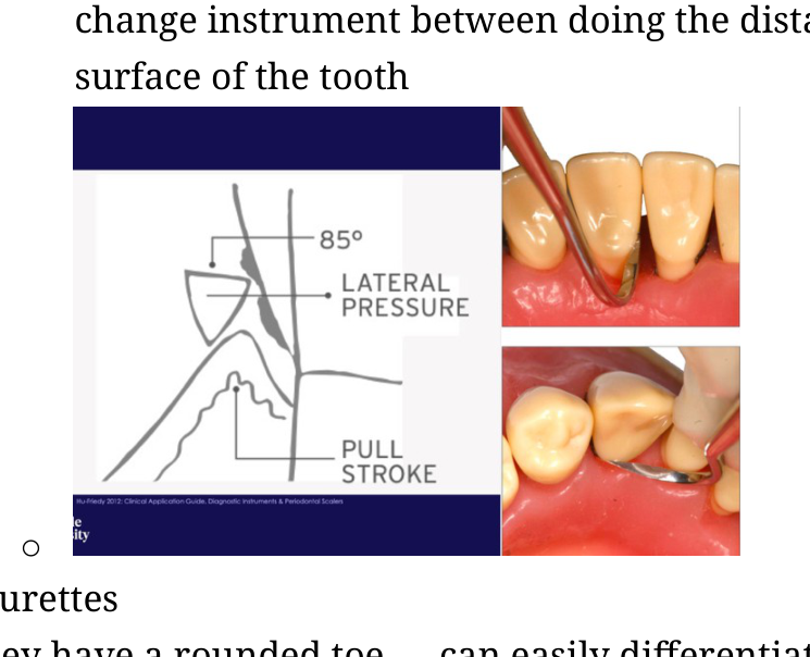
What is the recommended angulation between the tooth surface and the scaler blade to achieve effective deposit removal?
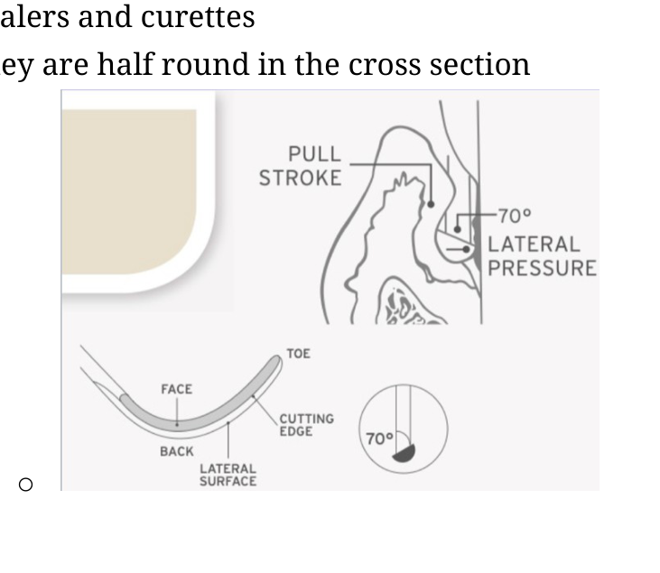
What are the characteristic features of Gracey curettes?
Rounded toe and half-round cross-section
Flat toe and triangular cross-section
Beveled edge and oval cross-section
Sharp pointed end and square cross-section
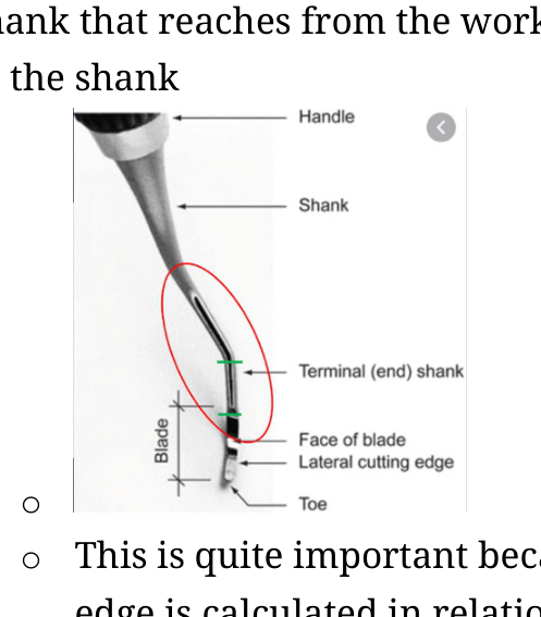
Which anatomical parts are identifiable on a standard curette blade cross-section?
What is the benefit of using a scaler with two cutting edges when removing biofilm and calculus?
Describe the proper application of lateral pressure when using a scaler for deposit removal.
How is the lower edge of a curette blade defined?
Which portion of the curette blade serves as the cutting edge?
What is the purpose of the rounded toe and half-round cross-section of a curette?
How is the terminal shank of a curette defined?
Which curette numbers are primarily used for anterior teeth in the ADH system?
For which surfaces are curette numbers 7 and 8 intended?
Which curette numbers are designated for use on the mesial surfaces of premolars and molars?
Which curette numbers are used on the distal surfaces of premolars and molars?
How can you identify the correct cutting edge of a curette?
What is the recommended angle for a curette blade against the root surface to achieve maximum cutting efficiency?
What happens if the terminal 2-3mm and the toe of the curette are not closely adapted to the root surface?
To ensure the blade maintains a correct \(70^{\circ}\) angulation against the root surface, how should the terminal shank be positioned?
Parallel to the gingival margin
Perpendicular to the long axis of the tooth
Parallel to the long axis of the tooth
At a \(45^{\circ}\) angle to the long axis
In dental instrumentation, where should the cutting edge be directed during use?
What is the correct adaptation for a curette when debriding a tooth surface?
What are the consequences of using a curette with incorrect angulation?
What are the primary effects of using blunt dental hand instruments?
How do ultrasonic and sonic scaling instruments function?
What mechanism produces the mechanical and biophysical forces used in a piezoelectric scaling device to remove deposits?
What component is contained within the piezoelectric handpiece that generates vibrations when an alternating electrical current is applied?
What is the typical frequency range of the metal tips in a piezoelectric scaling device?
25 to 50k hertz
100 to 150k hertz
10 to 20k hertz
60 to 80k hertz
5 to 10k hertz
Which type of piezoelectric scaling tip is designed specifically for large supragingival calculus deposits?
What is the primary application for the extra fine piezoelectric scaling tip?
How does the piezoelectric scaling tip move during operation?
Why is a water source connection required for piezoelectric scaling devices?
To prevent electrical shorts in the handpiece.
To lubricate the tooth surface.
To increase the frequency of vibrations.
To cool the tip due to heat generated by vibrational oscillation.
What auditory cue indicates that a dental scaler tip is incorrectly positioned?
Which sides of a dental scaler tip should be used during procedures?
What mechanism drives magnetostrictive ultrasonic scalers?
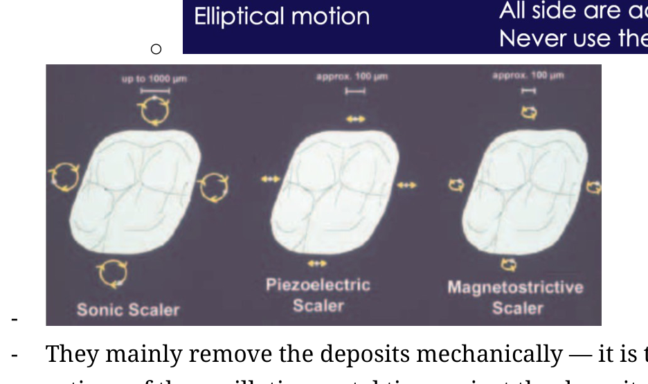
What type of motion pattern is produced by magnetostrictive ultrasonic scalers?
Rotational motion
Linear motion
Vibrational longitudinal motion only
Elliptical motion
How are sonic scalers powered?
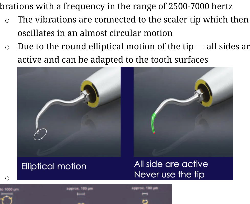
What is the frequency range of the vibrations generated by sonic scalers?
How does a sonic scaler tip move during operation?
Why are all sides of a sonic scaler tip considered active?
What is the primary mechanical mechanism by which power scalers remove deposits?
Why is a constant water coolant required when using high-speed power scalers?
What secondary function does the constant water flow provide during scaling?
Besides mechanical vibration and irrigation, what is another mode of action for power scalers?
What is defined as the formation and subsequent implosion of pulsating cavities or small bubbles?
How do cavitation bubbles contribute to the disruption of a biofilm?
What forces are generated in the water surrounding an oscillating tip that help disrupt biofilm?
What phenomenon occurs when the implosion of bubbles radiates a shockwave?
Hydrodynamic equilibrium
Cavitation
Laminar flow
Acoustic microstreaming
Which term describes the turbulent currents and large hydrodynamic shear stresses produced by an oscillating tip?
Surface tension
Cavitation
Acoustic microstreaming
Capillary action
Describe the lifecycle of the bubbles observed in cavitation.
What is the consequence of losing 1mm of length on an ultrasonic scaler tip?
At what point should an ultrasonic scaler tip be replaced?
What is the recommended angulation for ultrasonic scaler tips to prevent severe root or tooth damage?
How much hard substance is removed by a powered scaler when used at a zero degree angulation to the tooth surface?
Which parameter affects the depth of a defect created by a powered scaler the most?
Power setting
Tip material
Angulation
Lateral force
Why is the target for substance removal at zero degree angulation set to 50 microns?
Which dental instrument removes the least amount of hard tooth substance per working stroke?
Which instrument removes the most tooth hard substance per working stroke among ultrasonic scalers, fine curettes, and diamond burs?
Diamond burs
Air scalers
Fine curettes
Ultrasonic scalers
How does the tactile sensitivity of hand instruments compare to powered dental instruments?
What is a primary disadvantage of using hand instruments compared to powered instruments regarding operator physical strain?
Which of the following is a risk factor associated with powered dental instruments that is not listed for hand instruments?
High maintenance requirements
Cardiac pacemaker interference
Excessive operator fatigue
Difficulty with angulation
What is a major requirement for the efficient use of curettes during the debridement process?
Why might hand instruments be preferred over powered instruments in dental procedures?
What is a major clinical concern regarding the use of powered dental instruments on patients with infectious diseases?
What potential risk do magnetostrictive ultrasonic scalers pose for patients with implanted cardiac devices?
Which of the following materials should NOT be cleaned with a powered scaler?
Composite resin or porcelain
Gold crowns
Dentin
Enamel
What are two advantages of using powered dental instruments over hand instruments?
What is the primary mechanism of action of laser therapy in periodontal pockets?
What is the current evidence-based status of using laser therapy for routine periodontal debridement?
It is considered ineffective and should be avoided entirely.
It is recommended as the gold standard for removing subgingival calculus.
It provides significantly better clinical outcomes than other methods.
It provides similar results to conventional methods but lacks superior outcomes.
What are the two types of powders available for air polishing, and what are their respective particle sizes?
Which of the following describes the functional limitation of an air polishing machine in periodontal therapy?
It removes both biofilm and heavy calculus deposits.
It removes biofilm but cannot remove calculus.
It is exclusively used for polishing tooth structure rather than cleaning.
It can only be used on enamel and damages mucosal surfaces.
Beyond tooth surfaces, where else can air polishing be safely used?
Which powder is safe and effective for biofilm removal from both natural tooth structure and restorative materials?
What is a major clinical risk when using sodium bicarbonate powder for air polishing?
When using an air-polishing nozzle in a periodontal pocket, at what angle should it be held relative to the sulcus?
90 degrees
45 degrees
180 degrees
15 degrees
How does the effectiveness of glycine-powder air polishing compare to manual or ultrasonic instrumentation for cleaning periodontal pockets?
Why is air polishing particularly useful for patients with orthodontic appliances?
What is the primary challenge occurring during periodontal wound healing?
What biological process occurs within the periodontal pocket immediately following debridement during the first stage of wound healing?
Which specific immune cells migrate to the wound area following blood clot formation to help clean residual bacteria?
What is the primary function of the leukocyte band following subgingival instrumentation?
At what time point after subgingival instrumentation does the stage of epithelial proliferation typically start?
How do basal epithelial cells move during the proliferation phase of periodontal wound healing?
What is the primary driver of vascular proliferation during the wound healing process following instrumentation?
What substances do fibroblasts deposit into the free network during the proliferation stage?
How does the turnover rate of the junctional epithelium (JE) compare to other tissues in the body?
How long does the initial epithelialization and new junctional epithelium attachment take after subgingival instrumentation?
What is the clinical consequence of leaving residual biofilm and calculus at the base of a periodontal pocket?
How long can the maturation and remodeling phase of periodontal tissue healing last?
What processes contribute to the increasing resistance and functional strength of the gingiva during the healing process?
How long should a clinician typically wait after performing subgingival debridement before recharting or reprobing an area?
What are the clinical characteristics of healthy gingival tissue?
Which clinical signs indicate gingival inflammation?
What is the typical visual appearance of supragingival calculus?
What substance is used for the detection of supragingival biofilm?
Describe the clinical technique used for the supragingival detection of biofilm and calculus.
How much time is generally required for gingival tissue to fully heal after flap surgery?
What is the primary purpose of conducting a gingival assessment during a dental visit?
Does the absence of visible calculus on a radiograph confirm that no calculus is present on the tooth?
Why are tooth overhangs considered problematic during an oral assessment?
Which clinical conditions might be detected when using an 11/12 explorer probe on a tooth surface?
What is the correct alignment for an 11/12 explorer probe shank relative to the tooth during use?
How much of the 11/12 explorer probe's toe should be inserted underneath the gingiva when exploring the sulcus?
What does it indicate if dental floss gets stuck while sliding over a proximal tooth surface?
What is a systematic way to divide a tooth into its different surfaces for examination purposes?
Which curette is indicated for the area from the mesio-buccal line angle to the contact point and from the mesial palatal line to the mesial contact point?
Which curette is used for the area from the distobuccal line angle to the contact point and from the distopalatal line angle back to the contact point?
Which curette is used for the buccal and palatal surface from line angle to line angle?
What is the recommended width for instrumenting sections on a tooth surface?
When inserting a curette for instrumentation, what is the correct orientation of the terminal shank to the long axis of the tooth?
How many strokes are typically performed within a single section of the tooth surface during instrumentation?
How should lateral pressure be adjusted when transitioning from removing biofilm to removing calculus?
What is the primary challenge when instrumentation reaches the middle (3rd) division of a tooth surface?
How should an instrument be positioned when working on the middle (3rd) division to avoid the contact point?
What stroke types might be combined when vertical strokes cannot be performed on a tooth surface?
When using power scalers, what is the ideal angulation of the tip against the tooth surface?
30-45 degrees
0-5 degrees
0-15 degrees
45-90 degrees
What kind of water spray is required when using a power scaler?
Describe the technique for using power scaler strokes when cleaning a pocket.
What is the recommended angulation for power scaler tips during instrumentation?
What type of movement is recommended when using a power scaler?
How much pressure should be applied when using a power scaler tip?
At what speed should the power scaler tip be moved over the tooth surface?
What is the recommended path of movement for a power scaler tip during subgingival instrumentation?
What is the recommended width for the sections being instrumented with a power scaler?
Which tip is used for subgingival instrumentation in the clinic?
Which tip is used for subgingival instrumentation in the SimClinic?
Flashcards in this deck (143)
-
What is the primary definition of periodontal instrumentation?
It is the removal or disruption of dental deposits (biofilm) and plaque-retentive dental calculus from tooth surfaces and within the periodontal pocket space.
periodontics dentistry -
What is the primary goal of conserving dental cementum during periodontal instrumentation?
To help maintain or re-establish a healthy periodontal environment and eliminate periodontitis.
periodontics dentistry -
What occurs as biofilm matures?
The biofilm stops requiring an inflammatory response from the immune system.
The pH level of the biofilm becomes more alkaline.
The quality of the biofilm changes, with more gram-negative and anaerobic bacteria appearing.
The biofilm becomes exclusively composed of gram-positive aerobic bacteria.
biofilm periodontics -
How does an increase in mature biofilm affect the local environment in the periodontal pocket?
The pH level changes, allowing more pathogenic bacteria to thrive.
biofilm pathology -
Why does the immune system initiate an inflammatory response to bacterial biofilm?
It reacts because PMNs (polymorphonuclear leukocytes) are insufficient to fight the accumulating pathogenic bacteria.
immunology periodontics -
What is the first step in maintaining periodontal health regarding bacterial composition?
Directing the sub- and supra-gingival bacterial composition toward one that is more compatible with health.
periodontics prevention -
What are the primary benefits of mechanically disrupting and removing biofilm?
It permits interaction with the host defense system and reduces the bacterial load on the immune system.
biofilm dentistry -
Why does biofilm exhibit resistance to antibiotics?
Bacteria are incorporated into a protective matrix within the biofilm.
biofilm antibiotics -
How does professional debridement influence the subgingival environment?
It reduces microbiome product concentrations and GCF flow while promoting a more neutral biofilm pH level.
debridement biofilm -
Why must calculus be removed to effectively manage periodontal health?
Calculus creates a rough surface that facilitates microbial attachment and colonization; its removal eliminates these harbors for pathogenic bacteria.
calculus dentistry -
What is a key difference between supragingival and subgingival calculus in terms of appearance and composition?
Supragingival calculus is typically white or yellowish, while subgingival calculus is brown or black because blood hemoglobin is incorporated during its maturation.
calculus dentistry -
What are the two main types of cementum involved in PDL attachment to the root surface?
- Acellular extrinsic fibre cementum
- Cellular mixed fibre cementum
cementum anatomy -
What is the consequence for periodontal fibers if cementum is absent?
Fibers are unable to anchor or insert into the dentine, forcing the body to rely solely on epithelium attachment.
cementum anatomy -
What are the three main components that compose a dental hand instrument?
- Handle
- Shank
- Working end
dentistry instruments -
What is the mechanical action performed by hand instruments during debridement?
They involve manually stroking a bladed cutting edge across the tooth surface to mechanically break the bond between the deposit and the tooth.
dentistry debridement -
Which instruments are mentioned as being available for supragingival scaling?
Endo scaler, sickle scaler, McCall scaler, power scalers, and gracey curettes
Power scalers and explorers only
Only sickle scalers and endo scalers
Only gracey curettes
dentistry scaling -
What are the cross-sectional characteristics of a dental scaler?
Round in cross-section with 1 cutting edge
Oval in cross-section with no cutting edges
Triangular in cross-section with 2 cutting edges
Square in cross-section with 4 cutting edges
dentistry scalers -
Which specific instrument is identified for use on anterior teeth during supragingival scaling?
Sickle scaler
dentistry scaling -
Which instruments are indicated for subgingival scaling?
Endo scalers and power scalers with any tips
McCall scalers and sickle scalers
Only sickle scalers
Powered scalers with specific tips and gracey curettes
dentistry scaling -
Why are scalers with sharp pointed ends restricted to supra-gingival use?
Insertion into the sub-gingival area could damage the gingiva due to the sharp pointed tip.
dentistry instruments scaling -
What is the recommended angulation between the tooth surface and the scaler blade to achieve effective deposit removal?
Approximately an 85 degree angle.
 dentistry scaling technique
 -
What are the characteristic features of Gracey curettes?
Rounded toe and half-round cross-section
Flat toe and triangular cross-section
Beveled edge and oval cross-section
Sharp pointed end and square cross-section
dentistry instruments curettes -
Which anatomical parts are identifiable on a standard curette blade cross-section?
- Toe
- Face
- Back
- Cutting edges
dentistry anatomy instruments -
What is the benefit of using a scaler with two cutting edges when removing biofilm and calculus?
It allows the clinician to use both sides of the blade, eliminating the need to change instruments between the distal and mesial surfaces of the tooth.
dentistry scaling workflow -
Describe the proper application of lateral pressure when using a scaler for deposit removal.
Apply lateral pressure against the tooth and pull the scaler firmly in an upward direction to dislodge the deposit.
dentistry scaling technique -
How is the lower edge of a curette blade defined?
The side of the blade that tilts downwards at a \(70^{\circ}\) angle from the shank.
dentistry -
Which portion of the curette blade serves as the cutting edge?
The downwards tilting lower edge.
dentistry -
What is the purpose of the rounded toe and half-round cross-section of a curette?
They are designed to avoid harming the gingiva during insertion into the pocket space.
dentistry -
How is the terminal shank of a curette defined?
It is the section of the oval shank extending from the working end to the first bend in the shank.
 dentistry
 -
Which curette numbers are primarily used for anterior teeth in the ADH system?
Curettes numbers 3, 4, 5, and 6.
dentistry -
For which surfaces are curette numbers 7 and 8 intended?
The buccal and lingual surfaces of premolars and molars.
dentistry -
Which curette numbers are designated for use on the mesial surfaces of premolars and molars?
Curette numbers 11 and 12.
dentistry -
Which curette numbers are used on the distal surfaces of premolars and molars?
Curette numbers 13 and 14.
dentistry -
How can you identify the correct cutting edge of a curette?
Hold the terminal shank perpendicular to the floor and look at the face of the curette; the lower side is the sharp cutting edge.
dentistry instrumentation -
What is the recommended angle for a curette blade against the root surface to achieve maximum cutting efficiency?
The blade should be at an offset of \(70^{\circ}\) from the terminal shank.
dentistry instrumentation -
What happens if the terminal 2-3mm and the toe of the curette are not closely adapted to the root surface?
You lose tactile feedback and lack awareness of your position within the pocket space.
dentistry instrumentation -
To ensure the blade maintains a correct \(70^{\circ}\) angulation against the root surface, how should the terminal shank be positioned?
Parallel to the gingival margin
Perpendicular to the long axis of the tooth
Parallel to the long axis of the tooth
At a \(45^{\circ}\) angle to the long axis
dentistry instrumentation -
In dental instrumentation, where should the cutting edge be directed during use?
It should lie against the root surface, not towards the gingival tissue.
dentistry instrumentation -
What is the correct adaptation for a curette when debriding a tooth surface?
The terminal 2-3mm and the lower cutting edge should be against the tooth, with the terminal shank parallel to the tooth surface.
dentistry curette -
What are the consequences of using a curette with incorrect angulation?
Ineffective calculus removal, potential burnishing or polishing of deposits rather than removal, and the risk of cratering the tooth surface.
dentistry curette -
What are the primary effects of using blunt dental hand instruments?
- Reduced tactile sensitivity and instrument control
- Ineffective calculus removal
- Increased risk of damage to the tooth surface from added force
- Increased operator fatigue
dentistry instruments -
How do ultrasonic and sonic scaling instruments function?
They feature a blunt metal tip that swings at a high frequency, eliminating the need to create manual force to strike the blade against the tooth surface.
dentistry scalers -
What mechanism produces the mechanical and biophysical forces used in a piezoelectric scaling device to remove deposits?
The oscillation of the tip.
dentistry scaling -
What component is contained within the piezoelectric handpiece that generates vibrations when an alternating electrical current is applied?
A quartz crystal or a ceramic disc.
dentistry piezoelectric -
What is the typical frequency range of the metal tips in a piezoelectric scaling device?
25 to 50k hertz
100 to 150k hertz
10 to 20k hertz
60 to 80k hertz
5 to 10k hertz
dentistry piezoelectric -
Which type of piezoelectric scaling tip is designed specifically for large supragingival calculus deposits?
The regular tip.
dentistry tips -
What is the primary application for the extra fine piezoelectric scaling tip?
Subgingival debridement.
dentistry tips -
How does the piezoelectric scaling tip move during operation?
It swings in a linear, bi-directional (back and forth) way.
dentistry piezoelectric -
Why is a water source connection required for piezoelectric scaling devices?
To prevent electrical shorts in the handpiece.
To lubricate the tooth surface.
To increase the frequency of vibrations.
To cool the tip due to heat generated by vibrational oscillation.
dentistry safety -
What auditory cue indicates that a dental scaler tip is incorrectly positioned?
A change in the noise to a very pesky, high-pitched sound.
dentistry instruments -
Which sides of a dental scaler tip should be used during procedures?
The lateral sides only. Never use the tip.
dentistry instruments -
What mechanism drives magnetostrictive ultrasonic scalers?
An alternating electromagnetic field applied to a stack of nickel strips.
dentistry ultrasonic scalers -
What type of motion pattern is produced by magnetostrictive ultrasonic scalers?
Rotational motion
Linear motion
Vibrational longitudinal motion only
Elliptical motion
dentistry ultrasonic scalers -
How are sonic scalers powered?
They are operated by compressed air from the dental unit on the high speed fitting.
dentistry sonic scalers -
What is the frequency range of the vibrations generated by sonic scalers?
\(2500-7000 \text{ Hz}\)
dentistry instruments -
How does a sonic scaler tip move during operation?
It oscillates in an almost circular, elliptical motion.
 dentistry instruments
 -
Why are all sides of a sonic scaler tip considered active?
The elliptical motion allows the tip to adapt to tooth surfaces on all sides.
dentistry instruments -
What is the primary mechanical mechanism by which power scalers remove deposits?
The vibratory action of the oscillating metal tips breaks the bond between the deposit and the tooth surface.
 dentistry mechanisms
 -
Why is a constant water coolant required when using high-speed power scalers?
High-speed vibration generates heat, which must be dissipated to protect the tooth surface.
dentistry safety -
What secondary function does the constant water flow provide during scaling?
It acts as an irrigation system to flush out biofilm and remove debris.
dentistry procedure -
Besides mechanical vibration and irrigation, what is another mode of action for power scalers?
Cavitation.
dentistry mechanisms -
What is defined as the formation and subsequent implosion of pulsating cavities or small bubbles?
Cavitation
physics cavitation -
How do cavitation bubbles contribute to the disruption of a biofilm?
The implosion of the bubbles radiates a shockwave that disrupts the biofilm.
physics biofilm cavitation -
What forces are generated in the water surrounding an oscillating tip that help disrupt biofilm?
Turbulent currents of water and large hydrodynamic shear stresses.
physics microstreaming -
What phenomenon occurs when the implosion of bubbles radiates a shockwave?
Hydrodynamic equilibrium
Cavitation
Laminar flow
Acoustic microstreaming
physics cavitation -
Which term describes the turbulent currents and large hydrodynamic shear stresses produced by an oscillating tip?
Surface tension
Cavitation
Acoustic microstreaming
Capillary action
physics microstreaming -
Describe the lifecycle of the bubbles observed in cavitation.
The tiny individual bubbles go through a process of growing and then collapsing.
physics cavitation -
What is the consequence of losing 1mm of length on an ultrasonic scaler tip?
A 25% loss in efficiency.
dentistry scalers -
At what point should an ultrasonic scaler tip be replaced?
When the tip has lost more than 2mm of its original length.
dentistry scalers -
What is the recommended angulation for ultrasonic scaler tips to prevent severe root or tooth damage?
Close to 0 degrees (specifically 0-15 degrees).
dentistry scalers -
How much hard substance is removed by a powered scaler when used at a zero degree angulation to the tooth surface?
Less than 50 microns.
dentistry scalers -
Which parameter affects the depth of a defect created by a powered scaler the most?
Power setting
Tip material
Angulation
Lateral force
dentistry scalers -
Why is the target for substance removal at zero degree angulation set to 50 microns?
Because the goal is to avoid unnecessary removal of cementum, and 50 microns correlates with the thickness of the cementum.
dentistry scalers -
Which dental instrument removes the least amount of hard tooth substance per working stroke?
Ultrasonic scalers.
dentistry instruments -
Which instrument removes the most tooth hard substance per working stroke among ultrasonic scalers, fine curettes, and diamond burs?
Diamond burs
Air scalers
Fine curettes
Ultrasonic scalers
dentistry instruments -
How does the tactile sensitivity of hand instruments compare to powered dental instruments?
Hand instruments provide high tactile sensitivity, allowing for the detection of root surface quality, bumps, and ledges of calculus, whereas powered instruments provide decreased sensitivity.
dentistry instruments -
What is a primary disadvantage of using hand instruments compared to powered instruments regarding operator physical strain?
Hand instruments are associated with high operator fatigue, while powered instruments result in less operator fatigue.
dentistry ergonomics -
Which of the following is a risk factor associated with powered dental instruments that is not listed for hand instruments?
High maintenance requirements
Cardiac pacemaker interference
Excessive operator fatigue
Difficulty with angulation
dentistry safety -
What is a major requirement for the efficient use of curettes during the debridement process?
The clinician must maintain the correct instrument angulation.
dentistry curettes -
Why might hand instruments be preferred over powered instruments in dental procedures?
Patients may prefer hand instruments because powered instruments produce water, suction, vibrations, and a high-pitched noise.
dentistry instruments -
What is a major clinical concern regarding the use of powered dental instruments on patients with infectious diseases?
Powered instruments produce large aerosols, which could be a concern for patients with infectious diseases like HIV or hepatitis C.
dentistry safety -
What potential risk do magnetostrictive ultrasonic scalers pose for patients with implanted cardiac devices?
They may cause interference with some implanted cardiac devices.
dentistry safety -
Which of the following materials should NOT be cleaned with a powered scaler?
Composite resin or porcelain
Gold crowns
Dentin
Enamel
dentistry scalers -
What are two advantages of using powered dental instruments over hand instruments?
- Slim design allows for good access in narrow pockets.
- Less tiring for the clinician because they do not require applying pressure.
dentistry instruments -
What is the primary mechanism of action of laser therapy in periodontal pockets?
A dye is placed in the pocket and activated by a fibre optic diode laser, producing reactive single oxygen molecules that cause irreversible damage to bacteria.
dentistry laser periodontics -
What is the current evidence-based status of using laser therapy for routine periodontal debridement?
It is considered ineffective and should be avoided entirely.
It is recommended as the gold standard for removing subgingival calculus.
It provides significantly better clinical outcomes than other methods.
It provides similar results to conventional methods but lacks superior outcomes.
dentistry periodontics -
What are the two types of powders available for air polishing, and what are their respective particle sizes?
- Sodium bicarbonate: 250 microns
- Erythritol: 14 microns
dentistry air-polishing -
Which of the following describes the functional limitation of an air polishing machine in periodontal therapy?
It removes both biofilm and heavy calculus deposits.
It removes biofilm but cannot remove calculus.
It is exclusively used for polishing tooth structure rather than cleaning.
It can only be used on enamel and damages mucosal surfaces.
dentistry air-polishing -
Beyond tooth surfaces, where else can air polishing be safely used?
It can be used on mucosal surfaces such as the tongue or the palate.
dentistry air-polishing -
Which powder is safe and effective for biofilm removal from both natural tooth structure and restorative materials?
Glycine powder
dentistry biofilm -
What is a major clinical risk when using sodium bicarbonate powder for air polishing?
Soft tissue abrasion lesions
dentistry safety -
When using an air-polishing nozzle in a periodontal pocket, at what angle should it be held relative to the sulcus?
90 degrees
45 degrees
180 degrees
15 degrees
dentistry technique -
How does the effectiveness of glycine-powder air polishing compare to manual or ultrasonic instrumentation for cleaning periodontal pockets?
Glycine-powder air polishing is more effective at removing subgingival biofilm.
dentistry biofilm -
Why is air polishing particularly useful for patients with orthodontic appliances?
It is difficult to clean around the wires and brackets using conventional methods.
dentistry orthodontics -
What is the primary challenge occurring during periodontal wound healing?
The wound healing process occurs under constant bacterial contamination.
dentistry periodontics -
What biological process occurs within the periodontal pocket immediately following debridement during the first stage of wound healing?
A blood clot forms, representing the phase of haemostasis.
periodontology healing haemostasis -
Which specific immune cells migrate to the wound area following blood clot formation to help clean residual bacteria?
- Neutrophil granulocytes
- Monocytes
- Other immune cells
periodontology healing inflammation -
What is the primary function of the leukocyte band following subgingival instrumentation?
It sits over necrotic tissue to clean the area.
periodontology healing leukocytes -
At what time point after subgingival instrumentation does the stage of epithelial proliferation typically start?
Within the first 24 hours of wound healing.
periodontology healing epithelium -
How do basal epithelial cells move during the proliferation phase of periodontal wound healing?
They migrate through and along the fibrin clot.
periodontology healing epithelium -
What is the primary driver of vascular proliferation during the wound healing process following instrumentation?
Mediators produced by macrophages.
periodontology healing vascular -
What substances do fibroblasts deposit into the free network during the proliferation stage?
A loose extracellular matrix composed of collagen fibres and proteoglycans.
periodontology healing fibroblasts -
How does the turnover rate of the junctional epithelium (JE) compare to other tissues in the body?
It has one of the highest turnover rates in the body.
periodontology physiology -
How long does the initial epithelialization and new junctional epithelium attachment take after subgingival instrumentation?
2-7 days.
periodontology healing -
What is the clinical consequence of leaving residual biofilm and calculus at the base of a periodontal pocket?
- Chronic inflammation in the area
- Failure of epithelial attachment to the root surface
periodontology pathology -
How long can the maturation and remodeling phase of periodontal tissue healing last?
Up to 100 days.
periodontology healing -
What processes contribute to the increasing resistance and functional strength of the gingiva during the healing process?
- Maturation
- Remodeling
- Contraction of granulation tissue
periodontology healing -
How long should a clinician typically wait after performing subgingival debridement before recharting or reprobing an area?
3 months or approximately 100 days.
periodontology clinical -
What are the clinical characteristics of healthy gingival tissue?
- Firm consistency
- Salmon pink color
- Triangular and knife-edged papillae
periodontics gingiva -
Which clinical signs indicate gingival inflammation?
- Edema
- Loss of stippling
- Darker tissue color
- Increased probing depth
- Bleeding on probing (BOP)
periodontics inflammation -
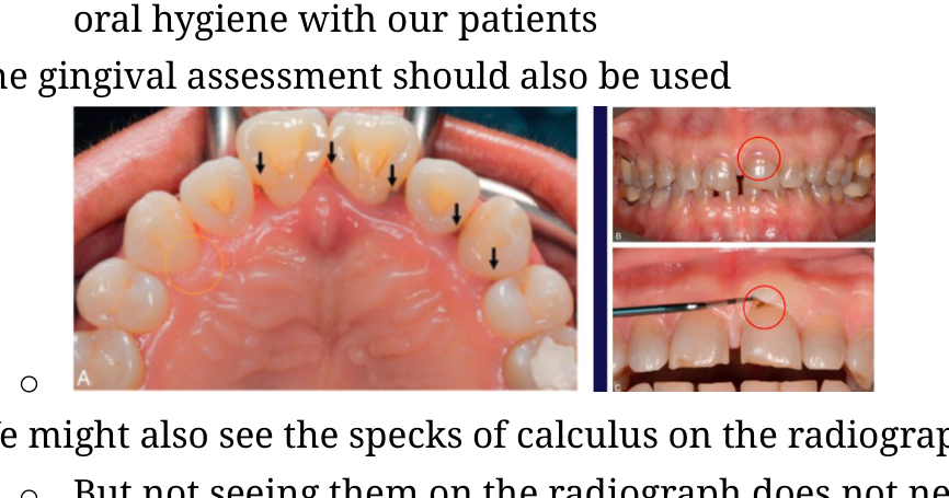
What is the typical visual appearance of supragingival calculus?
It appears as a chalky white area in comparison to the surrounding hard tissues.
dentistry calculus -
What substance is used for the detection of supragingival biofilm?
Triplaque ID gel.
biofilm dentistry -
Describe the clinical technique used for the supragingival detection of biofilm and calculus.
- Dry the teeth with a triplex syringe
- Observe with a mirror at different angles
- Use reflected light
dentistry diagnosis -
How much time is generally required for gingival tissue to fully heal after flap surgery?
3 months.
periodontics surgery -
What is the primary purpose of conducting a gingival assessment during a dental visit?
It serves to assess gingival tissue health and acts as an opportunity to discuss oral hygiene with the patient.
 assessment hygiene
 -
Does the absence of visible calculus on a radiograph confirm that no calculus is present on the tooth?
No. The absence of calculus on a radiograph does not guarantee it is not present, as smaller amounts may not be visible.
radiography calculus -
Why are tooth overhangs considered problematic during an oral assessment?
They act as plaque-retentive areas, leading one to expect a higher accumulation of biofilm or calculus in those specific locations.
restoration plaque -
Which clinical conditions might be detected when using an 11/12 explorer probe on a tooth surface?
- Supra-gingival calculus
- Sub-gingival calculus
- Insufficient filling margins
instrumentation periodontics -
What is the correct alignment for an 11/12 explorer probe shank relative to the tooth during use?
The shank of the instrument should be parallel to the long axis of the tooth.
instrumentation technique -
How much of the 11/12 explorer probe's toe should be inserted underneath the gingiva when exploring the sulcus?
Approximately 2-3 mm.
instrumentation technique -
What does it indicate if dental floss gets stuck while sliding over a proximal tooth surface?
The presence of calculus.
assessment calculus -
What is a systematic way to divide a tooth into its different surfaces for examination purposes?
Using line angles to divide the tooth.
examination anatomy -
Which curette is indicated for the area from the mesio-buccal line angle to the contact point and from the mesial palatal line to the mesial contact point?
11/12 curette
dentistry curettes -
Which curette is used for the area from the distobuccal line angle to the contact point and from the distopalatal line angle back to the contact point?
13/14 curette
dentistry curettes -
Which curette is used for the buccal and palatal surface from line angle to line angle?
7/8 curette
dentistry curettes -
What is the recommended width for instrumenting sections on a tooth surface?
\(1-2\text{ mm}\)
dentistry instrumentation -
When inserting a curette for instrumentation, what is the correct orientation of the terminal shank to the long axis of the tooth?
Parallel
dentistry technique -
How many strokes are typically performed within a single section of the tooth surface during instrumentation?
\(2-3\) strokes
dentistry technique -
How should lateral pressure be adjusted when transitioning from removing biofilm to removing calculus?
Use light strokes for biofilm; increase lateral pressure for calculus to effectively engage and dislodge it.
dentistry technique -
What is the primary challenge when instrumentation reaches the middle (3rd) division of a tooth surface?
The terminal shank often hits the embrasure around the contact point.
dentistry instrumentation -
How should an instrument be positioned when working on the middle (3rd) division to avoid the contact point?
The instrument must be angled to be below the contact point.
dentistry instrumentation -
What stroke types might be combined when vertical strokes cannot be performed on a tooth surface?
Oblique or horizontal strokes.
dentistry instrumentation -
When using power scalers, what is the ideal angulation of the tip against the tooth surface?
30-45 degrees
0-5 degrees
0-15 degrees
45-90 degrees
dentistry power-scalers -
What kind of water spray is required when using a power scaler?
A fine spraying mist generated around the tip.
dentistry power-scalers -
Describe the technique for using power scaler strokes when cleaning a pocket.
Use little, pressureless, feather-like strokes that overlap and meander down into the base of the pocket.
dentistry power-scalers -
What is the recommended angulation for power scaler tips during instrumentation?
\(0^\circ - 15^\circ\)
dentistry instrumentation -
What type of movement is recommended when using a power scaler?
Meandering movement
dentistry instrumentation -
How much pressure should be applied when using a power scaler tip?
Pressure less
dentistry instrumentation -
At what speed should the power scaler tip be moved over the tooth surface?
Slowly
dentistry instrumentation -
What is the recommended path of movement for a power scaler tip during subgingival instrumentation?
From the sulcus to the base of the pocket
dentistry instrumentation -
What is the recommended width for the sections being instrumented with a power scaler?
\(1 - 2\text{ mm}\)
dentistry instrumentation -
Which tip is used for subgingival instrumentation in the clinic?
XF tip
dentistry instrumentation -
Which tip is used for subgingival instrumentation in the SimClinic?
6a or 61/62 tip
dentistry instrumentation