덱이 사라지기 전에 저장해 둬
이 플래시카드는 아직 저장되지 않았어 — 페이지를 나가면 사라져. 무료 계정을 만들면 저장되고 아래 기능들도 모두 이용할 수 있어.
- Save this deck to your account
- Study with spaced repetition
- Export to Anki (.apkg) or PDF
- Process documents up to 100 pages
- Images extracted from your PDFs
- Sharper text extraction & a more advanced AI model
What is osteology?
What are the primary functions of bones?
What are examples of tubular bones?
What characterizes pneumatic bones?
What is the proximal epiphysis of a bone?
What is the distal epiphysis of a bone?
What is the diaphysis?
What is the function of the periosteum in bone anatomy?
What structure within a long bone houses bone marrow?
What is the role of articular cartilage in joint surfaces?
What defines surface markings on bones?
What is the characteristic organization of compact (cortical) bone?
What type of bone tissue uses a trabecular meshwork to provide strength with minimal weight?
What are the primary structures found in the head (caput) region of the skeleton?
Which specific vertebrae are located in the spinal (vertebral column) region?
What structures compose the chest (thorax) region of the skeletal system?
What bones make up the shoulder girdle?
The extremities of the skeleton primarily include which limbs?
Which bone is found in the arm region?
What bones make up the forearm?
Name the bones that constitute the human hand.
What bones form the pelvic girdle?
Which bone is located in the thigh?
What bone protects the knee?
What bones comprise the leg (region between knee and foot)?
List the bones that make up the human foot.
What are the characteristics of a fibrous (syndesmosis) joint?
What are the characteristics of a cartilaginous (synchondrosis) joint?
What are the characteristics of a bony (synostosis) joint?
What is defined as a synostotic connection?
Describe the characteristics and an example of a Uniaxial Hinge (ginglymus) joint.
What type of joint allows for rotational motion with a single axis, such as in the C1-C2 joint?
What kind of motion does a Biaxial Ellipsoid joint allow and where is it commonly found?
Which joint type permits opposition movement, exemplified by the thumb carpometacarpal joint?
What are the characteristics and examples of a Multiaxial Spheroid joint?
List the typical structural components of a synovial joint.
What are the two primary functions of muscle contraction?
What substance is released at the neuromuscular junction to initiate muscle contraction?
How does the size of a motor unit relate to movement precision?
What is the defining characteristic of a fusiform muscle?
What is the structural description of a flat muscle?
What distinguishes a multibellied muscle?
What are the characteristics of feather-shaped (pennatus) muscles?
How are muscles with multiple origins described?
What is the difference between muscle origin and insertion?
What are the three phases of bone healing?
What are the two primary components that make up a spinal disc? 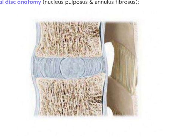
What are two structural characteristics of the human skull? 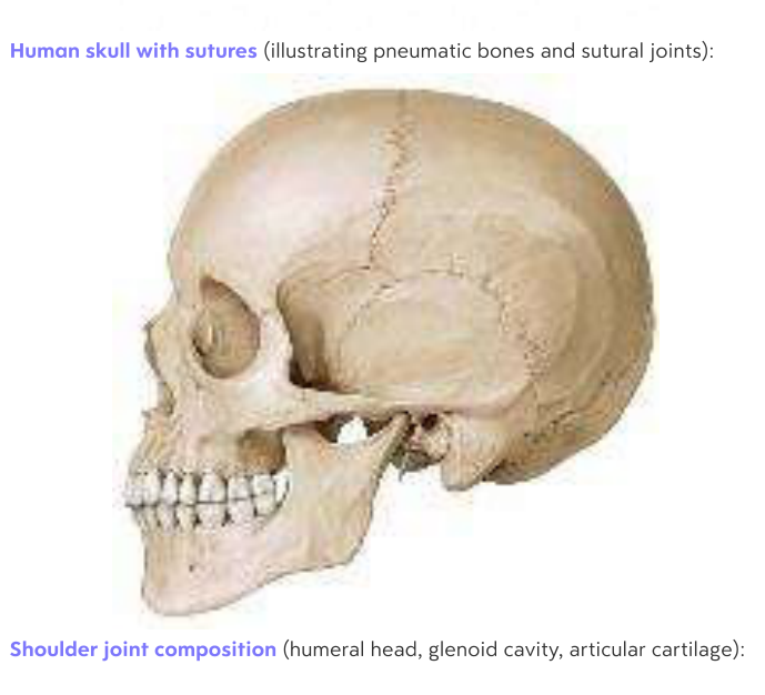
What are the three main components that compose the shoulder joint? 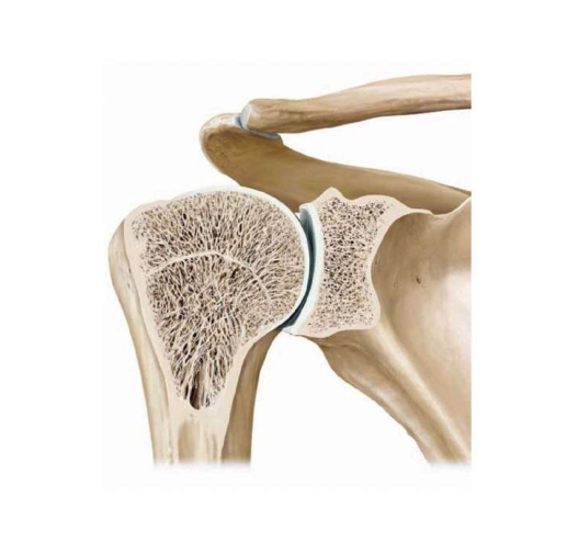
What is the central gel-like core of a spinal disc known as?
Which anatomical structure acts as a cushion between the bones in the shoulder joint?
What term describes the specialized joints that connect the bones of the skull?
What is the primary definition of epithelial tissue?
Which two stains are standard in histology and what colors do they produce?
What are the characteristics and common locations of simple epithelium?
What defines stratified epithelium and where is it typically found?
What is unique about the cellular structure of pseudostratified epithelium?
What are the primary functions and key sites of simple squamous epithelium?
What is the characteristic structure of simple cuboidal epithelium?
Where is simple cuboidal epithelium typically found in the body?
What is the primary structural characteristic of simple columnar epithelium?
What are the common surface specializations of columnar epithelium?
What are the functional and structural characteristics of microvilli?
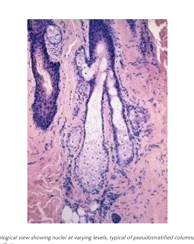
Why does pseudostratified columnar epithelium appear to be multilayered?
What is a key structural requirement for cells in pseudostratified columnar epithelium regarding the basal lamina?
What specialized cells are typically found within pseudostratified columnar epithelium?
What are two functional specializations of pseudostratified columnar epithelium and where are they located?
How is stratified epithelium classified?
What is the primary function and location of stratified squamous epithelium?
Where are stratified cuboidal and stratified columnar epithelia found?
What is the primary function of Zonula occludens (tight junctions)?
What are the main components and function of a Desmosome?
Which cell junction is responsible for direct electrical and chemical communication between cells?
What is the function of Hemidesmosomes?
What constitutes the structure of the basement membrane?
What is the fundamental difference between primary and secondary sensory epithelium?
What does Hematoxylin stain and what color does it produce?
What does Eosin stain and what color does it produce?
What is the primary function of HE staining in microscopy?
Name the three major parts of the small intestine.
List the components of the large intestine starting from the cecum.
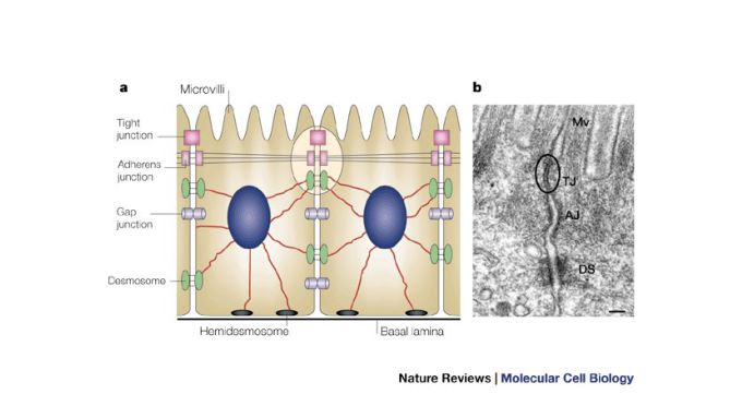
What are some examples of cellular junctions shown in the illustration?
List the organs located in the abdomen, excluding the alimentary tract.
Which pelvic organs are unique to the female anatomy?
Which pelvic organs are unique to the male anatomy?
Which pelvic organs are common to both biological sexes?
What major structures are located in the Right hypochondrium region?
Which organs are found in the Epigastrium?
What structures occupy the Left hypochondrium region?
Which structures are contained in the Right lateral region?
What is located in the Umbilical region?
Which structures are found in the Left lateral region?
What structures are located in the Vesical (suprapubic) region?
What structures are located in the left hypochondrium?
Which organs are found in the umbilical region?
What is the clinical significance of McBurney's point?
What is the peritoneum?
What type of tissue forms the peritoneum and what is its function?
What is the difference between the parietal and visceral peritoneum?
What are mesenteries (peritoneal reflections) and what is their role?
What is the primary function of the omentum majus?
What structures does the mesentery suspend?
Which organs are classified as primary retroperitoneal?
List the organs classified as secondary retroperitoneal.
What is the defining characteristic of an intraperitoneal organ?
How are secondary retroperitoneal organs defined regarding their developmental history?
What does the term infraperitoneal refer to?
What are the categories for organ classification within the abdomen?
What defines the peritoneal cavity?
What are the primary functions of peritoneal duplications, such as mesenteries and the omentum?
Which organs are classified as infraperitoneal in males?
Which organs are classified as infraperitoneal in females?
Does the female peritoneal cavity maintain a closed environment?
How does the lesser sac communicate with the greater sac?
What is the clinical significance of the 9 abdominal regions?
What is the continuous pathway of the urinary system for urine elimination?
What are the vertebral levels spanned by the right and left kidneys?
Which muscles do the kidneys rest against posteriorly?
Why does the right kidney sit slightly lower than the left kidney?
What is the function of the renal capsule?
What is the function of the adipose capsule?
What is the function of the renal fascia?
How are kidneys classified in relation to the peritoneum?
What is the primary functional tissue of the kidney?
What are the three main components found within the renal sinus?
What structures are contained within the renal cortex?
How is the renal medulla organized?
What components make up the medullary rays?
What is the function of the peritubular capillaries surrounding the nephron?
Which German term corresponds to the renal cortex in the provided diagram?
Which German term corresponds to the renal medulla in the provided diagram?
What is the primary composition of a nephron?
What are the two layers of the Bowman's capsule?
What is the function of the afferent arteriole in the kidney?
What is the function of the efferent arteriole?
What does the glomerulus consist of?
What is the pathway of blood flow through the renal cortex as part of renal circulation?
What structures compose the renal corpuscle?
What are the three glomerular filtration layers and their sizes?
What is the primary function of Erythropoietin (EPO) produced by the kidneys?
What is the primary function of Calcitriol produced by the kidneys?
What is the primary function of Renin produced by the kidneys?
What is the correct sequence of the urinary tract pathway?
What are the typical lengths of the urethra in females and males?
What lines the interior of the urinary bladder?
What is the primary function of blood?
What percentage of total body fluid is accounted for by intracellular fluid?
What are the two major components of whole blood and their respective percentages?
What are the main constituents of blood plasma?
What are the three main types of cellular elements found in blood?
Define the roles of plasma and cellular elements in the blood.
What is the primary function of water in plasma?
What is serum defined as in the context of blood components?
What are the three main proteins found in plasma and their primary functions?
What are the physical dimensions of a typical human red blood cell?
Why do red blood cells lack nuclei?
What is the structural composition of hemoglobin?
What is the primary function of red blood cells in the oxygen transport cycle?
What defines the ABO blood group system?
What are the RBC antigens and plasma antibodies present in an individual with type AB blood?
What are the RBC antigens and plasma antibodies present in an individual with type O blood?
Define anemia and its primary cause.
List common symptoms associated with anemia.
What is the primary function of platelets?
What is the typical count and size range of platelets?
What is the Blood Clotting Cascade?
What are the key components of the blood clotting process?
What condition results from a low platelet count?
What causes the red blood cells to deform into a sickle shape in sickle cell disease?
What are the common clinical consequences of sickle cell disease?
What antibodies and antigens are present in individuals with blood type A?
What is the typical percentage range of neutrophils in total leukocytes?
What is the primary function of eosinophil granulocytes?
Which granules are contained in basophil granulocytes?
What characterizes the nucleus and size of monocytes?
What is the function of neutrophils regarding bacterial infection?
What is the primary role of Cytotoxic T cells?
What function do Helper T cells perform in the immune system?
What is the primary role of B cells?
Define cyanosis.
Where does hematopoiesis occur?
What cells are produced from the Myeloid lineage?
What stimulates erythropoiesis?
What is the primary definition of lymphatic fluid?
How much interstitial fluid is typically formed daily, and what portion of it becomes lymph?
What structural characteristics allow lymphatic capillaries to transport substances like proteins and lipids?
What happens to the vast majority of interstitial fluid that is filtered through capillary walls?
What formula describes the effective filtration pressure (\(P_{eff}\)) in capillaries?
How is the filtration rate (\(Q_f\)) calculated according to the capillary pressure diagram?
What is the primary destination for substances in a lymphatic vessel?
How does the permeability of a lymphatic vessel compare to a capillary?
Describe the structural differences between capillary and lymphatic vessel openings.
What cells are contained within the primary lymphatic organ known as bone marrow?
What specific cells arise from multipotent hematopoietic stem cells in the bone marrow?
Which lymphatic organ is characterized by an epithelial reticulum framework of endodermal origin and lacks lymphatic follicles?
How is the Thymus organized structurally?
What is the primary function of the thymic cortex?
What processes occur in the thymic medulla?
What is the function of the capsule and septa in the thymus?
What is the role of High-Endothelial Venules (HEV) at the cortex-medulla border?
What is the purpose of T-cell double selection?
What is the function of the blood-thymus barrier?
What are the typical sizes for primary and secondary lymphatic follicles?
What are the primary components of a lymphoid follicle?
What cell types and structures are found in the lymph node cortex?
What is the primary function of the lymph node paracortex?
What components are found within the lymph node medulla?
What is the entry and exit point for blood and lymph vessels in a lymph node?
What is the correct sequence of lymph flow through a lymph node?
What are the structural characteristics of medullary sinuses?
What are the primary physiological functions of the spleen?
What is the composition and role of the capsule in the spleen?
What cell types are found within the white pulp of the spleen?
Which structure in the spleen does the white pulp typically surround?
What allows for the passage of blood cells through the sinuses in the red pulp?
What cells and components are hosted in the splenic cords?
What are the two pathways of circulation in the spleen?
What is the general term for mucosal-associated lymphoid tissue?
Provide examples of Gut-Associated (GALT) and Bronchus-Associated (BALT) lymphoid tissues.
What is Skin-Associated Lymphoid Tissue (SALT) and where is it located?
Describe the structure of tonsils.
List the four types of tonsils.
What type of epithelium covers the palatine tonsil?
What is the primary function of tonsillar crypts?
What is the primary cell type found in the interfollicular space of the tonsils?
What is the primary cell type found in tonsillar follicles?
Where does pus accumulate in cases of follicular tonsillitis?
What are the primary functions of fibroblasts in connective tissue?
How do fibrocytes differ from fibroblasts?
What are the primary roles of white adipocytes?
What is the specialized function of brown adipocytes?
What is the nature of mesenchymal cells?
Where are reticulum cells typically found?
List the three main types of connective tissue fibers.
How do fixed cells and mobile cells differ in connective tissue?
What is the primary function of histiocytes (macrophages) in connective tissue?
What substances are released by mast cells during an anaphylactic reaction?
Which specific vitamin is required for the hydroxylation process during collagen synthesis?
What are the physical properties of collagen fibers?
What is the historical significance of James Lind regarding collagen?
What components make up elastic fibers?
What morphological characteristics describe plasma cells?
What are the components of Mesenchyma tissue?
What is the primary composition of Wharton's jelly?
Which tissues are found in the Reticular category of mature cell-rich connective tissue?
Where is Spinocellular connective tissue found?
What are the key features and locations of Areolar tissue?
What is the function of Loose CT in mature fiber-rich connective tissue?
What is the characteristic fiber arrangement of Dense regular connective tissue?
What is the characteristic fiber arrangement of Dense irregular connective tissue?
Which stains are used to visualize reticular fibers?
What are the primary adhesive glycoproteins involved in cell-matrix adhesion?
What is the primary function of the perichondrium in cartilage?
Name the two types of cartilage and their distinct locations, as shown in the table.
What are the two major components of the bone matrix?
What is the bone cell lineage starting from the progenitor cell?
Which hormones and vitamin regulate calcium exchange via osteocytes and osteoclasts?
What are the primary cells that secrete matrix proteins in loose connective tissue?
What is the primary structural difference between compact bone and spongy bone?
Which microscopic structures are found within the compact bone system?
What is the tissue precursor for intramembranous ossification, and what are some examples of bones formed this way?
What is the tissue precursor for endochondral ossification?
From which embryonic tissue does Primary Angiogenic (Krompecher) ossification originate?
What are the three main zones of endochondral bone formation?
What is the cellular activity within the reserve cartilage zone?
What occurs during the proliferation stage of endochondral bone formation?
What is the primary activity in the hypertrophy zone?
What occurs during the 'Invasion' stage of bone formation?
What are the general characteristics of skeletal muscle?
What is the primary function of the sarcoplasmic reticulum in muscle cells?
What is the primary function of fixed cells in connective tissue?
What is the primary function of mobile cells in connective tissue?
What are the origin and representative bones of intramembranous ossification?
What are the origin and representative bones of endochondral ossification?
What structural differences distinguish thick skin from thin skin?
Which cellular structures in smooth muscle are analogous to Z-discs?
On which parts of the body are hair follicles absent?
How does the thickness of the epidermis differ between the palms/soles and the rest of the body?
What type of sweat glands are found on the palms and soles?
What is the function of the stratum basale in the epidermis?
How does the stratum spinosum contribute to the skin?
What significant biological process begins in the stratum granulosum?
What are the key characteristics of cells found in the stratum corneum?
What is the primary function of keratinocytes?
What is the physical appearance of the border between the epidermis and the underlying dermis?
What is the primary function of melanocytes in the epidermis?
What is the role of Langerhans cells in the skin?
What is the function of Merkel cells?
What are keratinocytes responsible for in the epidermis?
What structural component provides tensile strength to keratinocytes?
What factor primarily determines human skin colour?
What is the primary function of Langerhans cells in the epidermis?
What is the role of Merkel cells in the skin?
What is the purpose of the extended dendritic processes found on Langerhans cells?
What primary structures and tissues are contained within the dermis?
What is the primary composition of the papillary layer of the dermis?
What are the characteristics and functions of the reticular layer?
What is the primary function and composition of the subcutis (hypodermis)?
What is the function of the Vater-Pacini corpuscles?
What is the primary function and distribution of sweat glands?
Where are odoriferous (apocrine) glands located, and what is their secretion type?
What is the structural type and secretion of sebaceous glands?
What is the characteristic structure of apocrine glands?
How do merocrine sweat glands release their products?
What is the secretory mechanism of sebaceous glands?
Name the three concentric layers of a hair shaft.
What layers of the skin does the hair follicle traverse?
What is the primary function of the hair follicle bulge?
Which two types of glands are associated with the hair follicle?
What is the function of the outer root sheath of a hair follicle?
In which body cavity is the heart located?
Within the thoracic cavity, specifically where is the heart positioned?
What is the name of the membrane that encloses the heart?
What is the primary function of the pericardial sac?
Which structure lies adjacent to the heart?
What is the inferior relation of the heart?
What is the function of the Right atrium?
Which vessels return systemic blood to the heart?
What is the function of the Right ventricle?
What is the function of the Left atrium?
What is the function of the Left ventricle?
What is the role of the Aorta?
What is the function of the Pulmonary veins?
Which groove separates the atria from the ventricles?
Which valve is located at the right atrioventricular orifice?
What is the function of the chordae tendineae and papillary muscles?
What is the role of the coronary sinus in the heart?
Which valve is found at the left atrioventricular orifice?
Into which chamber of the heart do the four pulmonary veins empty?
From which heart chamber does the aorta originate?
Where are the atrioventricular valves located?
What is the primary function of the atrioventricular valves?
Where are the semilunar valves located?
What is the movement cycle of the semilunar valves?
What is the function of the papillary muscles and chordae tendineae?
Why is the obstruction of the LAD (anterior interventricular artery) clinically significant?
What is the full name for the cardiac abbreviation RA?
What is the full name for the cardiac abbreviation RV?
What is the full name for the cardiac abbreviation LA?
What is the full name for the cardiac abbreviation LV?
What is the full name for the cardiac abbreviation TP?
Define automaticity in the context of cardiac function.
How does sympathetic innervation affect the heart?
How does parasympathetic (vagus) innervation affect the heart?
What is the standard conduction pathway of the heart?
What is the primary function of the upper airways?
What is the function of the lower airways?
What occurs during the pulmonary circuit?
What is the effect of vasoconstriction on blood pressure?
What is the effect of vasodilation on blood pressure?
What is the role of lymph vessels?
What clinical condition results from impaired lymphatic drainage?
What is the primary function of the nasal conchae within the skull? 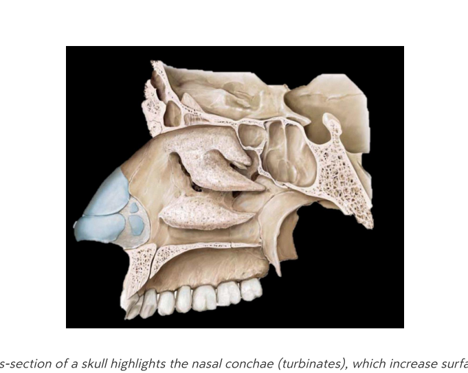
What are the structures collectively known as the respiratory tract? 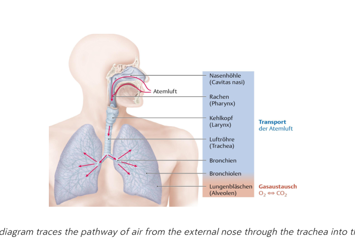
Which specific components comprise the upper and lower divisions of the respiratory tract?
What is the biological function of vibrissae in the nostrils? 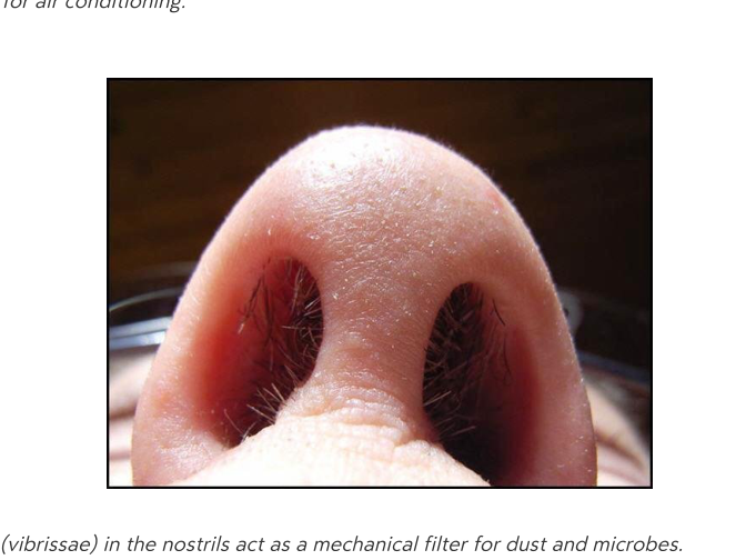
What defines the naris and the nasal septum?
What is the primary function of rudimentary nasal muscles like the levator labii superioris alaeque nasi?
Which type of epithelium lines the nasal vestibule, and what specialized structure does it contain?
What type of epithelium is characteristic of the proper nasal cavity?
What is the role of goblet cells in the nasal cavity?
What is the primary function of the nasal mucosa regarding inhaled air?
How are trapped particles transported through the nasal airway?
How are nasal sprays absorbed into the body?
What is the significance of the olfactory region in drug delivery?
What is the primary risk associated with the overuse of nasal sprays?
What is the anatomical function of the nasal turbinates (conchae)?
What is the choana?
What microscopic feature of the respiratory epithelium is visible as hair-like projections?
What are the four types of paranasal sinuses?
What are the primary functions of the paranasal sinuses?
What are the clinical signs of sinusitis?
What are the three parts of the ethmoid air cells?
What are the three main divisions of the pharynx?
Which structure connects the nasal cavity to the nasopharynx?
What are the three wall layers of the pharynx?
What are the two primary functions of the larynx?
What happens to the epiglottis during the swallowing sequence?
Name the primary cartilages of the larynx.
What is another name for the vestibular folds?
What is the function of the vocalis muscle?
What occurs when the glottis is partially open?
What is the function of the primary inspiratory muscle, the diaphragm?
What is the role of intercostal muscles in respiration?
What is a coniotomy?
What are the paired organs responsible for gas exchange in the respiratory tract?
Between which vertebral levels does the trachea extend?
Describe the structural composition of the tracheal cartilage rings.
What is the function of the ciliary rejection flow in the airways?
Why is the right main bronchus at a higher risk of foreign-body aspiration?
In the bronchial tree, which generation corresponds to the main (primary) bronchi?
What is the approximate size of terminal bronchioles?
What is the structural sequence of the bronchial tree following the respiratory bronchioles?
What is the first site of gas exchange in the respiratory tree?
What is the function of Type I pneumocytes in the alveoli?
What is the primary function of Type II pneumocytes in the alveoli?
How does parasympathetic stimulation affect the airway?
What is the effect of sympathetic stimulation on bronchial smooth muscle?
What are the two main types of pharmacological agents used as bronchodilators?
What is the role of glands and mucus cells in the airway?
What is the direction of gas diffusion between alveolar air and capillary blood?
What is the primary function of Type I pneumocytes?
What is the primary function of Type II pneumocytes?
What is the role of alveolar macrophages?
How does ciliary rejection flow protect the respiratory tract?
What is the purpose of the cough reflex?
Which biological components participate in the airway's immune responses?
What is the primary function of the pleura in the human body?
Describe the location and function of the visceral and parietal pleura.
What is the role of the pleural cavity?
What are the three subdivisions of the parietal pleura?
What occurs in the thoracic cavity during quiet inhalation?
What is the primary action of the external intercostal muscles during breathing?
What happens during quiet exhalation?
Which muscles are involved in forced expiration?
What is the principal muscle of quiet inspiration?
Which nerve provides innervation to the diaphragm?
What clinical consequence arises from a spinal cord injury at the C4 level or above?
What is the primary function of the external intercostal muscles?
What is the primary function of the internal intercostal muscles?
Which muscles act as accessory muscles to enhance thoracic expansion?
Which major structures pass through openings in the diaphragm?
What defines the boundaries of the oral vestibule?
What defines the boundaries of the oral cavity proper?
Where does the Stensen duct of the parotid gland open?
What is the primary secretory contribution of the submandibular gland?
Which gland produces mostly mucous, low-volume saliva through multiple minor ducts?
What is the name of the structure that acts as the opening for the parotid duct? 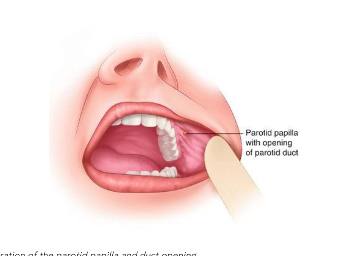
How is parotid inflammation (mumps) prevented in Hungary?
What are the primary components of saliva? 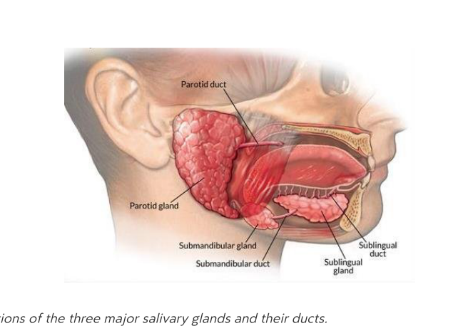
Which gland produces a more mucin-rich, enzyme-poor saliva under sympathetic stimulation?
What are the structural parts of a tooth? 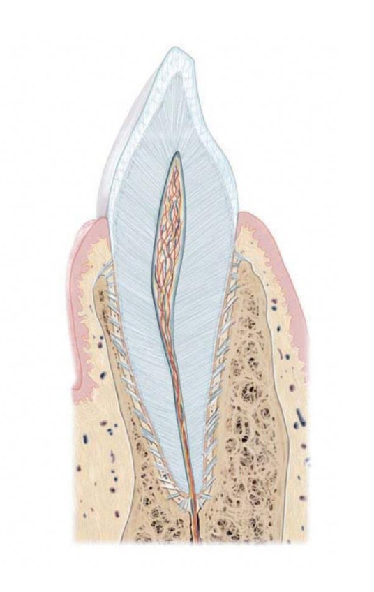
According to the dental formula per quadrant, how many incisors, canines, and premolars are in a permanent set?
Compare the number of premolars in a deciduous (primary) set versus a permanent set per quadrant.
What is the function of the intrinsic muscles of the tongue?
What is the primary function of extrinsic muscles of the tongue?
Which cranial nerve provides innervation to both intrinsic and extrinsic muscles of the tongue?
What lymphoid tissue is contained within the root of the tongue?
What is found on the body of the tongue?
Why is the sublingual area suitable for rapid drug absorption?
What anatomical structure serves as the boundary between the oral cavity and the oropharynx?
Which structures are responsible for flanking the palatine tonsil?
Where is the uvula located?
What is the alternative name for the Epipharynx?
What is the alternative name for the Mesopharynx?
What is the alternative name for the Hypopharynx?
What are the components of the mucous membrane (tunica mucosa)?
What is the primary function of the pharyngeal wall?
What kind of tissue is found in the submucosa layer of the pharynx?
What does the muscularis layer of the pharynx consist of?
What type of tissue constitutes the adventitia of the pharynx?
Identify the three sections of the pharynx shown in the sagittal cross-section.
What are the key pathophysiologic mechanisms involved in the development of gastro-esophageal reflux disease (GERD)?
What are the common clinical symptoms of GERD?
List the standard diagnostic work-up procedures for GERD.
What are the primary classes of pharmacologic agents used to manage GERD?
How does the epithelium differ between the nasopharynx and the oro-/laryngopharynx?
What type of epithelium covers the oral cavity?
What is the primary definition of the gastrointestinal (GI) tract?
At what vertebral levels is the stomach located?
What is the anatomical position of the stomach relative to the diaphragm?
What is the anatomical position of the stomach relative to the liver?
List the primary organs that make up the gastrointestinal system as shown in the diagram.
What is the function of the cardia region of the stomach?
What is the fundus of the stomach and what does it often contain?
What are the primary functions of the stomach?
What do parietal cells secrete in the stomach?
What do chief cells secrete, and what is the role of that substance?
What substance do G cells secrete?
What is the role of the gastric \(H^+\) produced by parietal cells?
What is the primary pathogenesis and typical treatment for a peptic ulcer?
What are the common causes and management for a drug-induced ulcer?
What is the trigger for a stress ulcer and how is it managed?
Which substances are received by the duodenum to facilitate digestion?
What is the role of the greater duodenal papilla?
What are the primary functions of the jejunum and ileum?
What are the components of the tunica mucosa layer in the small intestine?
What are the components of the tunica submucosa layer in the small intestine?
What are the components of the tunica muscularis in the small intestine?
What are the components of the tunica serosa/adventitia in the small intestine?
What are the three primary surface area enhancements in the small intestine?
What is the primary function of the large intestine?
Where is the vermiform appendix located?
How is the vermiform appendix classified in terms of organ type?
What are the common symptoms of vermiform appendix conditions?
What is the typical progression of pain in a patient experiencing early appendicitis?
From which anatomical structure does the rectum receive fecal matter?
What is the difference in control between the external and internal anal sphincters?
How does the mucosal lining of the large intestine differ from that of the small intestine regarding villi?
Which type of epithelium characterizes the Tunica mucosa of the large intestine?
What is the clinical significance of the venous drainage pattern in the lowest portion of the rectum?
What is the largest gland in the human body?
Name the primary metabolic processes of the liver.
What vitamins and substances are stored in the liver?
Which proteins does the liver synthesize?
What is the function of the Porta hepatis?
List the four surfaces of the liver.
Into how many functional segments is the human liver divided?
What is considered the smallest morphological or functional unit of the liver?
What is the drainage pathway of blood from the hepatic lobule?
What three structures make up the portal triad within a portal lobule?
From which vessels does the hepatic lobule receive blood?
Which structure within the hepatic lobule is responsible for draining bile?
What is the primary contribution of the portal vein to the liver's blood supply?
What is the primary contribution of the proper hepatic artery to the liver's blood supply?
What is the 'first-pass effect' in the context of liver circulation?
What is the pathway of blood flow through the liver?
How is the hepatic acinus organized?
What are the components of the bile duct system in the liver?
What is the definition of cholelithiasis?
What is icterus, also known as jaundice?
Which organ receives secretions via the pancreatic duct?
Into which part of the digestive tract does bile and pancreatic juice flow?
Name the anatomical structure that stores bile and is connected to the common bile duct.
What condition typically leads to the formation of collateral veins and portosystemic shunts?
Why are portosystemic shunts a significant clinical concern?
List the four major sites of porto-caval anastomoses.
What is the normal length of the pancreas?
What is the anatomical position of the pancreas in relation to the peritoneum?
Between which spinal levels does the pancreas span?
What are the two main types of functional activity of the pancreas?
What substances are secreted by serous acinar glands in the pancreas?
Which structure does the major pancreatic duct join at the duodenum?
What is the result of premature enzyme activation in the pancreas?
What condition is associated with chronic pancreatitis?
Where is the pancreas located in the body? 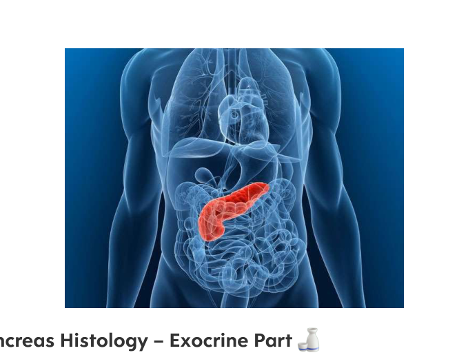
What does the provided histological micrograph represent? 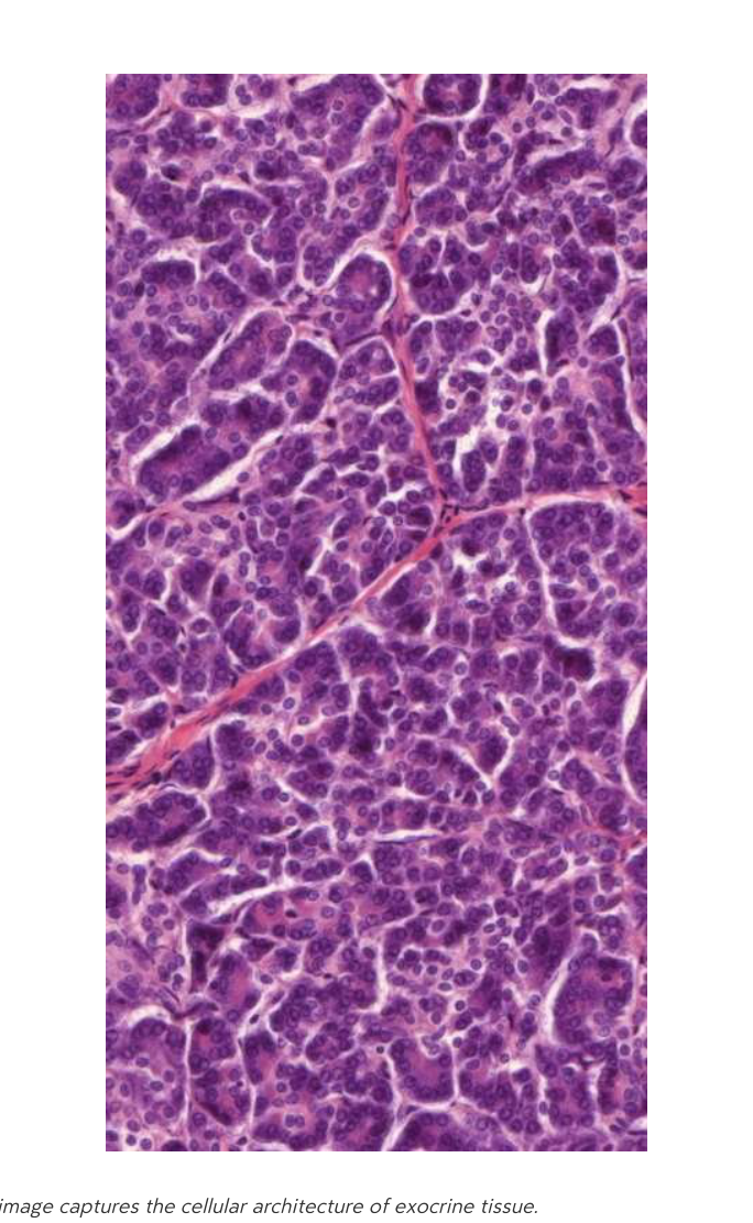
Where is the endocrine part of the pancreas located?
Which hormone is secreted by \(\alpha\)-cells in the pancreatic islets?
Which hormone is secreted by \(\beta\)-cells in the pancreatic islets?
What is the clinical consequence of an insulin deficiency?
Which hormone is secreted by \(\delta\)-cells in the pancreatic islets?
Identify the primary cell populations found within the endocrine pancreas.


Flashcards in this deck (546)
-
What is osteology?
The study of bone structure and function.
한글뜻: 골학. 뼈의 구조와 기능을 연구하는 학문입니다.
anatomy osteology -
What are the primary functions of bones?
- Provide a solid framework for the body.
- Support movement passively.
- Protect vital organs.
- Produce blood cells in red bone marrow.
- Store calcium and phosphate ions.
한글설명: 신체의 골격 제공, 움직임 보조, 장기 보호, 혈액 세포 생성, 칼슘과 인산염 저장 기능을 합니다.
bones functions -
What are examples of tubular bones?
Long bones such as the femur and humerus.
한글뜻: 관상골(대퇴골, 상완골 등 긴 뼈).
bones anatomy -
What characterizes pneumatic bones?
They contain air-filled sinuses.
한글뜻: 함기골. 뼈 내부에 공기가 차 있는 공간(부비동)을 포함합니다.
bones anatomy -
What is the proximal epiphysis of a bone?
The end of the bone located near the center of the body.
한글뜻: 근위 골단. 몸의 중심에 더 가까운 뼈의 끝부분입니다.
bones anatomy -
What is the distal epiphysis of a bone?
The end of the bone located farthest from the center of the body.
한글뜻: 원위 골단. 몸의 중심에서 가장 멀리 떨어진 뼈의 끝부분입니다.
bones anatomy -
What is the diaphysis?
The long shaft of a bone that connects the epiphyses.
한글뜻: 골간. 두 골단을 연결하는 뼈의 긴 몸통 부분입니다.
bones anatomy -
What is the function of the periosteum in bone anatomy?
The periosteum is a dense collagenous connective tissue that carries blood vessels and nerves.
한글 뜻: 골막 한글 설명: 뼈를 감싸고 있으며 혈관과 신경이 지나가는 치밀한 결합 조직입니다.
anatomy bone -
What structure within a long bone houses bone marrow?
The medullary cavity.
한글 뜻: 골수강 한글 설명: 뼈 내부에 있는 공간으로, 골수(적색 또는 황색)를 포함하고 있습니다.
anatomy bone -
What is the role of articular cartilage in joint surfaces?
It serves as a smooth tissue covering joint surfaces.
한글 뜻: 관절 연골 한글 설명: 관절면을 덮고 있는 매끄러운 조직으로, 마찰을 줄여주는 역할을 합니다.
anatomy joints -
What defines surface markings on bones?
They act as attachment sites for tendons and ligaments.
한글 뜻: 뼈 표면 표시 한글 설명: 힘줄(건)과 인대가 뼈에 붙는 지점입니다.
anatomy bone -
What is the characteristic organization of compact (cortical) bone?
It features lamellar organization in concentric tubes (Haversian systems).
한글 뜻: 치밀골 (피질골) 한글 설명: 하버스관계를 중심으로 층판 구조가 원형으로 배열되어 있습니다.
anatomy bone -
What type of bone tissue uses a trabecular meshwork to provide strength with minimal weight?
Spongy (cancellous) bone.
한글 뜻: 해면골 (망상골) 한글 설명: trabecula(잔기둥) 구조로 이루어져 있어 가벼우면서도 강도를 유지합니다.
anatomy bone -
What are the primary structures found in the head (caput) region of the skeleton?
The skull (cranium).
한글뜻: 머리(두개골) 부위 쉬운 설명: 우리 몸의 머리 부분을 보호하고 구성하는 핵심 뼈입니다.
anatomy skeleton -
Which specific vertebrae are located in the spinal (vertebral column) region?
Cervical, thoracic, and lumbar vertebrae.
한글뜻: 척추 부위 쉬운 설명: 목부터 허리까지 이어지는 등뼈들로 구성되어 있습니다.
anatomy skeleton -
What structures compose the chest (thorax) region of the skeletal system?
Ribs (costae) and sternum (breast bone).
한글뜻: 가슴 부위 쉬운 설명: 갈비뼈와 가슴 중앙의 납작한 뼈로, 장기를 보호합니다.
anatomy skeleton -
What bones make up the shoulder girdle?
Scapula and clavicle.
한글뜻: 어깨뼈 쉬운 설명: 팔을 몸통에 연결하는 어깨 부위의 뼈들입니다.
anatomy skeleton -
The extremities of the skeleton primarily include which limbs?
Upper and lower limbs.
한글뜻: 사지 쉬운 설명: 팔과 다리를 통칭하는 말입니다.
anatomy skeleton -
Which bone is found in the arm region?
Humerus
(한글 뜻: 위팔뼈 / 설명: 팔의 윗부분을 구성하는 뼈입니다.)
anatomy bones -
What bones make up the forearm?
- Radius
- Ulna
(한글 뜻: 노뼈, 자뼈 / 설명: 팔꿈치에서 손목까지 이어지는 아래팔의 두 뼈입니다.)
anatomy bones -
Name the bones that constitute the human hand.
- 8 carpal bones
- 5 metacarpals
- Phalanges (2–3 per digit)
(한글 뜻: 손목뼈, 손허리뼈, 손가락뼈 / 설명: 손을 구성하는 뼈들입니다.)
anatomy bones -
What bones form the pelvic girdle?
- Hip bone (os coxae = ilium + pubis + ischium)
- Sacrum
- Coccyx
(한글 뜻: 골반대, 엉덩뼈, 엉치뼈, 꼬리뼈 / 설명: 몸통과 다리를 연결하는 골반을 구성하는 뼈들입니다.)
anatomy bones -
Which bone is located in the thigh?
Femur
(한글 뜻: 넙다리뼈(대퇴골) / 설명: 허벅지를 구성하는 길고 강한 뼈입니다.)
anatomy bones -
What bone protects the knee?
Patella
(한글 뜻: 무릎뼈(슬개골) / 설명: 무릎 관절 앞쪽에 위치하여 관절을 보호하는 뼈입니다.)
anatomy bones -
What bones comprise the leg (region between knee and foot)?
- Tibia
- Fibula
(한글 뜻: 정강뼈, 종아리뼈 / 설명: 무릎 아래에서 발목까지 이어지는 종아리의 두 뼈입니다.)
anatomy bones -
List the bones that make up the human foot.
- 7 tarsal bones
- 5 metatarsals
- Phalanges (2–3 per digit)
(한글 뜻: 발목뼈, 발허리뼈, 발가락뼈 / 설명: 발을 구성하는 뼈들입니다.)
anatomy bones -
What are the characteristics of a fibrous (syndesmosis) joint?
- Characteristics: Minimal movement
- Example: Skull sutures, interosseous membrane
- 한글 뜻: 섬유성 관절 (신데스모시스)
- 한글 설명: 움직임이 거의 없는 관절입니다. 뼈들이 섬유 조직으로 단단히 연결되어 있습니다.
anatomy joints -
What are the characteristics of a cartilaginous (synchondrosis) joint?
- Characteristics: Slight flexibility
- Example: Intervertebral discs
- 한글 뜻: 연골성 관절 (신콘드로시스)
- 한글 설명: 약간의 유연성을 가진 관절입니다. 연골 조직이 뼈 사이를 연결합니다.
anatomy joints -
What are the characteristics of a bony (synostosis) joint?
- Characteristics: Bones essentially merged
- Example: Fusion of hip bone, sacrum
- 한글 뜻: 뼈 관절 (시노스토시스)
- 한글 설명: 뼈들이 완전히 합쳐져 하나로 융합된 상태의 관절입니다.
anatomy joints -
What is defined as a synostotic connection?
A connection where separate bones fuse together.
[한글 뜻: 유합성 관절] [한글 설명: 별개의 뼈들이 하나로 완전히 합쳐져 고정된 관절 형태를 말합니다.]
anatomy joints -
Describe the characteristics and an example of a Uniaxial Hinge (ginglymus) joint.
It has 1 axis of motion and allows for bending. Example: Phalanges.
[한글 뜻: 단축성 - 경첩 관절] [한글 설명: 축이 하나이며 주로 굽힘 운동을 합니다. 예: 손가락뼈.]
anatomy joints -
What type of joint allows for rotational motion with a single axis, such as in the C1-C2 joint?
Uniaxial Pivot (trochoid) joint.
[한글 뜻: 단축성 - 차축 관절] [한글 설명: 축이 하나이며 회전 운동을 담당합니다. 예: 경추 1-2번 관절.]
anatomy joints -
What kind of motion does a Biaxial Ellipsoid joint allow and where is it commonly found?
It has 2 axes for flexion-extension and ab-adduction. Example: Wrist.
[한글 뜻: 이축성 - 타원 관절] [한글 설명: 두 개의 축을 가지며 굽힘/폄 및 벌림/모음이 가능합니다. 예: 손목.]
anatomy joints -
Which joint type permits opposition movement, exemplified by the thumb carpometacarpal joint?
Biaxial Saddle joint.
[한글 뜻: 이축성 - 안장 관절] [한글 설명: 엄지손가락의 맞섬 운동처럼 두 축을 가진 관절입니다.]
anatomy joints -
What are the characteristics and examples of a Multiaxial Spheroid joint?
It has 3 axes and allows for a wide range of motion. Examples: Shoulder and hip.
[한글 뜻: 다축성 - 구상 관절] [한글 설명: 3개의 축을 가지며 아주 넓은 범위로 움직일 수 있습니다. 예: 어깨와 엉덩이.]
anatomy joints -
List the typical structural components of a synovial joint.
- Articular cartilage
- Synovial membrane
- Joint capsule
- Ligaments
- Bursae
[한글 뜻: 윤활 관절 구성 요소] [한글 설명: 관절을 이루는 핵심 부위들로 관절연골, 윤활막, 관절주머니, 인대, 윤활주머니 등이 있습니다.]
anatomy joints -
What are the two primary functions of muscle contraction?
- Production of active movement (shortening)
- Maintenance of posture (tone)
(한글 번역: 근육 수축의 두 가지 주요 기능은 무엇입니까? / 답: 1. 능동적 움직임 생성(단축) 2. 자세 유지(긴장) / 쉬운 설명: 근육은 몸을 움직이거나 일정한 자세를 유지하는 역할을 합니다.)
muscle physiology -
What substance is released at the neuromuscular junction to initiate muscle contraction?
Acetylcholine.
(한글 번역: 근육 수축을 시작하기 위해 신경근 접합부에서 방출되는 물질은 무엇입니까? / 답: 아세틸콜린 / 쉬운 설명: 신경에서 근육으로 신호를 전달하는 화학 물질입니다.)
muscle physiology -
How does the size of a motor unit relate to movement precision?
The size of a motor unit determines the precision of movement.
(한글 번역: 운동 단위의 크기는 움직임의 정밀도와 어떤 관련이 있습니까? / 답: 운동 단위의 크기가 움직임의 정밀도를 결정합니다. / 쉬운 설명: 하나의 신경이 조절하는 근육 섬유가 적을수록 더 정밀한 동작이 가능합니다.)
muscle physiology -
What is the defining characteristic of a fusiform muscle?
It has a round belly, typically seen in long, thin muscles like the biceps.
(한글 번역: 방추형 근육의 정의적인 특징은 무엇입니까? / 답: 이두박근과 같은 길고 얇은 근육에서 볼 수 있는 둥근 배 모양을 가지고 있습니다. / 쉬운 설명: 양끝은 가늘고 가운데가 볼록한 근육 모양입니다.)
muscle morphology -
What is the structural description of a flat muscle?
A broad, sheet-like muscle attached by a tendon-like fibrous sheet (aponeurosis).
(한글 번역: 편평근의 구조적 설명은 무엇입니까? / 답: 건막이라 불리는 힘줄 같은 섬유 시트를 통해 부착되는 넓고 판 모양의 근육입니다. / 쉬운 설명: 얇고 넓게 퍼진 형태의 근육입니다.)
muscle morphology -
What distinguishes a multibellied muscle?
It has two or more bellies, such as the quadriceps.
(한글 번역: 다복근은 무엇으로 구별됩니까? / 답: 대퇴사두근과 같이 두 개 이상의 근육 배를 가지고 있습니다. / 쉬운 설명: 하나의 근육에 볼록한 부분이 여러 개 있는 형태입니다.)
muscle morphology -
What are the characteristics of feather-shaped (pennatus) muscles?
Multiple slender fascicles radiating from a central tendon.
(한글 번역: 우상근의 특징은 무엇입니까? / 답: 중앙 힘줄에서 깃털 모양으로 갈라져 나오는 여러 개의 가느다란 근속들입니다. / 쉬운 설명: 깃털처럼 중앙 힘줄에 근육 섬유들이 비스듬히 붙어 있는 형태입니다.)
muscle morphology -
How are muscles with multiple origins described?
Muscles, such as the biceps or triceps, that possess several distinct attachment sites.
(한글 번역: 기시부가 여러 개인 근육은 어떻게 설명됩니까? / 답: 이두박근이나 삼두박근처럼 여러 개의 부착 부위를 가지고 있는 근육입니다. / 쉬운 설명: 근육이 뼈에 붙는 시작점이 여러 곳인 근육입니다.)
muscle morphology -
What is the difference between muscle origin and insertion?
The origin (punctum fixum) is the fixed attachment near the midline, while the insertion (punctum mobile) is the distal, movable attachment away from the midline.
(한글 번역: 근육의 기시와 정지의 차이는 무엇입니까? / 답: 기시부는 신체 중심 근처의 고정된 부착점이고, 정지부는 중심에서 멀리 떨어진 움직이는 부착점입니다.)
muscle physiology -
What are the three phases of bone healing?
- Inflammatory phase
- Soft callus formation
- Hard callus and remodeling
(한글 번역: 뼈 치유의 세 단계는 무엇입니까? / 답: 1. 염증기 2. 연성 가골 형성기 3. 경성 가골 및 재형성기 / 쉬운 설명: 뼈가 부러졌을 때 염증이 생기고, 연한 조직이 차오른 뒤, 점차 딱딱한 뼈로 변하며 다시 형태를 잡는 과정입니다.)
bone healing -
What are the two primary components that make up a spinal disc?
- Nucleus pulposus
- Annulus fibrosus
한글 의미: 척추 원반의 구성 요소 (수핵 및 섬유륜) 설명: 척추 원반은 중앙의 젤 같은 수핵과 이를 둘러싼 단단한 섬유륜으로 이루어져 있습니다.
anatomy spine -
What are two structural characteristics of the human skull?
- Pneumatic bones
- Sutural joints
한글 의미: 인간 두개골의 특징 (함기골 및 봉합 관절) 설명: 두개골에는 공기가 차 있는 공간인 함기골이 있고, 뼈와 뼈 사이는 봉합 관절로 연결되어 있습니다.
anatomy skull -
What are the three main components that compose the shoulder joint?
- Humeral head
- Glenoid cavity
- Articular cartilage
한글 의미: 어깨 관절의 구성 요소 (상완골두, 관절와, 관절 연골) 설명: 어깨 관절은 상완골의 머리 부분과 어깨뼈의 관절와가 만나며, 이 사이를 부드러운 관절 연골이 감싸고 있습니다.
anatomy shoulder -
What is the central gel-like core of a spinal disc known as?
Nucleus pulposus
한글 의미: 수핵 설명: 척추 원반의 가장 중심부에 위치한 젤 형태의 물질로, 충격을 흡수하는 역할을 합니다.
anatomy spine -
Which anatomical structure acts as a cushion between the bones in the shoulder joint?
Articular cartilage
한글 의미: 관절 연골 설명: 관절의 뼈 끝을 덮고 있는 부드러운 조직으로, 마찰을 줄이고 관절이 부드럽게 움직이도록 돕습니다.
anatomy shoulder -
What term describes the specialized joints that connect the bones of the skull?
Sutural joints
한글 의미: 봉합 관절 설명: 두개골의 뼈들을 단단하게 결합해 주는 고정된 관절의 한 형태입니다.
anatomy skull -
What is the primary definition of epithelial tissue?
It forms continuous coverings of body surfaces, lines internal passages, and forms glands. It is characterized by tightly packed cells with minimal intercellular substance.
[Korean: 상피 조직(Epithelial tissue)의 정의] - 한글 뜻: 몸의 표면을 덮고 내부 통로를 감싸며 분비샘을 형성하는 조직입니다. 세포가 매우 빽빽하게 밀집되어 있고 세포 사이 물질이 거의 없습니다.
biology histology -
Which two stains are standard in histology and what colors do they produce?
Haematoxylin, which stains blue, and Eosin, which stains pink.
[Korean: 조직학적 염색약] - 한글 뜻: 파란색을 띠는 헤마톡실린(Haematoxylin)과 분홍색을 띠는 에오신(Eosin)을 주로 사용합니다.
histology methods -
What are the characteristics and common locations of simple epithelium?
It consists of a single layer of cells that may be squamous, cuboidal, or columnar. It is found in locations such as the alveoli and kidney tubules.
[Korean: 단순 상피(Simple epithelium)의 특징] - 한글 뜻: 한 층의 세포로 이루어져 있으며 폐포나 신장 세관에서 발견됩니다.
epithelial histology -
What defines stratified epithelium and where is it typically found?
It is composed of two or more layers of cells. It is commonly found in the oral cavity, esophagus, and vagina.
[Korean: 중층 상피(Stratified epithelium)의 특징] - 한글 뜻: 두 개 이상의 세포 층으로 이루어져 있으며 구강, 식도, 질에서 발견됩니다.
epithelial histology -
What is unique about the cellular structure of pseudostratified epithelium?
Although it appears multilayered, all cells touch the basal lamina. It is composed of columnar cells and found in the respiratory tract and male reproductive ducts.
[Korean: 거짓중층 상피(Pseudostratified epithelium)의 구조] - 한글 뜻: 여러 층처럼 보이지만 사실 모든 세포가 바닥 막(기저판)에 닿아 있습니다. 호흡기 등에 존재합니다.
epithelial histology -
What are the primary functions and key sites of simple squamous epithelium?
It functions in diffusion, filtration, and secretion. It is found in the endothelium of blood vessels and the alveolar walls of the lungs.
[Korean: 단순 편평 상피(Simple squamous epithelium)의 기능] - 한글 뜻: 확산, 여과, 분비 기능을 하며 혈관 내피와 폐포 벽에서 발견됩니다.
epithelial histology -
What is the characteristic structure of simple cuboidal epithelium?
It consists of a single layer of cube-shaped cells with centrally located, round nuclei.
[한글 뜻: 단순 입방 상피] [쉬운 설명: 입방체 모양의 세포가 한 층으로 배열되어 있으며, 핵은 가운데에 위치합니다.]
histology epithelium -
Where is simple cuboidal epithelium typically found in the body?
- Outer surface of the lens of the eye
- Small ducts of many glands
- Proximal and distal convoluted tubules of the nephron
- Amniotic epithelium
[한글 뜻: 위치] [쉬운 설명: 눈의 수정체 표면, 분비샘의 작은 관, 신장의 세뇨관 등에서 발견됩니다.]
histology locations -
What is the primary structural characteristic of simple columnar epithelium?
It is composed of a single layer of column-shaped cells, where nuclei are often basal but may appear at various levels in pseudostratified variants.
[한글 뜻: 단순 원주 상피] [쉬운 설명: 기둥 모양의 세포가 한 층으로 되어 있으며, 핵은 보통 세포 아래쪽에 있습니다.]
histology epithelium -
What are the common surface specializations of columnar epithelium?
- Microvilli: Increase absorptive surface area (\(≈ 2μm\))
- Cilia: Motile structures composed of microtubules (\(≈ 10μm\))
- Stereocilia: Long microvilli (\(> 15μm\))
[한글 뜻: 특수 구조] [쉬운 설명: 미세융모(흡수), 섬모(운동), 그리고 긴 미세융모인 부동섬모가 있습니다.]
histology specializations -
What are the functional and structural characteristics of microvilli?
They are finger-like projections containing a core of actin filaments that dramatically expand the apical surface for absorption.
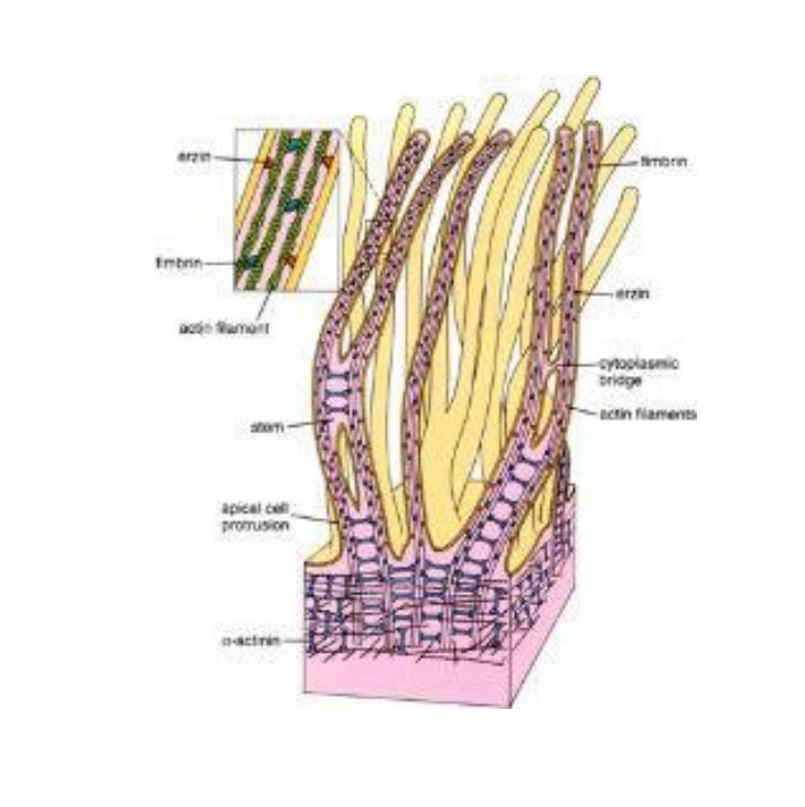
[한글 뜻: 미세융모] [쉬운 설명: 세포 표면의 손가락 모양 돌기로, 액틴 필라멘트가 들어있어 영양분 흡수를 돕습니다.]
histology microvilli -
Why does pseudostratified columnar epithelium appear to be multilayered?
It appears multilayered because the nuclei are positioned at different heights within the cells.
한글뜻: 위중층 원주 상피가 다층으로 보이는 이유는? 설명: 세포핵들이 서로 다른 높이에 위치하기 때문에 현미경으로 보면 층이 여러 개인 것처럼 보입니다.
histology epithelium -
What is a key structural requirement for cells in pseudostratified columnar epithelium regarding the basal lamina?
Every cell in the tissue contacts the basal lamina.
한글뜻: 위중층 원주 상피의 모든 세포가 갖는 구조적 특징은? 설명: 모든 세포가 바닥막(basal lamina)에 닿아 있습니다.
histology epithelium -
What specialized cells are typically found within pseudostratified columnar epithelium?
- Goblet cells: Secrete mucus
- Basal cells: Function as reserve cells
한글뜻: 위중층 원주 상피에서 볼 수 있는 특수 세포는? 설명: 점액을 분비하는 술잔세포(goblet cells)와 예비 역할을 하는 바닥세포(basal cells)가 포함되어 있습니다.
histology epithelium -
What are two functional specializations of pseudostratified columnar epithelium and where are they located?
- Respiratory tract: Contains cilia for mucus-propelling.
- Epididymis: Contains stereocilia for absorption.
한글뜻: 위중층 원주 상피의 특수화된 기능과 위치는? 설명: 호흡기에서는 섬모를 통해 점액을 밀어내고, 부고환에서는 부동섬모(stereocilia)를 통해 흡수를 합니다.
histology epithelium -
How is stratified epithelium classified?
It is classified based on the shape of the cells in the superficial (top) layer.
한글뜻: 중층 상피는 어떻게 분류되나요? 설명: 가장 바깥쪽(표면)에 있는 세포의 모양을 기준으로 분류합니다.
histology epithelium -
What is the primary function and location of stratified squamous epithelium?
- Function: Protects against abrasion.
- Location: Skin and oral mucosa.
한글뜻: 중층 편평 상피의 주된 기능과 위치는? 설명: 마찰로부터 보호하는 역할을 하며, 피부나 구강 점막 등에 분포합니다.
histology epithelium -
Where are stratified cuboidal and stratified columnar epithelia found?
- Stratified cuboidal: Lines some ducts of sweat glands and mammary glands.
- Stratified columnar: Rare; found in parts of the male urethra and certain glandular ducts.
한글뜻: 중층 입방 상피와 중층 원주 상피는 어디에 있나요? 설명: 중층 입방은 땀샘/유선관에, 중층 원주는 요도나 특정 분비샘 관에 드물게 존재합니다.
histology epithelium -
What is the primary function of Zonula occludens (tight junctions)?
They form a seal to prevent the passage of ions and water between cells.
(한글: 세포 사이의 물질 이동을 막는 밀착 연접. 세포 간의 틈을 봉쇄하여 이온과 물의 통과를 차단합니다.)
histology cell-junctions -
What are the main components and function of a Desmosome?
Components: Desmoglein, desmocollin, and intermediate filaments. Function: Provides mechanical strength to tissues.
(한글: 데스모솜. 주성분은 데스모글레인, 데스모콜린, 중간섬유이며, 조직에 기계적 강도를 제공합니다.)
histology cell-junctions -
Which cell junction is responsible for direct electrical and chemical communication between cells?
Gap junction. Its primary components are connexons (connexins).
(한글: 간극 연접. 코넥손(코넥신)으로 구성되며 세포 간 전기적, 화학적 신호 소통을 담당합니다.)
histology cell-junctions -
What is the function of Hemidesmosomes?
They anchor epithelial cells to the underlying basement membrane.
(한글: 반데스모솜. 상피 세포를 기저막에 고정시키는 역할을 합니다.)
histology cell-junctions -
What constitutes the structure of the basement membrane?
It consists of a \(50\text{ nm}\) basal lamina (produced by epithelium) and a reticular lamina (from connective tissue), totaling \(\approx 200\text{ nm}\) in thickness.
(한글: 기저막. 상피 유래의 기저판(\(50\text{ nm}\))과 결합조직 유래의 그물판으로 구성된 약 \(200\text{ nm}\) 두께의 구조물입니다.)
histology tissues -
What is the fundamental difference between primary and secondary sensory epithelium?
Primary sensory epithelium contains neurons with axons that transmit signals directly to the brain, whereas secondary sensory epithelium lacks axons and relies on receptors innervated by other neurons.
(한글: 일차 감각 상피는 직접 뇌로 신호를 보내는 신경세포를 가지지만, 이차 감각 상피는 축삭이 없으며 다른 신경의 지배를 받는 수용체를 가집니다.)
histology sensory-epithelium -
What does Hematoxylin stain and what color does it produce?
It stains acidic (basophilic) structures like the nucleus, RER, and ribosomes blue.
한글: 헤마톡실린(Haematoxylin)은 산성 구조(핵, 리보솜 등)를 파란색으로 염색합니다.
histology staining -
What does Eosin stain and what color does it produce?
It stains basic (acidophilic) components like cytoplasm and collagen fibers pink/red.
한글: 에오신(Eosin)은 염기성 구조(세포질, 콜라겐 섬유 등)를 분홍색/빨간색으로 염색합니다.
histology staining -
What is the primary function of HE staining in microscopy?
It provides contrast to reveal cellular morphology and tissue organization.
한글: HE 염색은 세포의 형태와 조직 구조를 명확하게 관찰할 수 있도록 대비(contrast)를 만들어 줍니다.
histology microscopy -
Name the three major parts of the small intestine.
- Duodenum
- Jejunum
- Ileum
한글: 소장의 세 부분은 십이지장(Duodenum), 공장(Jejunum), 회장(Ileum)입니다.
anatomy digestive -
List the components of the large intestine starting from the cecum.
- Cecum
- Vermiform appendix
- Ascending colon
- Transverse colon
- Descending colon
- Sigmoid colon
- Rectum
한글: 대장은 맹장(Cecum), 충수(Vermiform appendix), 상행/횡행/하행 결장, S상 결장, 직장(Rectum)으로 구성됩니다.
anatomy digestive -
What are some examples of cellular junctions shown in the illustration?
Tight junctions, adherens junctions, gap junctions, desmosomes, and hemidesmosomes.
한글: 세포 간 연결 구조로는 밀착연접, 부착연접, 간극연접, 데스모솜, 헤미데스모솜 등이 있습니다.
 biology cells
 -
List the organs located in the abdomen, excluding the alimentary tract.
- Liver (hepar)
- Gallbladder (vesica fellea)
- Spleen (lien, splen)
- Pancreas (pancreas)
- Kidney (ren)
- Adrenal gland (glandula suprarenalis)
- Ureter
(한글: 간, 쓸개, 지라(비장), 이자(췌장), 콩팥(신장), 부신, 요관. 소화관을 제외한 복부 장기들입니다.)
anatomy abdomen -
Which pelvic organs are unique to the female anatomy?
- Uterus
- Vagina
- Ovaries (ovarium)
- Fallopian tube (tuba uterina)
(한글: 자궁, 질, 난소, 난관. 여성에게만 있는 골반 장기입니다.)
anatomy pelvis female -
Which pelvic organs are unique to the male anatomy?
- Seminal vesicle (vesicula seminalis)
- Prostate (prostata)
(한글: 정낭, 전립선. 남성에게만 있는 골반 장기입니다.)
anatomy pelvis male -
Which pelvic organs are common to both biological sexes?
- Urinary bladder (vesica urinaria)
- Rectum
(한글: 방광, 직장. 남녀 공통 골반 장기입니다.)
anatomy pelvis -
What major structures are located in the Right hypochondrium region?
- Right liver lobe
- Gallbladder
- Right colic flexure
한글뜻: 우측 갈비밑부위 설명: 복부의 오른쪽 위 구역으로 간의 오른쪽 엽, 쓸개, 오른쪽 대장굽이가 위치합니다.
anatomy abdomen -
Which organs are found in the Epigastrium?
- Stomach (central)
- Pancreas
- Duodenum
한글뜻: 명치부위 설명: 복부 중앙 위쪽 구역으로 위, 췌장, 십이지장이 포함됩니다.
anatomy abdomen -
What structures occupy the Left hypochondrium region?
- Spleen
- Left colic flexure
- Stomach (left)
한글뜻: 좌측 갈비밑부위 설명: 복부 왼쪽 위 구역으로 지라(비장), 왼쪽 대장굽이, 위의 일부가 위치합니다.
anatomy abdomen -
Which structures are contained in the Right lateral region?
- Ascending colon
- Right kidney
한글뜻: 오른쪽 옆구리부위 설명: 복부 오른쪽 중간 구역으로 오름결장과 오른쪽 콩팥이 위치합니다.
anatomy abdomen -
What is located in the Umbilical region?
- Transverse colon
- Small intestine
한글뜻: 배꼽부위 설명: 복부 정중앙 구역으로 가로결장과 작은창자가 포함됩니다.
anatomy abdomen -
Which structures are found in the Left lateral region?
- Descending colon
- Left kidney
한글뜻: 왼쪽 옆구리부위 설명: 복부 왼쪽 중간 구역으로 내림결장과 왼쪽 콩팥이 위치합니다.
anatomy abdomen -
What structures are located in the Vesical (suprapubic) region?
- Urinary bladder
- Uterus (female)
- Prostate (male)
한글뜻: 방광위부위 설명: 복부 가장 아래 중앙 구역으로 방광, 자궁(여성), 전립선(남성)이 위치합니다.
anatomy abdomen -
What structures are located in the left hypochondrium?
- Spleen
- Left colic flexure
- Left portion of stomach
(한글뜻: 좌상복부 - 지라, 좌결장곡, 위장의 좌측 부분. 갈비뼈 아래 왼쪽 영역의 장기들입니다.)
anatomy abdominal -
Which organs are found in the umbilical region?
- Transverse colon
- Small intestine (jejunum, ileum)
(한글뜻: 배꼽 부위 - 가로결장, 소장. 배꼽을 중심으로 위치한 소화 기관들입니다.)
anatomy abdominal -
What is the clinical significance of McBurney's point?
It is an anatomical landmark used to diagnose appendicitis, where pain is often felt in the right iliac region.
(한글뜻: 맥버니점 - 맹장염을 진단하는 해부학적 위치입니다. 오른쪽 아랫배의 통증으로 나타납니다.)
medicine anatomy -
What is the peritoneum?
One of the three serous membranes (along with pleura and pericardium) that lines the abdominal cavity.
(한글뜻: 복막 - 복강 내부를 감싸고 있는 세 개의 장막 중 하나입니다.)
anatomy histology -
What type of tissue forms the peritoneum and what is its function?
Simple squamous epithelium (mesothelium); it secretes lubricating fluid.
(한글뜻: 복막 조직 - 단층 편평 상피(중피)로 구성되며, 장기들이 잘 움직이도록 윤활액을 분비합니다.)
histology anatomy -
What is the difference between the parietal and visceral peritoneum?
- Parietal peritoneum: Lines the abdominal wall.
- Visceral peritoneum: Covers the abdominal organs.
(한글뜻: 벽쪽복막과 내장쪽복막 - 벽쪽은 복벽을 덮고, 내장쪽은 장기 표면을 덮습니다.)
anatomy -
What are mesenteries (peritoneal reflections) and what is their role?
Double-layered folds of peritoneum that suspend organs and provide a pathway for vessels and nerves.
(한글뜻: 장간막 - 장기를 매달아 지지하고 혈관과 신경이 지나가는 통로 역할을 하는 복막의 이중 주름입니다.)
anatomy physiology -
What is the primary function of the omentum majus?
It hangs from the greater curvature of the stomach and covers the intestines.
(한글 뜻: 대망 - 위장의 큰 만곡부에서 늘어져 장을 덮고 있음.)
anatomy peritoneum -
What structures does the mesentery suspend?
It suspends the small intestine, specifically the jejunum and ileum.
(한글 뜻: 장간막 - 소장(공장과 회장)을 매달고 있음.)
anatomy peritoneum -
Which organs are classified as primary retroperitoneal?
- Kidney
- Adrenal gland
- Ureter
(한글 뜻: 일차 복막뒤기관 - 신장, 부신, 요관)
anatomy retroperitoneal -
List the organs classified as secondary retroperitoneal.
- Pancreas
- Duodenum (except the first part)
- Ascending colon
- Descending colon
- Middle part of the rectum
(한글 뜻: 이차 복막뒤기관 - 췌장, 십이지장(첫 부분 제외), 상행결장, 하행결장, 직장 중간부)
anatomy retroperitoneal -
What is the defining characteristic of an intraperitoneal organ?
It is completely surrounded by visceral peritoneum.
(한글 뜻: 복막내기관 - 장측 복막으로 완전히 둘러싸여 있음.)
anatomy peritoneum -
How are secondary retroperitoneal organs defined regarding their developmental history?
They are originally intraperitoneal but become secondarily fixed to the posterior wall.
(한글 뜻: 이차 복막뒤기관 - 원래는 복막내에 있었으나 이차적으로 고정된 기관.)
anatomy retroperitoneal -
What does the term infraperitoneal refer to?
Structures that lie below the peritoneal cavity, such as the bladder.
(한글 뜻: 복막하기관 - 방광처럼 복막강 아래에 위치하는 구조물.)
anatomy peritoneum -
What are the categories for organ classification within the abdomen?
Organs are classified as: - Intraperitoneal - Retroperitoneal - Infraperitoneal
[한글뜻: 장기 분류] [한글 설명: 복강 내 장기들은 위치에 따라 복막 내, 복막 후, 복막 하 장기로 나뉩니다.]
anatomy organs -
What defines the peritoneal cavity?
The peritoneal cavity is a continuous serous sac that exhibits anatomical differences between the sexes.
[한글뜻: 복막강] [한글 설명: 복막강은 연속된 장막 주머니이며, 남성과 여성의 해부학적 차이가 있습니다.]
anatomy peritoneum -
What are the primary functions of peritoneal duplications, such as mesenteries and the omentum?
They suspend organs and provide pathways for the transmission of vessels and nerves.
[한글뜻: 복막 중복(장간막/대망)] [한글 설명: 장간막이나 대망 같은 구조는 장기를 고정하고 혈관과 신경이 지나가는 통로 역할을 합니다.]
anatomy mesentery -
Which organs are classified as infraperitoneal in males?
- Urinary bladder
- Prostate
- Rectum (lowest portion)
- Rectovesical pouch (deepest point)
[한글뜻: 남성의 복막 하 장기] [한글 설명: 남성에게서 방광, 전립선, 직장의 하부, 직장방광오목이 이에 해당합니다.]
anatomy male -
Which organs are classified as infraperitoneal in females?
- Urinary bladder
- Uterus
- Rectum
- Vagina
- Associated pouches: Vesicouterine and Rectouterine (Douglas) pouch
[한글뜻: 여성의 복막 하 장기] [한글 설명: 여성에게서 방광, 자궁, 직장, 질 및 관련 복막 오목들이 해당합니다.]
anatomy female -
Does the female peritoneal cavity maintain a closed environment?
No, the female peritoneal cavity communicates with the exterior through the uterine tubes and the vagina.
[한글뜻: 여성 복막강의 개방성] [한글 설명: 여성의 복막강은 나팔관과 질을 통해 외부와 연결되어 완전히 닫혀 있지 않습니다.]
anatomy female physiology -
How does the lesser sac communicate with the greater sac?
It communicates via the omental foramen.
[한글뜻: 소망낭의 통로] [한글 설명: 소망낭은 대망공을 통해 복강의 큰 주머니와 연결됩니다.]
anatomy digestive -
What is the clinical significance of the 9 abdominal regions?
Each of the 9 abdominal regions contains characteristic organs, helping in clinical localization.
[한글뜻: 9개의 복부 영역] [한글 설명: 9개 복부 구역은 각각 특정 장기를 포함하고 있어 임상적 위치 파악에 유용합니다.]
anatomy abdomen -
What is the continuous pathway of the urinary system for urine elimination?
Kidney → Ureter → Bladder → Urethra
(소변 배출 경로: 신장 → 요관 → 방광 → 요도)
anatomy urinary -
What are the vertebral levels spanned by the right and left kidneys?
- Right kidney: T12-L3
- Left kidney: T11-L2
(척추 위치: 오른쪽 신장은 T12에서 L3, 왼쪽 신장은 T11에서 L2 척추뼈 사이에 위치합니다.)
anatomy kidney -
Which muscles do the kidneys rest against posteriorly?
The quadratus lumborum and psoas major muscles.
(신장 후방 근육: 신장은 요방형근과 대요근에 기대어 위치합니다.)
anatomy muscles -
Why does the right kidney sit slightly lower than the left kidney?
Because of the hepatic mass.
(오른쪽 신장이 더 낮은 이유: 간의 부피(hepatic mass) 때문입니다.)
anatomy liver -
What is the function of the renal capsule?
It acts as a thin, fibrous covering for the kidney.
(신장 피막: 신장을 얇게 덮고 있는 섬유질 막입니다.)
anatomy kidney -
What is the function of the adipose capsule?
It consists of perirenal fat surrounding the kidney.
(지방 피막: 신장 주위를 둘러싸고 있는 지방층입니다.)
anatomy kidney -
What is the function of the renal fascia?
It is connective tissue that anchors the kidney to surrounding structures.
(신장 근막: 신장을 주변 조직에 고정해주는 결합 조직입니다.)
anatomy kidney -
How are kidneys classified in relation to the peritoneum?
They are primary retroperitoneal organs.
(복막 위치: 신장은 복막 뒤쪽에 있는 후복막 장기입니다.)
anatomy kidney -
What is the primary functional tissue of the kidney?
Parenchyma.
한글뜻: 실질(기능성 조직) 설명: 신장의 주요 기능을 수행하는 조직입니다.
kidney anatomy -
What are the three main components found within the renal sinus?
- Cortex
- Medulla
- Calyx
한글뜻: 피질, 수질, 신배 설명: 신장 내부의 중심 공간인 신동에 위치한 구조들입니다.
kidney anatomy -
What structures are contained within the renal cortex?
Renal corpuscles, consisting of the glomerulus and Bowman's capsule.
한글뜻: 신소체 (사구체와 보우만 주머니) 설명: 신장의 바깥쪽 층인 피질에서 발견되는 주요 구조물입니다.
cortex nephron -
How is the renal medulla organized?
It is organized into renal pyramids.
한글뜻: 신추체 설명: 신장 내부의 안쪽 영역인 수질은 피라미드 모양의 구조로 정렬되어 있습니다.
medulla anatomy -
What components make up the medullary rays?
- Straight portions of the proximal convoluted tubule (PCT)
- Distal convoluted tubule (DCT)
- Henle's loop
- Collecting ducts
한글뜻: 수질 방사선 (근위세뇨관, 원위세뇨관, 헨레 고리, 집합관) 설명: 신장의 수질 영역을 구성하는 세뇨관들의 곧은 부분입니다.
medulla anatomy -
What is the function of the peritubular capillaries surrounding the nephron?
They facilitate reabsorption and secretion.
한글뜻: 재흡수와 분비 설명: 네프론을 감싸고 있는 모세혈관망은 혈액과 세뇨관 사이의 물질 교환을 돕습니다.
physiology nephron -
Which German term corresponds to the renal cortex in the provided diagram?
Rinde.
한글뜻: 피질 설명: 신장의 바깥쪽 층을 가리키는 독일어 의학 용어입니다.
terminology anatomy -
Which German term corresponds to the renal medulla in the provided diagram?
Mark.
한글뜻: 수질 설명: 신장의 안쪽 층을 가리키는 독일어 의학 용어입니다.
terminology anatomy -
What is the primary composition of a nephron?
A nephron consists of a renal corpuscle and a tubule system.
한글뜻: 네프론의 주요 구성 성분은 무엇인가요? 한글설명: 네프론은 신장의 기능적 단위로, 신소체와 세뇨관 시스템으로 이루어져 있습니다.
biology nephron -
What are the two layers of the Bowman's capsule?
- Visceral layer (podocytes)
- Parietal layer (simple epithelium)
한글뜻: 보우만 주머니(Bowman's capsule)의 두 층은 무엇인가요? 한글설명: 보우만 주머니는 장측층(족세포로 구성)과 벽측층(단층 상피로 구성)으로 나뉩니다.
biology kidney -
What is the function of the afferent arteriole in the kidney?
It serves as the input vessel that delivers blood to the glomerulus.
한글뜻: 신장에서 수입소동맥(afferent arteriole)의 역할은 무엇인가요? 한글설명: 수입소동맥은 사구체로 혈액을 공급하는 통로 역할을 합니다.
biology kidney -
What is the function of the efferent arteriole?
It drains filtered blood away from the glomerulus.
한글뜻: 수출소동맥(efferent arteriole)의 역할은 무엇인가요? 한글설명: 수출소동맥은 사구체에서 여과된 혈액을 밖으로 내보내는 역할을 합니다.
biology kidney -
What does the glomerulus consist of?
A tuft of capillaries.
한글뜻: 사구체(glomerulus)는 무엇으로 구성되어 있나요? 한글설명: 사구체는 모세혈관들이 실타래처럼 뭉쳐 있는 구조입니다.
biology kidney -
What is the pathway of blood flow through the renal cortex as part of renal circulation?
Cortical lobule \(\rightarrow\) interlobular artery \(\rightarrow\) afferent arteriole \(\rightarrow\) glomerulus \(\rightarrow\) efferent arteriole \(\rightarrow\) peritubular capillaries.
한글뜻: 신장 혈액 순환 과정에서 신피질을 통과하는 혈류 경로는 어떻게 되나요? 한글설명: 혈액은 엽간동맥에서 수입소동맥을 거쳐 사구체로 들어가고, 수출소동맥을 통해 세뇨관 주위 모세혈관으로 나갑니다.
biology circulation -
What structures compose the renal corpuscle?
The renal corpuscle is the combination of the glomerular capillary network and the enclosing Bowman's capsule.
(한글: 신소체(Renal corpuscle)는 무엇으로 구성되어 있나요? 한글 설명: 사구체 모세혈관망과 이를 감싸는 보우만 주머니의 결합체입니다.)
kidney anatomy -
What are the three glomerular filtration layers and their sizes?
- Fenestrated capillary endothelium: \(70-90 nm\) pores
- Basement membrane: acts as a size-and-charge barrier
- Podocyte slit diaphragms: \(\approx 25 nm\) gaps
(한글: 사구체 여과막의 세 가지 층과 그 크기는? 한글 설명: 구멍이 있는 내피세포(70-90nm), 기저막(여과 장벽), 족세포 여과열극(약 25nm)으로 구성됩니다.)
filtration kidney -
What is the primary function of Erythropoietin (EPO) produced by the kidneys?
It stimulates red blood cell production.
(한글: 신장에서 생성되는 에리스로포이에틴(EPO)의 주된 기능은? 한글 설명: 혈액 내 적혈구 생성을 촉진합니다.)
hormones kidney -
What is the primary function of Calcitriol produced by the kidneys?
It increases intestinal \(Ca^{2+}\) absorption.
(한글: 신장에서 생성되는 칼시트리올의 주된 기능은? 한글 설명: 장에서의 칼슘 흡수를 증가시킵니다.)
hormones kidney -
What is the primary function of Renin produced by the kidneys?
It initiates the renin-angiotensin-aldosterone system (RAAS) which helps to raise \(Ca^{2+}\).
(한글: 신장에서 생성되는 레닌의 주된 기능은? 한글 설명: RAAS 체계를 시작하여 칼슘 이온 농도를 조절하는 데 기여합니다.)
hormones kidney -
What is the correct sequence of the urinary tract pathway?
Minor calyx \(\rightarrow\) Major calyx \(\rightarrow\) Renal pelvis \(\rightarrow\) Ureter \(\rightarrow\) Urinary bladder \(\rightarrow\) Urethra.
(한글: 요로의 이동 경로 순서는? 한글 설명: 소신배에서 시작해 대신배, 신우, 요관, 방광을 거쳐 요도로 이어집니다.)
urinary anatomy -
What are the typical lengths of the urethra in females and males?
- Female: \(3-4 cm\)
- Male: \(\approx 20 cm\)
(한글: 여성과 남성의 요도 길이는 각각 얼마인가요? 한글 설명: 여성은 약 3-4cm, 남성은 약 20cm 정도입니다.)
anatomy urinary -
What lines the interior of the urinary bladder?
Urothelium, a specialized epithelium that protects underlying tissues from urine.
(한글: 방광 내벽은 무엇으로 덮여 있나요? 한글 설명: 요로상피(urothelium)라는 특수 상피세포가 소변으로부터 조직을 보호합니다.)
bladder histology -
What is the primary function of blood?
Blood serves as the body's transport medium for nutrients, gases, hormones, and cells, while maintaining temperature, pH, and overall homeostasis.
(한글: 혈액은 영양분, 가스, 호르몬, 세포의 운반체 역할을 하며, 체온과 pH를 조절하고 항상성을 유지합니다.)
biology blood -
What percentage of total body fluid is accounted for by intracellular fluid?
Intracellular fluid makes up approximately \(67\%\) of total body fluid.
(한글: 세포 내액은 전체 체액의 약 67%를 차지합니다.)
biology fluids -
What are the two major components of whole blood and their respective percentages?
- Plasma: \(55\%\)
- Cellular elements: \(45\%\)
(한글: 전혈의 주요 성분은 혈장(55%)과 세포 성분(45%)입니다.)
biology blood -
What are the main constituents of blood plasma?
- Water (~\(90\%\))
- Electrolytes (\(Na^+, K^+, Ca^{2+}, Cl^-, HCO_3^-\))
- Proteins (albumin, globulins, fibrinogen)
- Nutrients (glucose, amino acids)
- Waste products (urea, lactate)
(한글: 혈장의 주요 성분은 물, 전해질, 단백질, 영양소, 노폐물입니다.)
biology plasma -
What are the three main types of cellular elements found in blood?
- Red blood cells (RBCs)
- White blood cells (WBCs)
- Platelets (thrombocytes)
(한글: 혈액 내 세포 성분은 적혈구, 백혈구, 혈소판으로 구성됩니다.)
biology blood -
Define the roles of plasma and cellular elements in the blood.
Plasma acts as a liquid matrix for carrying dissolved substances, while cellular elements perform transport, immunity, and clotting functions.
(한글: 혈장은 물질을 운반하는 액체 매질이며, 세포 성분은 운반, 면역, 응고 기능을 수행합니다.)
biology blood -
What is the primary function of water in plasma?
Water makes up 90% of plasma and provides a solvent while maintaining osmotic pressure.
한글뜻: 혈장의 물은 90%를 차지하며 용매 역할을 하고 삼투압을 유지합니다.
blood plasma -
What is serum defined as in the context of blood components?
Serum is plasma without clotting proteins.
한글뜻: 혈청은 혈액 응고 단백질이 없는 혈장입니다.
blood serum -
What are the three main proteins found in plasma and their primary functions?
- Albumin: maintains oncotic pressure.
- Globulins: include immunoglobulins (immune defense).
- Fibrinogen: essential for clot formation.
한글뜻: 혈장의 주요 단백질은 알부민(교질삼투압 유지), 글로불린(면역 방어), 피브리노겐(혈액 응고)입니다.
blood proteins -
What are the physical dimensions of a typical human red blood cell?
Red blood cells are biconcave discs approximately \(7.5\mu m\) in diameter and \(2.0\mu m\) thick.
한글뜻: 적혈구는 지름 약 \(7.5\mu m\), 두께 \(2.0\mu m\)인 원반 모양입니다.
rbc anatomy -
Why do red blood cells lack nuclei?
They lack nuclei to maximize surface area for gas exchange and to accommodate hemoglobin.
한글뜻: 적혈구는 기체 교환을 위한 표면적을 극대화하고 헤모글로빈을 담기 위해 핵이 없습니다.
rbc biology -
What is the structural composition of hemoglobin?
Hemoglobin is a tetrameric protein consisting of two alpha chains, two beta chains, and four heme groups. Each heme group binds one \(O_2\) molecule.
한글뜻: 헤모글로빈은 4개의 사슬(알파 2개, 베타 2개)과 4개의 헴 그룹으로 구성된 사량체 단백질이며, 각 헴은 산소 분자 하나와 결합합니다.
hemoglobin proteins -
What is the primary function of red blood cells in the oxygen transport cycle?
They pick up oxygen (\(O_2\)) in the lungs, deliver it to tissues, and return carbon dioxide (\(CO_2\)) for exhalation.
(한글: 적혈구는 폐에서 산소를 받아 조직으로 전달하고, 이산화탄소를 폐로 되돌려 배출하는 역할을 합니다.)
physiology respiration -
What defines the ABO blood group system?
It is based on the presence of antigens A and B on the red blood cell membrane and corresponding antibodies in the plasma.
(한글: ABO 혈액형 시스템은 적혈구 세포막의 A, B 항원과 혈장의 항체 여부로 결정됩니다.)
immunology blood -
What are the RBC antigens and plasma antibodies present in an individual with type AB blood?
- RBC Antigens: A + B
- Plasma Antibodies: None
(한글: AB형 혈액은 적혈구에 A와 B 항원을 가지고 있으며, 혈장에 항체가 없습니다.)
immunology blood -
What are the RBC antigens and plasma antibodies present in an individual with type O blood?
- RBC Antigens: None
- Plasma Antibodies: Anti-A + Anti-B
(한글: O형 혈액은 적혈구에 항원이 없으며, 혈장에 Anti-A와 Anti-B 항체를 모두 가지고 있습니다.)
immunology blood -
Define anemia and its primary cause.
Anemia is a deficiency of functional red blood cells or hemoglobin, leading to inadequate tissue oxygenation. Its primary cause is often iron deficiency.
(한글: 빈혈은 적혈구나 헤모글로빈 부족으로 조직에 산소가 충분히 공급되지 않는 상태이며, 주로 철분 결핍으로 발생합니다.)
pathology anemia -
List common symptoms associated with anemia.
- Fatigue
- Weakness
- Pallor
- Dizziness
- Chest pain
- Cold extremities
- Headaches
(한글: 빈혈의 주요 증상으로는 피로, 쇠약, 창백, 현기증, 흉통, 수족냉증, 두통 등이 있습니다.)
pathology anemia -
What is the primary function of platelets?
Platelets form platelet plugs and release clotting factors to facilitate primary hemostasis.
(한글 뜻: 혈소판의 주된 기능은 일차 지혈을 위해 혈소판 마개를 형성하고 응고 인자를 방출하는 것입니다.)
biology blood hemostasis -
What is the typical count and size range of platelets?
- Count: \(250,000 - 300,000 / \mu L\)
- Size: \(2 - 5 \mu m\)
(한글 뜻: 혈소판의 일반적인 수치와 크기 범위입니다. 수치: \(250,000 - 300,000 / \mu L\), 크기: \(2 - 5 \mu m\))
biology blood platelets -
What is the Blood Clotting Cascade?
A series of proteolytic activations that results in the formation of a fibrin mesh.
(한글 뜻: 혈액 응고 연쇄 반응은 피브린 그물망 형성을 초래하는 일련의 단백질 분해 활성화 과정입니다.)
biology blood hemostasis -
What are the key components of the blood clotting process?
- Key protein: Fibrinogen is converted to fibrin via thrombin.
(한글 뜻: 혈액 응고의 핵심 요소입니다. 피브리노겐이 트롬빈을 통해 피브린으로 전환됩니다.)
biology blood -
What condition results from a low platelet count?
Thrombocytopenia, which is defined as a count below \(50,000 / \mu L\), leading to bleeding disorders.
(한글 뜻: 혈소판 수치가 낮을 때 발생하는 질환으로, \(50,000 / \mu L\) 미만일 때를 말하며 출혈 장애를 일으킵니다.)
medicine blood -
What causes the red blood cells to deform into a sickle shape in sickle cell disease?
A single \(\beta\)-globin mutation (HbS) causes polymerization under low \(O_2\) levels.
(한글 뜻: 겸상 적혈구 빈혈증에서 적혈구가 낫 모양으로 변형되는 원인은 \(\beta\)-글로빈 돌연변이(HbS)가 낮은 산소 농도에서 중합되기 때문입니다.)
genetics medicine -
What are the common clinical consequences of sickle cell disease?
- Vaso-occlusion
- Hemolysis
- Chronic pain
(한글 뜻: 겸상 적혈구 빈혈증의 일반적인 임상적 결과입니다. 혈관 폐쇄, 용혈, 만성 통증이 있습니다.)
medicine disease -
What antibodies and antigens are present in individuals with blood type A?
Individuals with blood type A possess Anti-B antibodies and A antigens.
한글뜻: A형 혈액을 가진 사람은 Anti-B 항체와 A 항원을 가지고 있습니다. 설명: A형 혈액은 B형에 대항하는 항체(Anti-B)를 가지고 있으며, 적혈구 표면에는 A항원이 있습니다.
blood abo antigens -
What is the typical percentage range of neutrophils in total leukocytes?
Neutrophils typically account for 60-70% of total leukocytes.
한글뜻: 호중구(Neutrophils)는 전체 백혈구의 60-70%를 차지합니다. 설명: 호중구는 백혈구 중에서 가장 많은 비중을 차지하며 감염에 대응하는 첫 번째 세포입니다.
leukocytes neutrophils blood -
What is the primary function of eosinophil granulocytes?
Eosinophils combat parasitic infections and mediate allergic responses by releasing major basic proteins.
한글뜻: 호산구(Eosinophil)는 기생충 감염을 막고 알레르기 반응을 매개하는 주요 단백질을 방출합니다. 설명: 호산구는 기생충이나 알레르기 반응 시 면역 체계에서 활발히 작동합니다.
eosinophils immune blood -
Which granules are contained in basophil granulocytes?
Basophils contain coarse blue granules that store heparin and histamine.
한글뜻: 호염기구(Basophil)는 헤파린(heparin)과 히스타민(histamine)이 포함된 굵은 청색 과립을 가지고 있습니다. 설명: 이 물질들은 염증 반응과 알레르기 반응에 관여합니다.
basophils granules blood -
What characterizes the nucleus and size of monocytes?
Monocytes are characterized by a kidney-shaped nucleus and a size of approximately 15-20 \(\mu\)m.
한글뜻: 단핵구(Monocytes)는 콩팥 모양의 핵을 가지며 크기는 약 15-20 \(\mu\)m입니다. 설명: 단핵구는 조직으로 이동하여 대식세포(macrophages)가 됩니다.
monocytes blood -
What is the function of neutrophils regarding bacterial infection?
Neutrophils act as primary first responders to bacterial infections through chemotaxis and phagocytosis.
한글뜻: 호중구(Neutrophils)는 화학주성과 식균 작용을 통해 세균 감염에 일차적으로 대응합니다. 설명: 세균이 침입했을 때 가장 먼저 현장에 도착하여 세균을 잡아먹고 고름을 형성합니다.
neutrophils immune infection -
What is the primary role of Cytotoxic T cells?
They kill virus-infected or tumor cells.
한글뜻: 세포독성 T세포의 주된 역할 한글설명: 바이러스에 감염된 세포나 암세포를 직접 제거하는 면역 세포입니다.
immunology lymphocytes -
What function do Helper T cells perform in the immune system?
They activate other immune cells.
한글뜻: 도움 T세포의 기능 한글설명: 다른 면역 세포들이 활동할 수 있도록 신호를 보내 활성화시키는 역할을 합니다.
immunology lymphocytes -
What is the primary role of B cells?
They differentiate into plasma cells to produce antibodies.
한글뜻: B세포의 주된 역할 한글설명: 형질세포로 분화하여 항체를 생성함으로써 면역 반응을 돕습니다.
immunology lymphocytes -
Define cyanosis.
Bluish skin discoloration due to increased deoxyhemoglobin.
한글뜻: 청색증 한글설명: 혈액 내 산소가 부족한 헤모글로빈(탈산소헤모글로빈)이 증가하여 피부가 푸르스름하게 변하는 현상입니다.
pathology blood -
Where does hematopoiesis occur?
In the red bone marrow (spongy bone).
한글뜻: 조혈 작용(혈액 생성)이 일어나는 곳 한글설명: 혈액 세포가 만들어지는 과정은 주로 뼈 속의 스펀지 형태인 적색 골수에서 일어납니다.
physiology hematopoiesis -
What cells are produced from the Myeloid lineage?
- Neutrophils
- Eosinophils
- Basophils
- Monocytes
- Platelets
한글뜻: 골수계 혈통에서 생성되는 세포들 한글설명: 호중구, 호산구, 호염기구, 단핵구, 혈소판 등이 이 계통에서 분화됩니다.
hematology lineage -
What stimulates erythropoiesis?
Erythropoietin (EPO) from kidneys in response to hypoxia.
한글뜻: 적혈구 생성의 자극 요인 한글설명: 몸에 산소가 부족한 저산소 상태가 되면 신장에서 '에리트로포이에틴'이라는 호르몬을 분비하여 적혈구 생성을 촉진합니다.
physiology erythropoiesis -
What is the primary definition of lymphatic fluid?
It is the extracellular fluid filtered through capillary walls.
(한글 뜻: 림프액은 모세혈관 벽을 통해 여과된 세포 외 액체를 말합니다.)
biology lymphatic -
How much interstitial fluid is typically formed daily, and what portion of it becomes lymph?
About \(20\,L\) is formed daily, and \(2–3\,L\) becomes lymph.
(한글 뜻: 매일 약 20리터의 간질액이 형성되며, 그중 2~3리터가 림프액이 됩니다.)
biology lymphatic -
What structural characteristics allow lymphatic capillaries to transport substances like proteins and lipids?
They have a wider, irregular lumen, lack a complete basal lamina, and possess a discontinuous endothelial lining.
(한글 뜻: 림프 모세혈관은 넓고 불규칙한 내강을 가지며, 기저막이 불완전하고 내피세포 층이 단절되어 있어 단백질과 지질이 통과할 수 있습니다.)
biology anatomy -
What happens to the vast majority of interstitial fluid that is filtered through capillary walls?
Approximately \(80–90\%\) of it is reabsorbed back into the blood vessels.
(한글 뜻: 모세혈관 벽을 통해 여과된 간질액의 약 80~90%는 다시 혈관으로 재흡수됩니다.)
biology physiology -
What formula describes the effective filtration pressure (\(P_{eff}\)) in capillaries?
\(P_{eff} = \Delta P - \Delta \pi\)
(한글 뜻: 유효 여과압은 정수압 차이(\(\Delta P\))에서 교질삼투압 차이(\(\Delta \pi\))를 뺀 값입니다.)
biology physiology math -
How is the filtration rate (\(Q_f\)) calculated according to the capillary pressure diagram?
\(Q_f = P_{eff} \cdot K_f\)
(한글 뜻: 여과율은 유효 여과압과 여과 계수의 곱으로 계산됩니다.)
biology physiology math -
What is the primary destination for substances in a lymphatic vessel?
Regional lymph nodes leading to the thoracic duct. (한국어 뜻: 림프관의 주 목적지는 흉관으로 이어지는 부위 림프절입니다.)
lymphatic anatomy -
How does the permeability of a lymphatic vessel compare to a capillary?
Lymphatic vessels have high permeability (allowing proteins and lipids), whereas capillaries have low permeability. (한국어 뜻: 림프관은 투과성이 높지만(단백질, 지질 통과), 모세혈관은 투과성이 낮습니다.)
lymphatic capillary physiology -
Describe the structural differences between capillary and lymphatic vessel openings.
Capillaries are open to blood, while lymphatic vessels are blind-ending. (한국어 뜻: 모세혈관은 혈액 쪽으로 열려 있고, 림프관은 끝이 막혀 있습니다.)
lymphatic capillary -
What cells are contained within the primary lymphatic organ known as bone marrow?
Pluripotent hematopoietic stem cells (HSC) that develop all blood cell lineages. (한국어 뜻: 골수에는 모든 혈구 계통을 발달시키는 다능성 조혈모세포가 포함되어 있습니다.)
bone_marrow histology -
What specific cells arise from multipotent hematopoietic stem cells in the bone marrow?
Lymphoid stem cells, which serve as precursors for T-lymphocytes and naïve B cells. (한국어 뜻: 골수의 다능성 조혈모세포는 T세포와 순진 B세포의 전구체인 림프계 줄기세포를 생성합니다.)
bone_marrow lymphocytes -
Which lymphatic organ is characterized by an epithelial reticulum framework of endodermal origin and lacks lymphatic follicles?
The Thymus. (한국어 뜻: 흉선입니다. 내배엽 유래의 상피 세망 틀로 구성되며 림프 소절이 없습니다.)
thymus histology -
How is the Thymus organized structurally?
It is structured into lobules separated by capsules and septa containing vessels. (한국어 뜻: 흉선은 혈관이 포함된 피막과 격벽에 의해 소엽 구조로 나누어져 있습니다.)
thymus anatomy -
What is the primary function of the thymic cortex?
The cortex is the site of T-cell precursor proliferation.
한글뜻: 흉선 피질의 주요 기능은 무엇인가? 한글 설명: T세포 전구체가 증식하는 곳입니다.
thymus anatomy -
What processes occur in the thymic medulla?
Maturation and selection of T-cells.
한글뜻: 흉선 수질에서는 어떤 과정이 일어나는가? 한글 설명: T세포의 성숙과 선택이 일어나는 곳입니다.
thymus anatomy -
What is the function of the capsule and septa in the thymus?
They provide structural support and house vessels.
한글뜻: 흉선의 피막과 중격의 기능은 무엇인가? 한글 설명: 구조적 지지를 제공하고 혈관을 포함합니다.
thymus anatomy -
What is the role of High-Endothelial Venules (HEV) at the cortex-medulla border?
They permit naive lymphocytes to enter circulation.
한글뜻: 피질-수질 경계에 있는 고내피 세정맥(HEV)의 역할은 무엇인가? 한글 설명: 미성숙 림프구가 순환계로 들어갈 수 있게 해줍니다.
thymus immunology -
What is the purpose of T-cell double selection?
To recognize self-MHC (positive selection) and eliminate autoreactive cells (negative selection).
한글뜻: T세포 이중 선택의 목적은 무엇인가? 한글 설명: 자가 MHC를 인식하고(양성 선택), 자가 반응성 세포를 제거합니다(음성 선택).
thymus immunology -
What is the function of the blood-thymus barrier?
It protects developing thymocytes from antigens.
한글뜻: 혈액-흉선 장벽의 기능은 무엇인가? 한글 설명: 발달 중인 흉선 세포를 항원으로부터 보호합니다.
thymus immunology -
What are the typical sizes for primary and secondary lymphatic follicles?
Primary: \(50–100 \mu m\). Secondary: \(200–400 \mu m\).
한글뜻: 1차 및 2차 림프 소절의 일반적인 크기는 어느 정도인가? 한글 설명: 1차는 50~100 마이크로미터, 2차는 200~400 마이크로미터입니다.
lymphatic anatomy -
What are the primary components of a lymphoid follicle?
- Mantle (cap)
- Germinal centre
- Follicular dendritic cells (FDC)
(Korean: 림프 소절의 주요 구성 요소는 외투층(mantle), 배중심(germinal centre), 여포 수지상 세포(FDC)입니다.)
histology lymph -
What cell types and structures are found in the lymph node cortex?
The cortex contains follicles with B cells and a few interfollicular T cells.
(Korean: 림프절 피질에는 B세포가 있는 림프 소절과 소수의 소절 사이 T세포가 포함되어 있습니다.)
histology lymph -
What is the primary function of the lymph node paracortex?
It serves as the T cell zone and contains interdigitating dendritic cells for antigen presentation.
(Korean: 부피질은 T세포 구역으로, 항원 제시를 위한 수지상 세포들이 분포해 있습니다.)
histology lymph -
What components are found within the lymph node medulla?
- Cords and sinuses
- B cells
- Plasma cells
- Macrophages
(Korean: 림프절 수질에는 림프삭(cords), 림프동(sinuses), B세포, 형질세포, 대식세포가 포함됩니다.)
histology lymph -
What is the entry and exit point for blood and lymph vessels in a lymph node?
The hilum.
(Korean: 림프절에서 혈관과 림프관이 드나드는 통로를 림프문(hilum)이라고 합니다.)
anatomy lymph -
What is the correct sequence of lymph flow through a lymph node?
- Afferent lymph vessels
- Subcortical (marginal) sinuses
- Cortical sinuses
- Paracortical sinuses
(Korean: 림프 흐름 경로는 구심성 림프관 → 피질하동 → 피질동 → 부피질동 순서입니다.)
physiology lymph -
What are the structural characteristics of medullary sinuses?
- Wide, pool-like spaces
- Discontinuous endothelium
- Lack a basal lamina
(Korean: 수질동은 넓은 웅덩이 형태의 공간으로, 불연속적인 내피세포로 되어 있으며 기저판이 없습니다.)
histology lymph -
What are the primary physiological functions of the spleen?
The spleen acts as an immunological filter for blood-borne antigens, removes aged red blood cells (RBCs), stores platelets, and supports fetal hematopoiesis.
(한글 뜻: 비장의 주요 기능. 혈액 매개 항원에 대한 면역 필터 역할, 노후 적혈구 제거, 혈소판 저장, 태아기 조혈 작용 지원.)
spleen biology physiology -
What is the composition and role of the capsule in the spleen?
It is composed of dense connective tissue with minimal smooth muscle. Its role is to provide protection and give rise to trabeculae.
(한글 뜻: 비장 캡슐의 구성 및 역할. 조밀 결합 조직과 최소한의 평활근으로 이루어져 보호 기능을 하며 비장 기둥(trabeculae)을 형성함.)
spleen histology anatomy -
What cell types are found within the white pulp of the spleen?
White pulp contains small and intermediate lymphocytes, dendritic cells, and macrophages.
(한글 뜻: 백색수질에 포함된 세포. 소형/중형 림프구, 수지상세포, 대식세포가 있음.)
spleen white_pulp histology -
Which structure in the spleen does the white pulp typically surround?
The white pulp typically surrounds the central arterioles (PALS).
(한글 뜻: 백색수질이 둘러싸고 있는 구조. 중심동맥을 둘러싸고 있음.)
spleen white_pulp histology -
What allows for the passage of blood cells through the sinuses in the red pulp?
The sinuses have a discontinuous basal membrane which permits the passage of blood cells.
(한글 뜻: 적색수질 동양혈관의 혈구 통과 원리. 불연속적인 기저막을 가지고 있어 혈구가 통과할 수 있음.)
spleen red_pulp histology -
What cells and components are hosted in the splenic cords?
Splenic cords host macrophages, granulocytes, platelets, and abundant red blood cells (RBCs).
(한글 뜻: 비장 끈의 주요 구성 요소. 대식세포, 과립구, 혈소판, 그리고 다량의 적혈구를 포함함.)
spleen red_pulp histology -
What are the two pathways of circulation in the spleen?
- Open: artery → red pulp → sinusoids → veins
- Closed: artery → sinusoids → vein
(한글 뜻: 비장의 두 가지 혈액 순환 경로. 개방형 순환과 폐쇄형 순환이 있음.)
spleen circulation physiology -
What is the general term for mucosal-associated lymphoid tissue?
Mucosa-Associated Lymphoid Tissue (MALT).
한글뜻: 점막 연관 림프 조직 쉬운 설명: 신체의 점막 부위에 분포하여 외부 침입자를 방어하는 면역 조직입니다.
immunology histology -
Provide examples of Gut-Associated (GALT) and Bronchus-Associated (BALT) lymphoid tissues.
- GALT: Peyer patches (소장 림프 소절)
- BALT: Airways (기도)
한글뜻: GALT 및 BALT의 예시 쉬운 설명: GALT는 장관에, BALT는 호흡기에 위치한 면역 조직입니다.
immunology anatomy -
What is Skin-Associated Lymphoid Tissue (SALT) and where is it located?
SALT refers to lymphoid aggregates located within the dermis and epidermis of the skin.
한글뜻: 피부 연관 림프 조직 쉬운 설명: 피부의 진피와 표피층에 모여 있는 면역 세포들의 집합입니다.
immunology histology -
Describe the structure of tonsils.
Tonsils are composed of lymphoid follicles surrounded by an incomplete capsule that connects to the overlying mucous epithelium.
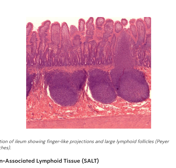
한글뜻: 편도체의 구조 쉬운 설명: 편도는 불완전한 피막으로 둘러싸여 있고, 표면의 점막 상피와 연결된 림프 소절을 포함합니다.
anatomy immunology -
List the four types of tonsils.
- Palatine (Tonsilla pharyngea)
- Tubal (Tonsilla tubaria)
- Pharyngeal (Tonsilla palatina)
- Lingual (Tonsilla lingualis)
한글뜻: 네 가지 종류의 편도 쉬운 설명: 위치에 따라 구개편도, 관편도, 인두편도, 설편도로 나뉩니다.
anatomy immunology -
What type of epithelium covers the palatine tonsil?
Stratified non-keratinizing squamous epithelium.
한글뜻: 중층 비각질 편평상피 쉬운 설명: 편도 표면을 덮고 있는 층이 여러 개인 세포층입니다.
histology tonsils -
What is the primary function of tonsillar crypts?
They are invaginations that increase the surface area of the tissue.
한글뜻: 편도 소와 (편도 구멍) 쉬운 설명: 편도의 표면적을 넓혀 면역 반응을 더 효율적으로 만들게 도와주는 주름진 구조입니다.
histology tonsils -
What is the primary cell type found in the interfollicular space of the tonsils?
Predominantly T-lymphocytes.
한글뜻: 소포간 공간 쉬운 설명: 림프 소포 사이의 공간으로, 주로 T-림프구가 모여 있는 곳입니다.
histology tonsils -
What is the primary cell type found in tonsillar follicles?
Predominantly B-lymphocytes.
한글뜻: 림프 소포 쉬운 설명: 편도 내에서 B-림프구가 집중되어 면역 기능을 수행하는 주머니 모양 구조입니다.
histology tonsils -
Where does pus accumulate in cases of follicular tonsillitis?
It accumulates in the tonsillar crypts.
한글뜻: 여포성 편도염 쉬운 설명: 편도염 발생 시 고름이 편도 표면의 구멍(소와) 속에 고이게 됩니다.
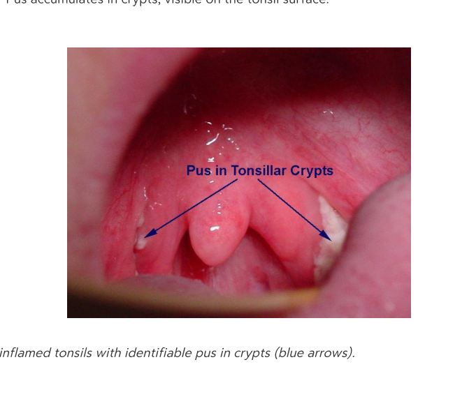 pathology tonsillitis
 -
What are the primary functions of fibroblasts in connective tissue?
Fibroblasts are large cells that synthesize fibers and ground substance.
[한국어 설명] 섬유아세포는 결합 조직 내에서 섬유와 기질 성분을 합성하는 큰 세포입니다.
histology cells -
How do fibrocytes differ from fibroblasts?
Fibrocytes are the inactive, smaller form of fibroblasts.
[한국어 설명] 섬유세포는 섬유아세포의 비활성 상태이자 더 작은 형태입니다.
histology cells -
What are the primary roles of white adipocytes?
They are unilocular fat droplets that store energy, provide insulation, and offer cushioning.
[한국어 설명] 백색 지방세포는 에너지를 저장하고 단열 및 완충 역할을 하는 단일방 지방세포입니다.
histology adipocytes -
What is the specialized function of brown adipocytes?
They are multilocular droplets specialized in thermogenesis.
[한국어 설명] 갈색 지방세포는 열 발생(체온 조절)을 전문으로 하는 다방 지방세포입니다.
histology adipocytes -
What is the nature of mesenchymal cells?
They are multipotent stem cells capable of differentiating into diverse cell types such as osteoblasts and chondrocytes.
[한국어 설명] 중간엽세포는 조골세포나 연골세포 등 다양한 세포로 분화할 수 있는 다능성 줄기세포입니다.
histology cells -
Where are reticulum cells typically found?
They are located at the cross-points of reticular fibers in tissues like bone marrow, lymph nodes, and the spleen.
[한국어 설명] 세망세포는 골수, 림프절, 비장 등의 세망섬유가 교차하는 지점에 위치합니다.
histology cells -
List the three main types of connective tissue fibers.
- Collagen
- Elastic
- Reticular
[한국어 설명] - 콜라겐(교원) 섬유 - 탄력 섬유 - 세망 섬유
histology fibers -
How do fixed cells and mobile cells differ in connective tissue?
Fixed cells remain in place within the matrix, while mobile cells can migrate and are involved in immunity and repair.
[한국어 설명] 고정 세포는 기질 내에 고정되어 있고, 이동성 세포는 자유롭게 이동하며 면역과 수복 작용을 합니다.
histology cells -
What is the primary function of histiocytes (macrophages) in connective tissue?
They perform phagocytosis and antigen presentation.
(한글 뜻: 조직구(대식세포)의 주요 기능은 식세포 작용과 항원 제시입니다.)
histology connective_tissue -
What substances are released by mast cells during an anaphylactic reaction?
Mast cells release heparin, histamine, and serotonin.
(한글 뜻: 비만세포는 아나필락시스 반응 시 헤파린, 히스타민, 세로토닌을 방출합니다.)
histology connective_tissue -
Which specific vitamin is required for the hydroxylation process during collagen synthesis?
Vitamin C is required for hydroxylation.
(한글 뜻: 콜라겐 합성 중 수산화 반응에는 비타민 C가 필요합니다.)
histology collagen -
What are the physical properties of collagen fibers?
They are the most abundant mammalian protein (~30%), inelastic, eosinophilic, and provide great tensile strength.
(한글 뜻: 콜라겐 섬유는 가장 풍부한 포유류 단백질이며, 비탄력적이고, 호산성이며, 강한 인장 강도를 가집니다.)
histology collagen -
What is the historical significance of James Lind regarding collagen?
He identified scurvy as a collagen deficiency, which was historically treated with Hungarian jelly.
(한글 뜻: 제임스 린드는 괴혈병이 콜라겐 결핍이라는 것을 밝혀냈으며, 이는 역사적으로 헝가리 젤리로 치료되었습니다.)
histology collagen -
What components make up elastic fibers?
They are composed of elastic microfibrils (\(8-10 \text{ nm}\)) and amorphous elastin.
(한글 뜻: 탄력 섬유는 탄력 미세섬유(\(8-10 \text{ nm}\))와 무정형 엘라스틴으로 구성됩니다.)
histology elastic_fibers -
What morphological characteristics describe plasma cells?
They possess an eccentric nucleus, radial chromatin cords, and basophilic cytoplasm rich in rough ER.
(한글 뜻: 형질세포는 편재된 핵, 방사형 염색질 끈, 조면소포체가 풍부한 호염기성 세포질을 가집니다.)
histology plasma_cells -
What are the components of Mesenchyma tissue?
Mesenchymal cells, fibers, and GAGs.
한글뜻: 간엽조직 (Mesenchyma). 쉬운 설명: 배아 결합조직의 일종으로, 세포와 섬유, 당단백질(GAG)이 섞여 있는 초기 형태입니다.
connective tissue embryonic -
What is the primary composition of Wharton's jelly?
Fibroblasts, fibers, and GAGs, found in the umbilical cord.
한글뜻: 와튼 젤리 (Wharton's jelly). 쉬운 설명: 탯줄에 존재하는 결합조직으로, 세포와 섬유가 점액질 속에 들어 있는 형태입니다.
connective tissue embryonic -
Which tissues are found in the Reticular category of mature cell-rich connective tissue?
Reticulum cells and reticular fibers, found in bone marrow and the spleen.
한글뜻: 그물 결합조직 (Reticular connective tissue). 쉬운 설명: 뼈 속의 골수나 비장 같은 곳에서 그물망 구조를 형성하여 세포를 지지합니다.
connective tissue mature -
Where is Spinocellular connective tissue found?
In the ovary and the uterus (decidua).
한글뜻: 방추세포성 결합조직 (Spinocellular connective tissue). 쉬운 설명: 난소나 자궁 탈락막에서 발견되는 세포가 풍부한 조직입니다.
connective tissue mature -
What are the key features and locations of Areolar tissue?
It is a type of loose connective tissue found in the mesentery and lymph nodes.
한글뜻: 성긴 결합조직 (Areolar tissue). 쉬운 설명: 우리 몸의 장간막이나 림프절에 존재하며 빈 공간을 채우고 지지하는 느슨한 조직입니다.
connective tissue mature -
What is the function of Loose CT in mature fiber-rich connective tissue?
It fills spaces and supports vessels and nerves.
한글뜻: 느슨한 결합조직 (Loose CT). 쉬운 설명: 신체 내부의 빈틈을 메워주고 혈관과 신경이 지나가는 통로를 지지합니다.
connective tissue mature -
What is the characteristic fiber arrangement of Dense regular connective tissue?
Parallel collagen fibers; found in tendons and ligaments.
한글뜻: 규칙 치밀 결합조직 (Dense regular connective tissue). 쉬운 설명: 콜라겐 섬유가 나란히 배열되어 힘을 잘 견디며, 힘줄이나 인대를 구성합니다.
connective tissue mature -
What is the characteristic fiber arrangement of Dense irregular connective tissue?
Interwoven collagen fibers; found in the dermis and intestines.
한글뜻: 불규칙 치밀 결합조직 (Dense irregular connective tissue). 쉬운 설명: 콜라겐 섬유가 엉켜 있어 모든 방향에서 가해지는 힘에 저항할 수 있으며, 피부 진피층 등에 있습니다.
connective tissue mature -
Which stains are used to visualize reticular fibers?
Silver impregnation or PAS reaction.
한글뜻: 그물 섬유 염색법. 쉬운 설명: 그물 섬유를 현미경으로 관찰하기 위해 은염색이나 PAS 반응을 사용합니다.
histology reticular -
What are the primary adhesive glycoproteins involved in cell-matrix adhesion?
Fibronectin, laminin, and tenascin.
한글뜻: 부착 당단백질 (Adhesive glycoproteins). 쉬운 설명: 세포가 조직의 바탕질에 잘 달라붙을 수 있게 도와주는 접착제 같은 단백질입니다.
glycoproteins cell-matrix -
What is the primary function of the perichondrium in cartilage?
It supplies nutrients and facilitates growth.
한글뜻: 연골막(perichondrium)의 주요 기능은 무엇인가요? 쉬운 설명: 연골에 영양분을 공급하고 연골이 자랄 수 있도록 돕습니다.
histology cartilage -
Name the two types of cartilage and their distinct locations, as shown in the table.
- Hyaline: Trachea, articular surfaces
- Fibrous: Intervertebral discs, menisci
한글뜻: 표에 제시된 두 가지 연골의 종류와 위치를 말하세요. 쉬운 설명: 초자연골은 기관과 관절면에, 섬유연골은 척추 사이 원반과 반월상 연골에 있습니다.
histology cartilage -
What are the two major components of the bone matrix?
- Organic: Type I collagen, proteoglycans (GAG)
- Inorganic: Hydroxyapatite crystals (\(Ca_{10}(PO_4)_6(OH)_2\))
한글뜻: 뼈 바탕질(bone matrix)의 두 가지 주요 구성 성분은 무엇인가요? 쉬운 설명: 유기 성분인 콜라겐과 당단백질, 무기 성분인 수산화인회석 결정으로 이루어져 있습니다.
histology bone -
What is the bone cell lineage starting from the progenitor cell?
Osteoprogenitor \(\rightarrow\) Osteoblast \(\rightarrow\) Osteocyte
한글뜻: 뼈 조상세포에서 시작하는 뼈 세포의 분화 과정은 어떻게 되나요? 쉬운 설명: 뼈 조상세포가 분화하여 조골세포가 되고, 이후 성숙한 뼈세포가 됩니다.
histology bone -
Which hormones and vitamin regulate calcium exchange via osteocytes and osteoclasts?
- Parathyroid hormone
- Calcitonin
- Vitamin D
한글뜻: 뼈세포와 파골세포를 통한 칼슘 교환을 조절하는 호르몬과 비타민은 무엇인가요? 쉬운 설명: 부갑상선 호르몬, 칼시토닌, 그리고 비타민 D가 칼슘 농도를 조절합니다.
histology bone physiology -
What are the primary cells that secrete matrix proteins in loose connective tissue?
Fibroblasts
한글뜻: 성긴 결합조직에서 바탕질 단백질을 분비하는 주요 세포는 무엇인가요? 쉬운 설명: 섬유모세포가 결합조직의 바탕질을 만드는 역할을 합니다.
histology connective-tissue -
What is the primary structural difference between compact bone and spongy bone?
Compact bone consists of densely packed osteons providing strength, while spongy bone is a lighter trabecular network containing marrow. 한글뜻: 치밀골은 강도를 제공하는 조밀한 오스테온으로 구성되며, 해면골은 골수를 포함하는 더 가벼운 그물망 구조입니다. 설명: 치밀골은 뼈의 외곽에서 단단하게 지탱해주고, 해면골은 뼈 내부에서 무게를 줄이고 골수를 담는 역할을 합니다.
anatomy bone -
Which microscopic structures are found within the compact bone system?
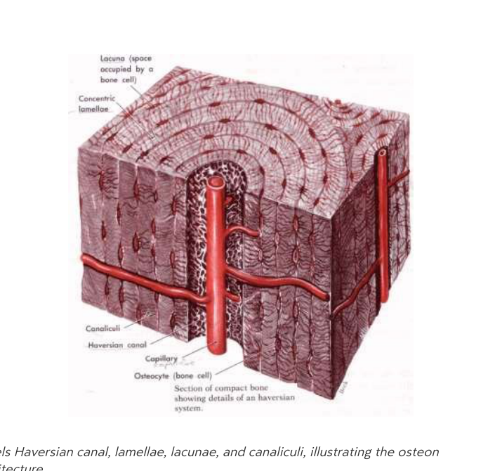 - Haversian canal
- Lamellae
- Lacunae
- Canaliculi
- Haversian canal
- Lamellae
- Lacunae
- Canaliculi한글뜻: 치밀골의 주요 미세 구조물은 하버스관, 층판, 골소강, 골세관입니다. 설명: 이 구조들은 뼈 세포가 영양분을 공급받고 서로 연결될 수 있도록 돕는 통로와 공간들입니다.
anatomy histology -
What is the tissue precursor for intramembranous ossification, and what are some examples of bones formed this way?
The precursor is connective tissue (CT). Examples include flat skull bones, the mandible, and the clavicle. 한글뜻: 막내골화는 결합 조직(CT)에서 시작됩니다. 예로는 평평한 두개골, 하악골, 쇄골이 있습니다. 설명: 연골을 거치지 않고 결합 조직이 직접 뼈 조직으로 바뀌는 방식입니다.
development ossification -
What is the tissue precursor for endochondral ossification?
Hyaline cartilage. This process typically forms long bones. 한글뜻: 연골내골화는 유리 연골에서 시작됩니다. 이 과정은 주로 긴 뼈를 형성합니다. 설명: 뼈가 자라기 위해 먼저 연골로 된 틀을 만들고, 그 후 연골이 점차 뼈로 교체되는 방식입니다.
development ossification -
From which embryonic tissue does Primary Angiogenic (Krompecher) ossification originate?
Mesenchyme. 한글뜻: 일차 혈관형성(크롬페허) 골화는 중간엽에서 유래합니다. 설명: 중간엽은 배아기에 뼈나 근육 등 신체의 다양한 조직으로 분화할 수 있는 세포 집단입니다.
embryology ossification -
What are the three main zones of endochondral bone formation?
- Reserve cartilage
- Proliferation
- Hypertrophy
(한글: 내연골 골화의 세 단계 - 예비 연골, 증식, 비대)
biology bones -
What is the cellular activity within the reserve cartilage zone?
Quiescent chondrocytes.
(한글: 예비 연골 구역 - 휴지기 연골세포)
biology bones -
What occurs during the proliferation stage of endochondral bone formation?
Chondrocytes divide and form columns.
(한글: 증식 단계 - 연골세포가 분열하여 기둥을 형성함)
biology bones -
What is the primary activity in the hypertrophy zone?
Cells enlarge and secrete matrix.
(한글: 비대 구역 - 세포가 커지고 기질을 분비함)
biology bones -
What occurs during the 'Invasion' stage of bone formation?
Mesenchyme infiltrates and becomes osteogenic.
(한글: 침윤 단계 - 간엽세포가 침투하여 골 형성 세포가 됨)
biology bones -
What are the general characteristics of skeletal muscle?
- Location: Skeletal muscles, tongue, esophagus
- Structure: Striated, under somatic nervous control
- Dimensions: \(10–100 \mu m\) diameter, \(1–70 cm\) length
(한글: 골격근의 특징 - 위치, 구조, 섬유 크기)
biology muscles -
What is the primary function of the sarcoplasmic reticulum in muscle cells?
\(Ca^{2+}\) storage.
(한글: 근형질세망 - 칼슘 이온 저장)
biology muscles -
What is the primary function of fixed cells in connective tissue?
Matrix production and structural support.
한글뜻: 결합 조직 내 고정 세포의 주요 기능 한글설명: 세포 기질을 생성하고 구조적 지지를 제공합니다.
histology cells -
What is the primary function of mobile cells in connective tissue?
Immune surveillance, inflammation, and repair.
한글뜻: 결합 조직 내 유동 세포의 주요 기능 한글설명: 면역 감시, 염증 반응 및 조직 복구 역할을 합니다.
histology cells -
What are the origin and representative bones of intramembranous ossification?
- Origin: Connective tissue (mesenchyme)
- Typical Bones: Skull (calvaria), mandible, clavicle
한글뜻: 막내골화의 기원 및 대표 뼈 한글설명: 간엽 조직에서 시작되며 두개골, 하악골, 쇄골이 여기에 해당합니다.
bone histology -
What are the origin and representative bones of endochondral ossification?
- Origin: Hyaline cartilage
- Typical Bones: Long bones (femur, humerus)
한글뜻: 연골내골화의 기원 및 대표 뼈 한글설명: 초자 연골에서 시작되며 대퇴골, 상완골과 같은 긴 뼈가 형성됩니다.
bone histology -
What structural differences distinguish thick skin from thin skin?
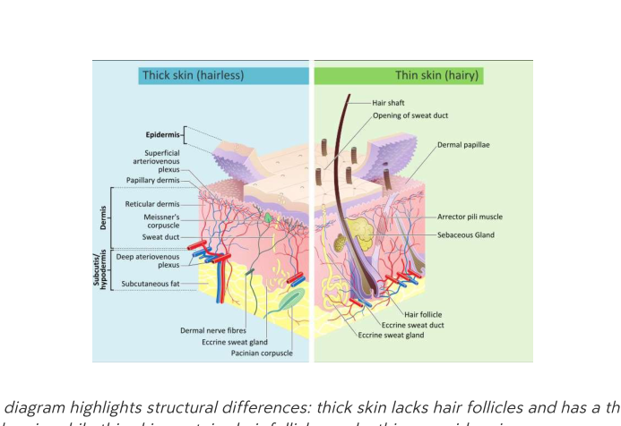 Thick skin lacks hair follicles and has a thicker epidermis compared to thin skin.
한글뜻: 두꺼운 피부와 얇은 피부의 구조적 차이 한글설명: 두꺼운 피부는 모낭이 없으며 얇은 피부보다 표피가 더 두껍습니다.
anatomy integument -
Which cellular structures in smooth muscle are analogous to Z-discs?
Dense bodies.
한글뜻: 평활근에서 Z-원반과 유사한 세포 구조 한글설명: 밀집체(Dense bodies)는 골격근의 Z-원반과 비슷한 기능을 합니다.
muscle histology -
On which parts of the body are hair follicles absent?
Hair follicles are absent on the palms and soles.
한글 의미: 털집(모낭) 부재 부위 쉬운 설명: 손바닥과 발바닥에는 털이 자라지 않습니다.
anatomy integumentary -
How does the thickness of the epidermis differ between the palms/soles and the rest of the body?
The epidermis is thicker on the palms and soles compared to the rest of the body.
한글 의미: 표피 두께 쉬운 설명: 손바닥과 발바닥의 피부 표피층은 몸의 다른 부위보다 더 두껍습니다.
anatomy integumentary -
What type of sweat glands are found on the palms and soles?
Only eccrine sweat glands are found on the palms and soles.
한글 의미: 땀샘 분포 쉬운 설명: 손바닥과 발바닥에는 에크린 땀샘만 존재하며, 아포크린 땀샘은 없습니다.
anatomy integumentary -
What is the function of the stratum basale in the epidermis?
It acts as the basal layer of dividing cells and serves as the source for all upper-layer cells.
한글 의미: 기저층 쉬운 설명: 피부의 가장 아래층으로, 세포 분열이 일어나 새로운 피부 세포를 공급하는 곳입니다.
anatomy epidermis -
How does the stratum spinosum contribute to the skin?
It provides strength to the skin through desmosomes.
한글 의미: 가시층 쉬운 설명: 세포들이 서로 단단하게 결합하여 피부가 튼튼하게 유지되도록 돕습니다.
anatomy epidermis -
What significant biological process begins in the stratum granulosum?
The onset of keratinisation begins here.
한글 의미: 과립층 쉬운 설명: 피부 세포가 딱딱한 각질로 변하는 과정이 시작되는 단계입니다.
anatomy epidermis -
What are the key characteristics of cells found in the stratum corneum?
- They are rich in keratin
- They undergo apoptosis
- They are eventually exfoliated
한글 의미: 각질층 쉬운 설명: 케라틴이 가득 찬 죽은 세포들로 이루어져 있으며, 결국 피부 밖으로 떨어져 나갑니다.
anatomy epidermis -
What is the primary function of keratinocytes?
They form a protective barrier for the skin and undergo keratinisation.
한글 의미: 각질형성세포 쉬운 설명: 피부를 보호하는 장벽을 만들고 각질을 형성하는 주된 세포입니다.
anatomy cells -
What is the physical appearance of the border between the epidermis and the underlying dermis?
The border has a wavy appearance.
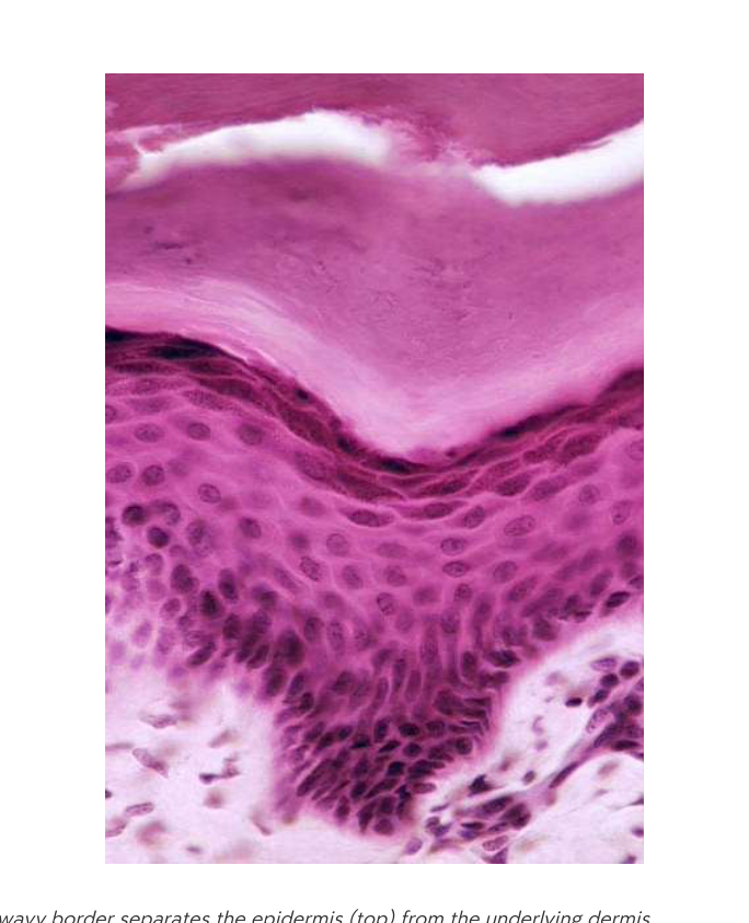
한글 의미: 진피-표피 경계 쉬운 설명: 현미경으로 보면 표피와 진피가 맞닿은 경계면이 물결처럼 굽어 있습니다.
anatomy histology -
What is the primary function of melanocytes in the epidermis?
Melanocytes synthesize melanin and transfer melanosomes to neighboring keratinocytes.
한글뜻: 멜라닌 세포는 멜라닌을 합성하여 주변 각질 세포로 멜라노좀을 전달합니다. 설명: 피부색을 결정하는 멜라닌 색소를 만들어 각질 세포로 보내주는 세포입니다.
biology dermatology -
What is the role of Langerhans cells in the skin?
They act as antigen-presenting cells of the skin immune system.
한글뜻: 랑게르한스 세포는 피부 면역 체계의 항원 제시 세포 역할을 합니다. 설명: 외부에서 침입한 이물질을 감지하고 면역 반응을 일으키는 세포입니다.
biology dermatology -
What is the function of Merkel cells?
Merkel cells act as mechanoreceptors for light touch.
한글뜻: 메르켈 세포는 가벼운 접촉에 반응하는 기계적 수용체 역할을 합니다. 설명: 피부에 닿는 섬세한 촉각을 감지하는 세포입니다.
biology dermatology -
What are keratinocytes responsible for in the epidermis?
They are the predominant epidermal cells responsible for the formation of the keratin barrier.
한글뜻: 각질 세포는 각질 장벽 형성을 담당하는 주요 표피 세포입니다. 설명: 피부의 가장 바깥 층에서 방어막 역할을 하는 핵심 세포입니다.
biology dermatology -
What structural component provides tensile strength to keratinocytes?
Abundant intermediate filaments composed of keratin.
한글뜻: 풍부한 중간 필라멘트(케라틴)가 인장 강도를 제공합니다. 설명: 케라틴 단백질 섬유가 세포 구조를 튼튼하게 유지해 줍니다.
biology dermatology -
What factor primarily determines human skin colour?
Skin colour is determined by the amount of melanosomes, not the number of melanocytes.
한글뜻: 피부색은 멜라닌 세포의 수가 아닌 멜라노좀의 양에 의해 결정됩니다. 설명: 사람마다 세포 수는 비슷해도 멜라닌 색소 덩어리(멜라노좀)의 양이 달라 피부색 차이가 납니다.
biology dermatology -
What is the primary function of Langerhans cells in the epidermis?
Langerhans cells act as antigen-presenting cells.
한글뜻: 랑게르한스 세포는 표피에서 항원 제시 세포 역할을 합니다. 쉬운 설명: 우리 몸의 피부에서 외부 침입자(항원)를 잡아 면역 체계에 알려주는 역할을 합니다.
dermatology biology -
What is the role of Merkel cells in the skin?
Merkel cells act as low-threshold mechanoreceptors that detect light touch.
한글뜻: 메르켈 세포는 가벼운 접촉을 감지하는 저역치 기계적 수용체 역할을 합니다. 쉬운 설명: 피부에 닿는 아주 가벼운 자극을 느끼게 해주는 감각 세포입니다.
dermatology biology -
What is the purpose of the extended dendritic processes found on Langerhans cells?
They increase the surface area for antigen capture.
한글뜻: 랑게르한스 세포의 확장된 수지상 돌기는 항원 포획을 위한 표면적을 증가시킵니다. 쉬운 설명: 나뭇가지 모양의 돌기가 넓게 퍼져 있어 외부 세균이나 물질을 더 잘 붙잡을 수 있게 합니다.
dermatology biology -
What primary structures and tissues are contained within the dermis?
The dermis contains connective tissue, blood vessels, nerves, and appendages.
한글뜻: 진피는 결합 조직, 혈관, 신경 및 부속기관을 포함합니다.
dermis anatomy -
What is the primary composition of the papillary layer of the dermis?
It is made of loose connective tissue and contains capillaries and nerve endings.
한글뜻: 유두층은 느슨한 결합 조직으로 이루어져 있으며 모세혈관과 신경 말단을 포함합니다.
dermis papillary-layer -
What are the characteristics and functions of the reticular layer?
The reticular layer is composed of dense connective tissue that provides tensile strength and houses larger vessels and glands.
한글뜻: 망상층은 치밀 결합 조직으로 구성되어 인장 강도를 제공하며 큰 혈관과 샘을 포함합니다.
dermis reticular-layer -
What is the primary function and composition of the subcutis (hypodermis)?
It mainly consists of white adipose tissue and serves for nutrient storage and insulation.
한글뜻: 피하 조직은 주로 백색 지방 조직으로 구성되며 영양 저장과 단열 역할을 합니다.
subcutis hypodermis -
What is the function of the Vater-Pacini corpuscles?
They are deep-lying mechanoreceptors that detect vibration.
한글뜻: 파터-파치니 소체는 진동을 감지하는 심부 기계적 수용기입니다.
mechanoreceptors subcutis -
What is the primary function and distribution of sweat glands?
Sweat glands are simple tubular merocrine glands that produce watery sweat for thermoregulation. They are distributed throughout the skin.
한글뜻: 땀샘 (Sweat gland) 쉬운 설명: 피부 전체에 분포하며, 체온 조절을 위해 수분 위주의 땀을 분비하는 관 모양의 샘입니다.
histology glands skin -
Where are odoriferous (apocrine) glands located, and what is their secretion type?
Odoriferous glands are located in the axillae, nipple, and perineum. They produce a milky, pheromonal secretion and discharge into hair follicles.
한글뜻: 취선/아포크린샘 (Odoriferous/Apocrine gland) 쉬운 설명: 겨드랑이, 유두, 회음부에 위치하며, 페로몬 역할을 하는 뿌연 분비물을 털구멍(모낭)으로 배출합니다.
histology glands skin -
What is the structural type and secretion of sebaceous glands?
Sebaceous glands are branched, alveolar, holocrine glands that secrete sebum (oil) and are associated with hair follicles.
한글뜻: 피지샘 (Sebaceous gland) 쉬운 설명: 털구멍과 연결된 가지 모양의 샘으로, 기름 성분인 피지를 분비하여 피부를 보호합니다.
histology glands skin -
What is the characteristic structure of apocrine glands?
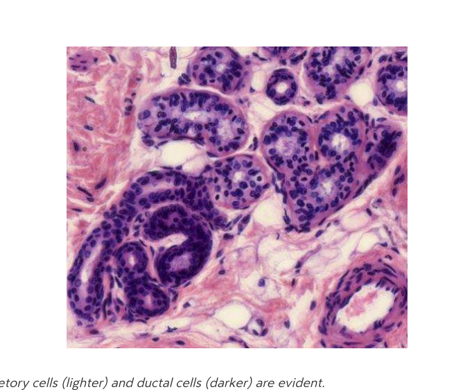 Apocrine glands possess a wide lumen and discharge their secretions into hair follicles.
한글뜻: 아포크린샘의 구조 쉬운 설명: 아포크린샘은 내부 공간(내강)이 넓으며, 모낭을 통해 분비물을 내보냅니다.
histology glands -
How do merocrine sweat glands release their products?
Sweat glands are classified as merocrine, meaning they release their secretory product via exocytosis.
한글뜻: 메로크린 분비 방식 쉬운 설명: 세포가 분비물을 주머니(소낭)에 담아 세포 밖으로 내보내는 방식으로 땀을 배출합니다.
histology glands -
What is the secretory mechanism of sebaceous glands?
Sebaceous glands are holocrine glands, meaning whole cells disintegrate to release sebum. (한글: 피지샘은 분비 방식이 홀로크라인(전분비) 방식으로, 세포 전체가 파괴되면서 피지를 배출합니다.)
histology integumentary -
Name the three concentric layers of a hair shaft.
- Cuticle: Outermost, protective scales.
- Cortex: Middle layer containing keratin fibers.
- Medulla: Central core (often absent in fine hair).
(한글: 머리카락을 구성하는 3개의 층입니다. - 큐티클: 가장 바깥쪽의 보호용 비늘 - 피질(코텍스): 케라틴 섬유가 포함된 중간층 - 수질(메듈라): 중심부 (가는 머리카락에는 종종 없음))
anatomy hair -
What layers of the skin does the hair follicle traverse?
The hair follicle traverses the epidermis, dermis, and subcutis. (한글: 모낭은 표피, 진피, 피하조직을 관통합니다.)
anatomy hair -
What is the primary function of the hair follicle bulge?
The bulge acts as a reservoir of stem cells. (한글: 벌지(팽대부)는 줄기세포의 저장소 역할을 합니다.)
anatomy hair -
Which two types of glands are associated with the hair follicle?
- Sebaceous glands (open into the hair follicle)
- Sweat glands (adjacent to the hair follicle)
(한글: 모낭과 관련된 두 가지 샘입니다. - 피지샘 (모낭으로 열림) - 땀샘 (모낭 옆에 위치))
anatomy glands -
What is the function of the outer root sheath of a hair follicle?
It is continuous with the epidermis. (한글: 모낭의 바깥뿌리집은 표피와 연속되어 있습니다.)
anatomy hair -
In which body cavity is the heart located?
The heart is located in the thoracic cavity.
한글: 흉강 (Thoracic cavity). 심장은 가슴 안쪽 공간인 흉강에 위치합니다.
anatomy heart -
Within the thoracic cavity, specifically where is the heart positioned?
It is positioned within the mediastinum (specifically the cardiac mediastinum).
한글: 종격동 (Mediastinum). 심장은 가슴 안의 종격동이라는 공간에 자리 잡고 있습니다.
anatomy heart -
What is the name of the membrane that encloses the heart?
The pericardium.
한글: 심낭 (Pericardium). 심장을 감싸고 보호하는 주머니 모양의 막입니다.
anatomy heart -
What is the primary function of the pericardial sac?
It protects the heart and limits its motion within the thorax.
한글: 심낭은 심장을 보호하고 가슴(흉강) 안에서 심장이 과도하게 움직이지 않도록 제한합니다.
anatomy heart -
Which structure lies adjacent to the heart?
The pleura.
한글: 흉막 (Pleura). 폐를 감싸고 있으며 심장과 인접해 있는 막입니다.
anatomy heart -
What is the inferior relation of the heart?
The peritoneum.
한글: 복막 (Peritoneum). 심장의 아래쪽과 관련이 있는 복부의 막입니다.
anatomy heart -
What is the function of the Right atrium?
It receives deoxygenated blood via the superior and inferior vena cava.
한글뜻: 우심방. 한글설명: 상대정맥과 하대정맥을 통해 들어오는 산소가 부족한 혈액을 받는 곳입니다.
cardiology anatomy -
Which vessels return systemic blood to the heart?
The Superior/Inferior vena cava.
한글뜻: 상대정맥/하대정맥. 한글설명: 온몸에서 심장으로 산소가 부족한 혈액을 되돌려 보내는 큰 정맥입니다.
cardiology anatomy -
What is the function of the Right ventricle?
It pumps deoxygenated blood to the pulmonary trunk.
한글뜻: 우심실. 한글설명: 폐동맥으로 산소가 부족한 혈액을 펌프질하여 보냅니다.
cardiology anatomy -
What is the function of the Left atrium?
It receives oxygenated blood from the pulmonary veins.
한글뜻: 좌심방. 한글설명: 폐에서 산소가 풍부해진 혈액을 폐정맥을 통해 받아들이는 곳입니다.
cardiology anatomy -
What is the function of the Left ventricle?
It pumps oxygenated blood into the aorta.
한글뜻: 좌심실. 한글설명: 산소가 풍부한 혈액을 대동맥으로 펌프질하여 온몸으로 보냅니다.
cardiology anatomy -
What is the role of the Aorta?
It serves as the main systemic artery.
한글뜻: 대동맥. 한글설명: 심장에서 나가는 혈액을 온몸으로 전달하는 가장 큰 혈관입니다.
cardiology anatomy -
What is the function of the Pulmonary veins?
They return oxygenated blood from the lungs.
한글뜻: 폐정맥. 한글설명: 폐에서 가스 교환을 마치고 산소가 풍부해진 혈액을 심장(좌심방)으로 되돌려 보냅니다.
cardiology anatomy -
Which groove separates the atria from the ventricles?
The Coronary groove (atrioventricular sulcus).
한글뜻: 관상구(방실구). 한글설명: 심장의 심방과 심실 경계를 나누는 고랑으로, 심장 혈관이 지나갑니다.
cardiology anatomy -
Which valve is located at the right atrioventricular orifice?
Tricuspid valve.
(삼첨판: 우심방과 우심실 사이의 통로를 여닫는 판막입니다.)
heart anatomy -
What is the function of the chordae tendineae and papillary muscles?
They prevent regurgitation.
(건삭과 유두근: 심장 판막이 뒤집히지 않게 잡아주어 혈액이 거꾸로 흐르는 역류를 막습니다.)
heart anatomy -
What is the role of the coronary sinus in the heart?
It collects cardiac venous blood.
(관상정맥동: 심장 근육에서 사용한 정맥혈을 모아 우심방으로 보내는 혈관입니다.)
heart anatomy -
Which valve is found at the left atrioventricular orifice?
Mitral valve.
(승모판/이첨판: 좌심방과 좌심실 사이를 여닫는 판막입니다.)
heart anatomy -
Into which chamber of the heart do the four pulmonary veins empty?
Left atrium.
(좌심방: 폐에서 산소가 풍부해진 혈액이 심장으로 들어오는 방입니다.)
heart anatomy -
From which heart chamber does the aorta originate?
Left ventricle.
(좌심실: 대동맥을 통해 산소가 많은 혈액을 온몸으로 내보내는 가장 강한 심장 방입니다.)
heart anatomy -
Where are the atrioventricular valves located?
They are located between the atria and the ventricles. (한글: 심방과 심실 사이. 심장의 윗부분인 심방과 아랫부분인 심실 사이의 통로에 위치합니다.)
cardiology anatomy -
What is the primary function of the atrioventricular valves?
To prevent blood backflow during ventricular contraction. (한글: 심실 수축기 동안 혈액 역류 방지. 심실이 짤 때 혈액이 이전 방으로 거꾸로 흐르지 않게 막아줍니다.)
cardiology physiology -
Where are the semilunar valves located?
At the bases of the great arteries. (한글: 대동맥과 폐동맥 기저부. 심장에서 혈액이 나가는 큰 혈관의 시작 지점에 있습니다.)
cardiology anatomy -
What is the movement cycle of the semilunar valves?
They open during systole and close during diastole. (한글: 수축기에 열리고 이완기에 닫힘. 심장이 수축하여 혈액을 보낼 때는 열리고, 쉴 때는 혈액이 다시 돌아오지 않게 닫힙니다.)
cardiology physiology -
What is the function of the papillary muscles and chordae tendineae?
They pull to keep valves closed during systole. (한글: 수축기 동안 판막을 닫힌 상태로 유지. 판막이 뒤집히지 않도록 강한 힘으로 꽉 잡아당겨 줍니다.)
cardiology anatomy -
Why is the obstruction of the LAD (anterior interventricular artery) clinically significant?
It is a common cause of myocardial infarction, often called the 'widow-maker'. (한글: 좌전하행지 폐쇄는 심근경색의 주된 원인. 심장 앞쪽의 큰 혈관이 막히면 치명적인 심장마비가 발생할 위험이 커 '위도우 메이커'라 불립니다.)
cardiology medicine -
What is the full name for the cardiac abbreviation RA?
Right atrium
(한글: 우심방 - 심장의 오른쪽 위 부분)
cardiology anatomy -
What is the full name for the cardiac abbreviation RV?
Right ventricle
(한글: 우심실 - 심장의 오른쪽 아래 부분)
cardiology anatomy -
What is the full name for the cardiac abbreviation LA?
Left atrium
(한글: 좌심방 - 심장의 왼쪽 위 부분)
cardiology anatomy -
What is the full name for the cardiac abbreviation LV?
Left ventricle
(한글: 좌심실 - 심장의 왼쪽 아래 부분)
cardiology anatomy -
What is the full name for the cardiac abbreviation TP?
Pulmonary trunk
(한글: 폐동맥 간 - 심장에서 폐로 혈액을 보내는 혈관)
cardiology anatomy -
Define automaticity in the context of cardiac function.
The heart generates its own rhythm via the sinoatrial (SA) node.
(한글: 자동능 - 동방결절(SA node)을 통해 심장이 스스로 리듬을 생성하는 능력)
cardiology physiology -
How does sympathetic innervation affect the heart?
It increases heart rate and contractility.
(한글: 교감신경 지배 - 심박수와 심근 수축력을 증가시킴)
cardiology physiology -
How does parasympathetic (vagus) innervation affect the heart?
It decreases heart rate and contractility.
(한글: 부교감신경(미주신경) 지배 - 심박수와 심근 수축력을 감소시킴)
cardiology physiology -
What is the standard conduction pathway of the heart?
SA node → atrial myocardium → AV node → Bundle of His → right & left bundle branches (Bundle of Tawara) → Purkinje fibers → ventricular myocardium
(한글: 심장 전도 경로 - 전기 신호가 심장 근육을 따라 흐르는 순서)
cardiology physiology -
What is the primary function of the upper airways?
They condition inhaled air by filtering, warming, and humidifying it.
(한글: 상기도는 흡입된 공기를 여과, 가온, 가습하여 상태를 조절하는 역할을 합니다.)
respiratory physiology -
What is the function of the lower airways?
They conduct air to the gas-exchange units, known as alveoli.
(한글: 하기도는 공기를 가스 교환 단위인 폐포로 전달합니다.)
respiratory physiology -
What occurs during the pulmonary circuit?
Blood travels from the right ventricle through the pulmonary arteries to the lungs for gas exchange, then returns via pulmonary veins to the left atrium.
(한글: 폐순환은 우심실에서 폐동맥을 통해 폐로 혈액을 보내 가스 교환을 한 뒤, 폐정맥을 통해 좌심방으로 돌아오는 과정입니다.)
cardiovascular circulation -
What is the effect of vasoconstriction on blood pressure?
It increases blood pressure because the muscle contracts.
(한글: 혈관 수축은 근육이 수축하여 혈압을 높입니다.)
cardiovascular physiology -
What is the effect of vasodilation on blood pressure?
It decreases blood pressure because the muscle relaxes.
(한글: 혈관 확장은 근육이 이완되어 혈압을 낮춥니다.)
cardiovascular physiology -
What is the role of lymph vessels?
They collect interstitial fluid.
(한글: 림프관은 조직 사이에 있는 간질액을 수집합니다.)
lymphatic physiology -
What clinical condition results from impaired lymphatic drainage?
Edema.
(한글: 림프 배액이 제대로 되지 않으면 부종이 발생합니다.)
lymphatic pathology -
What is the primary function of the nasal conchae within the skull?
They increase the surface area for air conditioning.
한글뜻: 비갑개 한글설명: 코 안쪽의 뼈 구조로, 공기가 폐로 들어가기 전 온도와 습도를 조절하는 면적을 넓혀줍니다.
anatomy respiratory -
What are the structures collectively known as the respiratory tract?
- Nose (nasus)
- Throat (pharynx)
- Larynx
- Trachea
- Bronchus
- Bronchiolus
- Alveolus
한글뜻: 호흡기계 한글설명: 공기가 지나가는 통로로, 코부터 폐포까지의 구조물들을 의미합니다.
anatomy respiratory -
Which specific components comprise the upper and lower divisions of the respiratory tract?
- Upper: Nose, pharynx, larynx
- Lower: Trachea, bronchi, bronchioles, alveoli
한글뜻: 호흡기 상부와 하부 한글설명: 후두를 기준으로 위쪽은 상기도, 아래쪽은 하기도로 나뉩니다.
anatomy respiratory -
What is the biological function of vibrissae in the nostrils?
They are coarse hairs that act as a mechanical filter for dust and microbes.
한글뜻: 코털 한글설명: 콧구멍 입구에 있는 굵은 털로, 외부 공기의 먼지나 세균이 들어오지 못하게 걸러주는 역할을 합니다.
anatomy respiratory -
What defines the naris and the nasal septum?
- Naris: External inlet of the nose.
- Nasal septum: Midline wall separating the two nasal cavities.
한글뜻: 콧구멍과 비중격 한글설명: Naris는 콧구멍 입구이고, Nasal septum은 코를 양쪽으로 나누는 중간 벽입니다.
anatomy respiratory -
What is the primary function of rudimentary nasal muscles like the levator labii superioris alaeque nasi?
They enable nasal flaring, a sign often associated with labored breathing. (코의 근육들: 코를 벌렁거리게 하며 주로 호흡 곤란 시 나타남.)
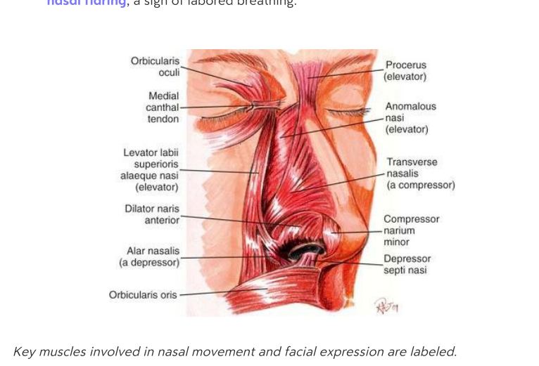 anatomy muscles
 -
Which type of epithelium lines the nasal vestibule, and what specialized structure does it contain?
It is lined by stratified squamous keratinized epithelium and contains vibrissae. (코안뜰: 중층편평상피로 덮여 있으며 콧털인 vibrissae가 있음.)
histology nasal -
What type of epithelium is characteristic of the proper nasal cavity?
It is lined by pseudostratified columnar ciliated epithelium. (고유 코안: 거짓중층원주섬모상피로 이루어져 있음.)
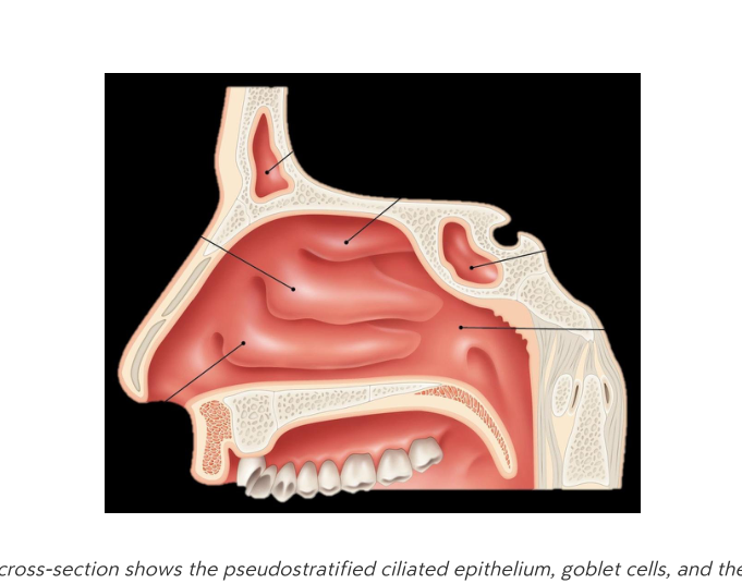 histology respiratory
 -
What is the role of goblet cells in the nasal cavity?
They produce mucus that traps inhaled particles. (술잔세포: 점액을 생성하여 흡입된 입자를 포획함.)
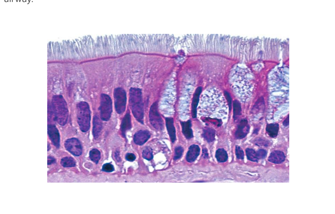 histology cells
 -
What is the primary function of the nasal mucosa regarding inhaled air?
The nasal mucosa warms, humidifies, and filters inhaled air. (코점막: 흡입된 공기를 따뜻하고 촉촉하게 하며 여과함.)
physiology respiratory -
How are trapped particles transported through the nasal airway?
Through mucociliary clearance, where kinocilia move the mucus toward the throat. (점액섬모청소: 섬모가 움직여 점액을 목구멍 쪽으로 이동시킴.)
physiology respiratory -
How are nasal sprays absorbed into the body?
They are absorbed rapidly through the highly vascularized nasal mucosa.
한글뜻: 비강 스프레이는 어떻게 체내로 흡수되나요? 설명: 코 점막에는 혈관이 풍부하여 스프레이가 빠르게 혈류로 흡수됩니다.
pharmacology nasal absorption -
What is the significance of the olfactory region in drug delivery?
It provides a direct route to the central nervous system by bypassing the blood-brain barrier.
한글뜻: 약물 전달에서 후각 영역의 의의는 무엇인가요? 설명: 혈액-뇌 장벽을 거치지 않고 중추신경계로 바로 약물을 보낼 수 있는 경로입니다.
pharmacology olfactory nervous-system -
What is the primary risk associated with the overuse of nasal sprays?
Overuse can cause rhinitis medicamentosa, which is known as rebound congestion.
한글뜻: 비강 스프레이의 과도한 사용과 관련된 주요 위험은 무엇인가요? 설명: 약물성 비염(반동성 충혈)을 유발할 수 있으므로 3-5일 이상 사용을 제한해야 합니다.
pharmacology side-effects rhinitis -
What is the anatomical function of the nasal turbinates (conchae)?
They create turbulent airflow and increase contact time for inhaled substances.
한글뜻: 코선반(비갑개)의 해부학적 기능은 무엇인가요? 설명: 공기의 흐름을 소용돌이치게 만들어 공기가 점막과 닿는 시간을 늘려줍니다.
anatomy nasal turbinates -
What is the choana?
It is the posterior opening to the nasopharynx.
한글뜻: 후비공이란 무엇인가요? 설명: 비강에서 인두로 넘어가는 뒤쪽의 통로를 말합니다.
anatomy nasopharynx -
What microscopic feature of the respiratory epithelium is visible as hair-like projections?
Cilia.
한글뜻: 호흡기 상피세포에서 머리카락처럼 보이는 미세한 구조는 무엇인가요? 설명: 섬모라고 하며, 점액과 함께 호흡기를 보호하는 역할을 합니다.
histology cilia anatomy -
What are the four types of paranasal sinuses?
- Frontal sinus
- Ethmoid air cells (anterior, middle, posterior)
- Maxillary sinus
- Sphenoidal sinus
(한글: 4가지 부비동의 종류는 무엇인가요? 답: 전두동, 사골동, 상악동, 접형동. 부비동은 얼굴 뼈 내부의 공기가 찬 공간입니다.)
anatomy sinuses -
What are the primary functions of the paranasal sinuses?
- Lighten skull weight
- Resonance for voice
- Communicating with the nasal meatus
(한글: 부비동의 주요 기능은 무엇인가요? 답: 두개골 무게 감소, 목소리 공명, 비강과 연결된 공기 통로 역할입니다.)
anatomy function -
What are the clinical signs of sinusitis?
- Thickened mucosa
- Fluid accumulation
- Facial pain that worsens on forward bending
(한글: 부비동염의 임상적 특징은 무엇인가요? 답: 점막 두꺼워짐, 액체 고임, 앞으로 숙일 때 악화되는 얼굴 통증입니다.)
pathology sinusitis -
What are the three parts of the ethmoid air cells?
- Anterior
- Middle
- Posterior
(한글: 사골동의 세 부분은 무엇인가요? 답: 전사골동, 중사골동, 후사골동입니다.)
anatomy ethmoid -
What are the three main divisions of the pharynx?
- Nasopharynx
- Oropharynx
- Laryngopharynx
한글뜻: 인두의 세 부분 한글 설명: 인두는 위치에 따라 비인두, 구인두, 후인두의 세 가지 구역으로 나뉩니다.
anatomy pharynx -
Which structure connects the nasal cavity to the nasopharynx?
The Choana.
한글뜻: 후비공 한글 설명: 후비공은 비강과 비인두를 연결해 주는 통로입니다.
anatomy pharynx -
What are the three wall layers of the pharynx?
- Tunica mucosa
- Tunica submucosa
- Tunica muscularis
한글뜻: 인두벽의 세 층 한글 설명: 인두벽은 점막층, 점막하층, 근육층의 세 가지 구조적 층으로 이루어져 있습니다.
anatomy histology -
What are the two primary functions of the larynx?
- Phonation (voice production)
- Airway protection (as a sphincter)
한글뜻: 후두의 두 가지 주요 기능 한글 설명: 후두는 소리를 내는 발성 기능과 기도로 이물질이 들어가지 않게 막는 기도 보호 기능을 합니다.
anatomy larynx -
What happens to the epiglottis during the swallowing sequence?
It folds down to seal the laryngeal inlet.
한글뜻: 삼킴 과정 중 후두덮개의 역할 한글 설명: 음식을 삼킬 때 후두덮개가 아래로 접혀 후두 입구를 막아 기도를 보호합니다.
anatomy physiology -
Name the primary cartilages of the larynx.
- Hyoid bone
- Thyroid cartilage
- Cricoid cartilage
- Arytenoid cartilages
- Epiglottis
한글뜻: 후두를 구성하는 연골들 한글 설명: 후두는 설골을 포함하여 갑상연골, 윤상연골, 피열연골, 후두덮개 등으로 이루어져 구조를 유지합니다.
anatomy larynx -
What is another name for the vestibular folds?
False vocal cords. (한글: 가성대. 진짜 성대가 아닌 조직으로, 발성보다는 보호 기능을 합니다.)
anatomy larynx -
What is the function of the vocalis muscle?
It adjusts the tension of the vocal cords. (한글: 성대 근육. 성대의 긴장도를 조절하여 음의 높낮이를 결정합니다.)
anatomy phonation -
What occurs when the glottis is partially open?
Phonation (vocal folds vibrate). (한글: 성문이 부분적으로 열리면 성대가 진동하여 소리가 납니다.)
phonation physiology -
What is the function of the primary inspiratory muscle, the diaphragm?
It is the primary muscle responsible for inhalation. (한글: 횡격막. 숨을 들이마시는 데 주된 역할을 하는 근육입니다.)
respiration anatomy -
What is the role of intercostal muscles in respiration?
External muscles aid in inspiration, while internal muscles are used for forced expiration. (한글: 늑간근. 바깥쪽 근육은 숨을 들이마실 때, 안쪽 근육은 강제로 숨을 내쉴 때 사용됩니다.)
respiration anatomy -
What is a coniotomy?
A surgical incision into the laryngeal cartilage. (한글: 윤상갑상막 절개술. 후두 연골을 절개하여 기도를 확보하는 응급 수술입니다.)
surgery larynx -
What are the paired organs responsible for gas exchange in the respiratory tract?
Lungs (Pulmo). (한글: 폐. 호흡기에서 산소와 이산화탄소를 교환하는 기관입니다.)
anatomy respiration -
Between which vertebral levels does the trachea extend?
The trachea extends from C7 to T4. (한글: 기도(trachea)는 제7경추(C7)에서 제4흉추(T4)까지 이어집니다. 설명: 목 아래부터 가슴 중앙까지 위치하는 공기 통로입니다.)
anatomy trachea -
Describe the structural composition of the tracheal cartilage rings.
They consist of C-shaped hyaline cartilage rings that are open posteriorly. (한글: 기도의 연골은 C자 모양의 초자연골(hyaline cartilage)로, 뒤쪽이 열려 있습니다. 설명: 이 구조는 기도를 지지하면서도 유연하게 만듭니다.)
anatomy trachea -
What is the function of the ciliary rejection flow in the airways?
It acts as a self-clearing mechanism where the coordinated beating of kinocilia moves mucus upward. (한글: 섬모 배출 흐름(ciliary rejection flow)은 점액을 위로 이동시켜 기도를 청소하는 자가 정화 기전입니다. 설명: 기도의 섬모가 움직여 이물질과 점액을 밖으로 밀어냅니다.)
physiology trachea -
Why is the right main bronchus at a higher risk of foreign-body aspiration?
It is wider, shorter, and more vertical than the left main bronchus. (한글: 오른쪽 주기관지는 왼쪽보다 더 넓고 짧으며 수직에 가깝기 때문에 이물질 흡입 위험이 높습니다. 설명: 이물질이 폐로 들어가는 경로가 더 곧기 때문입니다.)
anatomy bronchi -
In the bronchial tree, which generation corresponds to the main (primary) bronchi?
Generation 1. (한글: 기관지 나무 구조에서 제1세대(Generation 1)는 주기관지(Main bronchi)입니다. 설명: 기도(trachea)에서 처음으로 갈라져 나오는 가지입니다.)
anatomy bronchial-tree -
What is the approximate size of terminal bronchioles?
Less than 1 mm. (한글: 종말세기관지(Terminal bronchioles)의 크기는 1mm 미만입니다. 설명: 기관지가 나뭇가지처럼 아주 작게 갈라진 말단 부분 중 하나입니다.)
anatomy bronchioles -
What is the structural sequence of the bronchial tree following the respiratory bronchioles?
- Respiratory bronchioles
- Alveolar ducts
- Alveolar sacs (한글: 호흡세기관지에서 이어지는 순서는 호흡세기관지 → 폐포관 → 폐포낭입니다. 설명: 이 구조들은 폐에서 가스 교환이 일어나는 최종 단계들입니다.)
anatomy bronchial-tree -
What is the first site of gas exchange in the respiratory tree?
The respiratory bronchiole.
(한글: 호흡세기관지 - 가스 교환이 처음 일어나는 곳)
anatomy respiratory -
What is the function of Type I pneumocytes in the alveoli?
They are thin cells that facilitate gas diffusion.
(한글: 제1형 폐세포 - 가스 확산을 돕는 얇은 세포)
histology lungs -
What is the primary function of Type II pneumocytes in the alveoli?
They produce surfactant, which reduces surface tension.
(한글: 제2형 폐세포 - 계면활성제를 생산하여 표면장력을 줄임)
histology lungs -
How does parasympathetic stimulation affect the airway?
It causes bronchoconstriction.
(한글: 부교감 신경 자극 - 기관지 수축을 유발함)
physiology regulation -
What is the effect of sympathetic stimulation on bronchial smooth muscle?
It causes bronchial relaxation.
(한글: 교감 신경 자극 - 기관지 이완을 유발함)
physiology regulation -
What are the two main types of pharmacological agents used as bronchodilators?
- Sympathomimetics (\(\beta_2\)-agonists)
- Parasympatholytics (anticholinergics)
(한글: 기관지 확장제 종류 - 교감신경 효능제와 부교감신경 차단제)
pharmacology respiratory -
What is the role of glands and mucus cells in the airway?
They secrete mucus to trap particles.
(한글: 샘과 점액 세포 - 입자를 걸러내는 점액을 분비)
histology airway -
What is the direction of gas diffusion between alveolar air and capillary blood?
\(O_2\) diffuses from alveolar air into the capillary blood, while \(CO_2\) diffuses in the opposite direction.
[한글 설명] \(O_2\)는 폐포에서 혈관으로 이동하고, \(CO_2\)는 혈관에서 폐포로 이동합니다.
physiology respiration -
What is the primary function of Type I pneumocytes?
They provide a thin barrier to facilitate gas diffusion.
[한글 설명] 제1형 폐포상피세포는 폐포벽을 얇게 구성하여 가스 교환이 잘 일어나도록 돕습니다.
physiology respiration -
What is the primary function of Type II pneumocytes?
They secrete surfactant, which prevents the alveoli from collapsing.
[한글 설명] 제2형 폐포상피세포는 계면활성제(서팩턴트)를 분비하여 폐포가 찌그러지지 않게 합니다.
physiology respiration -
What is the role of alveolar macrophages?
They phagocytose (engulf and digest) inhaled particles.
[한글 설명] 폐포 대식세포는 폐로 들어온 이물질을 잡아먹어 제거합니다.
physiology immunity -
How does ciliary rejection flow protect the respiratory tract?
It uses coordinated ciliary motion to move mucus upward out of the tract.
[한글 설명] 섬모 운동을 통해 점액을 위쪽으로 밀어내어 이물질을 제거합니다.
physiology protection -
What is the purpose of the cough reflex?
The sudden expulsion of air clears larger inhaled particles from the airways.
[한글 설명] 갑작스러운 기침을 통해 큰 이물질을 밖으로 배출합니다.
physiology protection -
Which biological components participate in the airway's immune responses?
- Resident macrophages
- Neutrophils
- IgA
- Lymphocytes
[한글 설명] 면역 반응은 대식세포, 호중구, IgA 항체, 림프구 등이 관여합니다.
physiology immunity -
What is the primary function of the pleura in the human body?
The pleura is a serous membrane consisting of two layers that enclose the lungs and line the thoracic cavity.
(한글: 흉막은 폐를 감싸고 흉강을 둘러싸는 두 층으로 된 장막입니다.)
anatomy respiratory -
Describe the location and function of the visceral and parietal pleura.
- Visceral pleura: Adheres directly to the lung surface.
- Parietal pleura: Lines the thoracic wall, mediastinum, and diaphragm.
(한글: 내장흉막은 폐 표면에 붙어 있고, 벽쪽흉막은 흉벽, 종격동, 횡격막을 덮습니다.)
anatomy pleura -
What is the role of the pleural cavity?
It is a thin, fluid-filled space that allows the lungs to glide smoothly during respiration.
(한글: 흉막강은 액체로 채워진 얇은 공간으로, 호흡 시 폐가 부드럽게 움직이도록 돕습니다.)
anatomy pleura -
What are the three subdivisions of the parietal pleura?
- Costal pleura: Lines the lateral thoracic wall.
- Mediastinal pleura: Covers the mediastinum.
- Diaphragmatic pleura: Covers the diaphragmatic surface.
(한글: 벽쪽흉막은 늑골흉막, 종격흉막, 횡격흉막의 세 부분으로 나뉩니다.)
anatomy pleura -
What occurs in the thoracic cavity during quiet inhalation?
The diaphragm contracts and moves caudally (downward), enlarging the vertical thoracic dimension.
(한글: 조용한 흡기 시, 횡격막이 수축하여 아래로 내려가며 흉곽의 수직 크기를 넓힙니다.)
respiration physiology -
What is the primary action of the external intercostal muscles during breathing?
They lift the ribs, which increases the transverse diameter of the thoracic cavity.
(한글: 외늑간근은 갈비뼈를 들어 올려 흉곽의 가로 지름을 넓히는 역할을 합니다.)
respiration muscles -
What happens during quiet exhalation?
The diaphragm relaxes, and the ribs return to their resting position due to elastic recoil.
(한글: 조용한 호기 시, 횡격막이 이완되고 탄성 반동에 의해 갈비뼈가 원래 위치로 돌아옵니다.)
respiration physiology -
Which muscles are involved in forced expiration?
The internal intercostal muscles and the abdominal muscles, which compress the thoracic cavity.
(한글: 강제 호기 시에는 내늑간근과 복근이 사용되어 흉곽을 압박합니다.)
respiration muscles -
What is the principal muscle of quiet inspiration?
The diaphragm.
한글뜻: 가로막(횡격막). 설명: 조용한 호흡 시 숨을 들이마시는 주된 근육입니다.
respiration anatomy -
Which nerve provides innervation to the diaphragm?
The phrenic nerve (C3-C5).
한글뜻: 횡격막신경. 설명: 가로막의 움직임을 조절하는 신경으로, 경추 3번에서 5번 사이에서 기시합니다.
innervation anatomy -
What clinical consequence arises from a spinal cord injury at the C4 level or above?
Diaphragmatic palsy (paralysis of the diaphragm).
한글뜻: 가로막 마비. 설명: 척수 손상이 C4 또는 그 이상에서 발생하면 가로막이 마비되어 정상적인 환기가 어려워집니다.
clinical injury -
What is the primary function of the external intercostal muscles?
To elevate the ribs during inspiration.
한글뜻: 바깥갈비사이근(외늑간근). 설명: 숨을 들이마실 때 갈비뼈를 들어 올려 흉곽을 확장합니다.
respiration muscles -
What is the primary function of the internal intercostal muscles?
To depress the ribs during forced expiration.
한글뜻: 속갈비사이근(내늑간근). 설명: 강제적으로 숨을 내쉴 때 갈비뼈를 내려 흉곽을 수축시킵니다.
respiration muscles -
Which muscles act as accessory muscles to enhance thoracic expansion?
- Sternocleidomastoid
- Scalenes
- Pectoralis minor
한글뜻: 보조 호흡근. 설명: 흉곽 확장을 돕는 근육들로, 목빗근(흉쇄유돌근), 목갈비근(사각근), 작은가슴근(소흉근)이 포함됩니다.
respiration muscles -
Which major structures pass through openings in the diaphragm?
- Aorta
- Inferior vena cava (IVC)
- Esophagus
한글뜻: 가로막의 개구부. 설명: 대동맥, 하대정맥, 식도가 가로막을 관통합니다.
anatomy diaphragm -
What defines the boundaries of the oral vestibule?
It is the space between the lips/cheeks (bucca) and the teeth. (구강 전정: 입술과 볼, 치아 사이의 공간)
anatomy oral -
What defines the boundaries of the oral cavity proper?
It is the space bounded by the teeth anteriorly and the oropharyngeal isthmus posteriorly. (구강 본체: 앞쪽으로는 치아, 뒤쪽으로는 구강인두 협부로 경계 지어지는 공간)
anatomy oral -
Where does the Stensen duct of the parotid gland open?
It opens into the oral vestibule opposite the upper 2nd molar. (귀밑샘관: 위쪽 두 번째 어금니 맞은편의 구강 전정으로 열림)
anatomy glands -
What is the primary secretory contribution of the submandibular gland?
It contributes 65-70% of total saliva, consisting of mixed serous-mucous secretion. (턱밑샘: 전체 침의 65-70%를 분비하며 장액과 점액이 섞인 형태임)
anatomy glands -
Which gland produces mostly mucous, low-volume saliva through multiple minor ducts?
The sublingual gland. (혀밑샘: 여러 개의 작은 관을 통해 주로 점액성이며 양이 적은 침을 분비함)
anatomy glands -
What is the name of the structure that acts as the opening for the parotid duct?
The parotid papilla. (귀밑샘 유두: 귀밑샘관의 입구가 위치한 작은 돌기)
anatomy glands -
How is parotid inflammation (mumps) prevented in Hungary?
It is prevented by mandatory MMR vaccination. (볼거리 예방: 헝가리에서는 MMR 백신 접종이 의무화되어 있음)
medicine public-health -
What are the primary components of saliva?
- 99.5% water
- Ions, enzymes (\(\alpha\)-amylase, lipase)
- Mucin (0.5–1 L/day)
한글뜻과 설명: - 침의 구성 성분: 물 99.5%, 이온, 효소(알파-아밀라아제, 리파아제), 뮤신(점액)으로 이루어져 있습니다.
saliva physiology -
Which gland produces a more mucin-rich, enzyme-poor saliva under sympathetic stimulation?
The submandibular gland.
한글뜻과 설명: - 악하선(턱밑샘): 교감신경 자극을 받으면 뮤신이 많고 효소가 적은 침을 분비합니다.
glands saliva -
What are the structural parts of a tooth?
- Crown: covered by enamel
- Neck: transition zone between enamel and cementum
- Root: encased by cementum, dentin, and contains the pulp
- Periodontium: supporting tissues (gingiva, periodontal ligament, alveolar bone)
한글뜻과 설명: - 치아 구조: 치관(법랑질), 치경(경계부), 치근(상아질, 치수 포함), 치주조직(잇몸, 치주인대, 치조골).
teeth anatomy -
According to the dental formula per quadrant, how many incisors, canines, and premolars are in a permanent set?
- Incisors: 2
- Canines: 1
- Premolars: 2
한글뜻과 설명: - 영구치 치식(한 사분면 기준): 앞니 2개, 송곳니 1개, 작은어금니(소구치) 2개.
teeth dentistry -
Compare the number of premolars in a deciduous (primary) set versus a permanent set per quadrant.
- Deciduous: 0
- Permanent: 2
한글뜻과 설명: - 유치(젖니)에는 작은어금니(소구치)가 없고, 영구치에는 2개가 있습니다.
teeth comparison -
What is the function of the intrinsic muscles of the tongue?
They change the shape of the tongue (e.g., flatten, curl).
한글뜻: 혀의 내재근은 혀의 모양을 변형(납작하게 하거나 둥글게 마는 등)시키는 역할을 합니다.
anatomy musculature -
What is the primary function of extrinsic muscles of the tongue?
They move the tongue within the oral cavity.
한글뜻: 혀의 외재근은 혀를 구강 내에서 이동시키는 역할을 합니다.
anatomy musculature -
Which cranial nerve provides innervation to both intrinsic and extrinsic muscles of the tongue?
Hypoglossal nerve (XII).
한글뜻: 설하신경(제12뇌신경)이 혀의 모든 근육에 신경을 분포시킵니다.
anatomy innervation -
What lymphoid tissue is contained within the root of the tongue?
Lingual tonsil.
한글뜻: 혀뿌리(설근)에는 설편도(혀편도)가 포함되어 있습니다.
anatomy tongue -
What is found on the body of the tongue?
Papillae housing taste buds.
한글뜻: 혀 몸통은 맛봉오리(미뢰)를 포함한 유두로 덮여 있습니다.
anatomy tongue -
Why is the sublingual area suitable for rapid drug absorption?
The tongue's rich capillary network allows for rapid absorption.
한글뜻: 혀 아래(설하) 영역은 모세혈관망이 풍부하여 약물이 빠르게 흡수될 수 있습니다.
pharmacology tongue -
What anatomical structure serves as the boundary between the oral cavity and the oropharynx?
The soft palate.
한글뜻: 연구개 설명: 입안(구강)과 목구멍 중간 부분(구인두)을 나누는 부드러운 입천장입니다.
anatomy pharynx -
Which structures are responsible for flanking the palatine tonsil?
The palatoglossal and palatopharyngeal arches.
한글뜻: 구개설궁 및 구개인두궁 설명: 편도선을 양옆에서 감싸고 있는 근육 구조물입니다.
anatomy pharynx -
Where is the uvula located?
It hangs from the posterior edge of the soft palate.
한글뜻: 목젖 설명: 입천장 뒤쪽 끝부분에 매달려 있는 구조물입니다.
anatomy -
What is the alternative name for the Epipharynx?
Nasopharynx.
한글뜻: 비인두 설명: 인두의 가장 윗부분으로 코 뒤쪽에 위치합니다.
anatomy pharynx -
What is the alternative name for the Mesopharynx?
Oropharynx.
한글뜻: 구인두 설명: 인두의 중간 부분으로 입 뒤쪽에 위치합니다.
anatomy pharynx -
What is the alternative name for the Hypopharynx?
Laryngopharynx.
한글뜻: 후두인두 설명: 인두의 가장 아래쪽 부분으로 후두와 연결됩니다.
anatomy pharynx -
What are the components of the mucous membrane (tunica mucosa)?
- Epithelium
- Lamina propria
- Muscularis mucosae
한글뜻: 점막 설명: 상피층, 고유층, 점막근층으로 이루어진 조직입니다.
histology anatomy -
What is the primary function of the pharyngeal wall?
The pharyngeal wall acts as a muscular tube that coordinates swallowing and airway protection.
한글뜻: 인두벽의 주요 기능은 무엇인가? 한글설명: 인두벽은 근육으로 이루어진 관으로서 음식물을 삼키는 작용을 조절하고 호흡기를 보호하는 역할을 합니다.
anatomy pharynx -
What kind of tissue is found in the submucosa layer of the pharynx?
Loose to dense connective tissue containing glands and vessels.
한글뜻: 인두의 점막하층(submucosa)에는 어떤 조직이 있는가? 한글설명: 샘(gland)과 혈관을 포함하는 느슨하거나 조밀한 결합 조직으로 구성되어 있습니다.
anatomy pharynx -
What does the muscularis layer of the pharynx consist of?
Skeletal muscle, specifically constrictors and elevators.
한글뜻: 인두의 근육층(muscularis)은 무엇으로 구성되는가? 한글설명: 수축근과 거상근을 포함하는 골격근으로 이루어져 있습니다.
anatomy pharynx -
What type of tissue constitutes the adventitia of the pharynx?
Loose connective tissue.
한글뜻: 인두의 외막(adventitia)은 어떤 조직으로 구성되는가? 한글설명: 느슨한 결합 조직으로 이루어져 있습니다.
anatomy pharynx -
Identify the three sections of the pharynx shown in the sagittal cross-section.
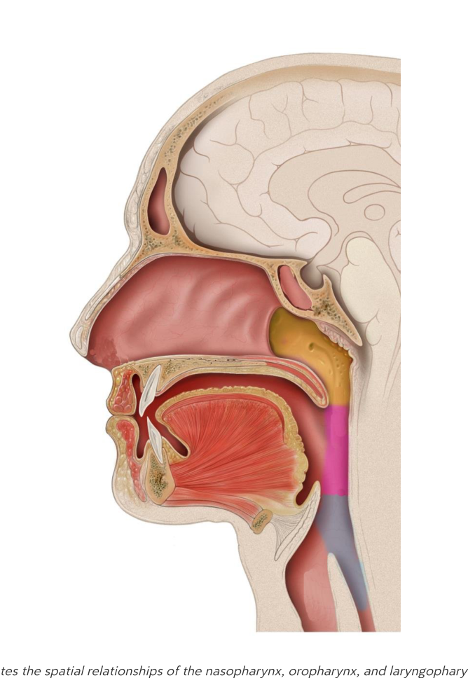 - Nasopharynx
- Oropharynx
- Laryngopharynx
- Nasopharynx
- Oropharynx
- Laryngopharynx한글뜻: 그림에 표시된 인두의 세 부분은 무엇인가? 한글설명: 비인두(코인두), 구인두(입인두), 후인두(후두인두)입니다.
anatomy pharynx -
What are the key pathophysiologic mechanisms involved in the development of gastro-esophageal reflux disease (GERD)?
- Increase in transient lower esophageal sphincter relaxations (TLESR)
- Decreased esophageal clearance
- Decreased gastric emptying
[한국어 설명] GERD의 병태생리: 하부 식도 괄약근의 일시적 이완 증가, 식도 청소율 저하, 위 배출 지연.
gerd pathology gastroenterology -
What are the common clinical symptoms of GERD?
- Heartburn
- Chest pain
- Regurgitation
- Dysphagia
- Extra-esophageal symptoms (e.g., cough, asthma)
[한국어 설명] GERD의 증상: 속쓰림, 흉통, 역류, 연하곤란, 식도 외 증상(기침, 천식).
gerd symptoms -
List the standard diagnostic work-up procedures for GERD.
- Detailed history
- Endoscopy
- 24-hour pH monitoring
- Manometry
- Barium swallow
[한국어 설명] GERD 진단 검사: 상세 병력 청취, 내시경, 24시간 pH 모니터링, 내압 검사, 바륨 삼킴 검사.
gerd diagnosis -
What are the primary classes of pharmacologic agents used to manage GERD?
- Proton pump inhibitors (PPIs)
- Potassium-competitive acid blockers (PCAB)
- H2-receptor antagonists
- Antacids
- Alginates
[한국어 설명] GERD 약물 치료: 양성자 펌프 억제제(PPI), 칼륨 경쟁적 위산 차단제(PCAB), H2 수용체 길항제, 제산제, 알긴산염.
gerd pharmacology management -
How does the epithelium differ between the nasopharynx and the oro-/laryngopharynx?
- Nasopharynx: pseudostratified columnar epithelium with cilia
- Oro- and laryngopharynx: stratified squamous non-keratinized epithelium
[한국어 설명] 인두 조직학: 비인두는 섬모가 있는 거짓중층원주상피, 구인두와 후인두는 중층편평상피(무각질)입니다.
histology pharynx anatomy -
What type of epithelium covers the oral cavity?
Stratified squamous non-keratinized epithelium.
[한국어 설명] 구강 점막 상피: 중층편평상피(무각질)로 덮여 있습니다.
histology oral anatomy -
What is the primary definition of the gastrointestinal (GI) tract?
The GI tract is a continuous tube extending from the mouth to the anus, containing specialized regions dedicated to digestion, absorption, and the formation of waste.
Korean: 소화관은 입에서 항문까지 이어지는 연속적인 관으로, 소화, 흡수 및 노폐물 형성을 담당하는 특수 영역으로 구성됩니다.
anatomy gi-tract -
At what vertebral levels is the stomach located?
The stomach is located at the vertebral levels of T11 through L1.
Korean: 위는 척추 T11번에서 L1번 위치에 자리 잡고 있습니다.
anatomy stomach -
What is the anatomical position of the stomach relative to the diaphragm?
The stomach is positioned inferior to the diaphragm.
Korean: 위는 횡격막의 아래쪽에 위치합니다.
anatomy stomach -
What is the anatomical position of the stomach relative to the liver?
The stomach is positioned posterior to the liver.
Korean: 위는 간의 뒤쪽에 위치합니다.
anatomy stomach -
List the primary organs that make up the gastrointestinal system as shown in the diagram.
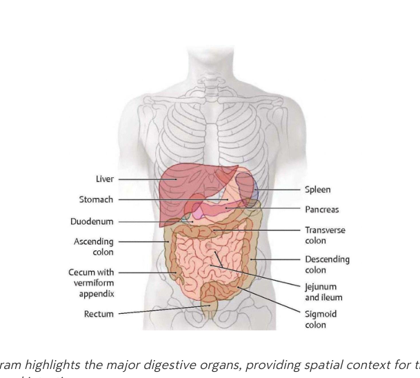 - Liver
- Stomach
- Duodenum
- Ascending colon
- Caecum and appendix vermiformis
- Rectum
- Spleen
- Pancreas
- Transverse colon
- Descending colon
- Jejunum and ileum
- Sigmoid colon
- Liver
- Stomach
- Duodenum
- Ascending colon
- Caecum and appendix vermiformis
- Rectum
- Spleen
- Pancreas
- Transverse colon
- Descending colon
- Jejunum and ileum
- Sigmoid colon Korean: 소화계의 주요 장기: 간, 위, 십이지장, 상행결장, 맹장 및 충수, 직장, 비장, 췌장, 횡행결장, 하행결장, 공장 및 회장, S상결장.
anatomy digestive-system -
What is the function of the cardia region of the stomach?
It serves as the transition zone where the esophagus enters the stomach.
(한글 뜻: 분문부 - 식도가 위로 연결되는 이행 구역)
stomach anatomy -
What is the fundus of the stomach and what does it often contain?
It is the upper curved portion of the stomach, which often contains gas bubbles.
(한글 뜻: 위저부 - 위의 상단 굽은 부분으로, 종종 가스 거품을 포함함)
stomach anatomy -
What are the primary functions of the stomach?
Mechanical churning, protein digestion, and creating an acidic environment (\(pH \approx 1-2\)) for enzyme activation.
(한글 뜻: 위의 주 기능 - 기계적 연동 운동, 단백질 소화, 효소 활성화를 위한 산성 환경 조성)
stomach physiology -
What do parietal cells secrete in the stomach?
Hydrochloric acid and intrinsic factor.
(한글 뜻: 벽세포 - 염산과 내인성 인자를 분비함)
stomach histology -
What do chief cells secrete, and what is the role of that substance?
They secrete pepsinogen, which is converted to pepsin in the acidic lumen to aid digestion.
(한글 뜻: 주세포 - 펩시노겐을 분비하며, 이는 산성 위 내강에서 펩신으로 전환되어 소화를 도움)
stomach histology -
What substance do G cells secrete?
The hormone gastrin.
(한글 뜻: G세포 - 가스트린 호르몬을 분비함)
stomach histology -
What is the role of the gastric \(H^+\) produced by parietal cells?
It is necessary for pepsin activation.
(한글 뜻: 벽세포가 생성하는 수소 이온(\(H^+\))은 펩신을 활성화하는 데 필수적임)
stomach physiology -
What is the primary pathogenesis and typical treatment for a peptic ulcer?
- Pathogenesis: Imbalance between aggressive factors (gastric acid, pepsin) and defensive factors (mucus, bicarbonate).
- Treatment: Eradication of Helicobacter pylori (antibiotics), PPI therapy, lifestyle modification.
[한글: 소화성 궤양] 위산/펩신 등 공격 인자와 점액/중탄산염 등 방어 인자의 불균형으로 발생하며, 제균 요법 및 PPI 투여로 치료합니다.
stomach pathology medicine -
What are the common causes and management for a drug-induced ulcer?
- Causes: NSAIDs, aspirin, SSRIs, steroids.
- Management: Discontinue offending drug and PPI support.
[한글: 약물 유발성 궤양] NSAID 등 특정 약물이 점막 보호를 저해하여 발생합니다. 원인 약물을 중단하고 PPI를 지원하여 치료합니다.
stomach pathology medicine -
What is the trigger for a stress ulcer and how is it managed?
- Cause: Extreme physiological stress increases acid secretion.
- Management: Prophylactic PPI or \(H_2\)-receptor antagonists.
[한글: 스트레스성 궤양] 심한 신체적 스트레스로 위산이 과다 분비될 때 발생하며, PPI나 H2 차단제로 예방합니다.
stomach pathology medicine -
Which substances are received by the duodenum to facilitate digestion?
- Bile (via common bile duct)
- Pancreatic secretions (amylase, DNase, lipase, trypsin)
[한글: 십이지장 분비물] 담즙과 췌장액(아밀라아제, 리파아제 등)이 십이지장으로 유입되어 음식물 소화를 돕습니다.
digestion anatomy -
What is the role of the greater duodenal papilla?
It serves as the common opening for bile and pancreatic ducts, facilitating coordinated digestion.
[한글: 대십이지장유두] 담관과 췌관이 만나는 통로로, 담즙과 췌장액을 십이지장으로 보내 소화를 돕습니다.
digestion anatomy -
What are the primary functions of the jejunum and ileum?
Digestion of nutrients and resorption of amino acids, sugars, fatty acids, and vitamins.
[한글: 공장 및 회장의 기능] 영양소를 소화하고 아미노산, 당분, 지방산, 비타민 등을 흡수하는 주요 장소입니다.
digestion anatomy -
What are the components of the tunica mucosa layer in the small intestine?
- Simple columnar epithelium with brush border
- Lamina propria (connective tissue)
- Lamina muscularis (smooth muscle)
[한글: 점막층] 단순 원주상피, 고유판(결합조직), 근육판으로 구성된 소장의 내벽입니다.
histology anatomy -
What are the components of the tunica submucosa layer in the small intestine?
Connective tissue and the submucosal (Meissner) plexus.
[한글: 점막하층] 결합조직과 소화기관 조절을 담당하는 마이스너 신경총이 포함되어 있습니다.
histology anatomy -
What are the components of the tunica muscularis in the small intestine?
- Inner circular smooth muscle
- Outer longitudinal smooth muscle
- Interposed myenteric (Auerbach) plexus
[한글: 근육층] 속 돌림근(내륜근), 겉 세로근(외종근)과 근육 사이의 아우어바흐 신경총으로 구성됩니다.
histology anatomy -
What are the components of the tunica serosa/adventitia in the small intestine?
Serosa on intraperitoneal portions (mesothelium) and adventitia on extraperitoneal sections.
[한글: 장막층 및 외막층] 복강 내부는 장막(중피)으로, 복강 외부는 외막으로 싸여 소장을 보호합니다.
histology anatomy -
What are the three primary surface area enhancements in the small intestine?
- Folds (plicae circulares): Primary macroscopic folds.
- Villi: Finger-like projections that increase absorptive area.
- Microvilli: Brush border on epithelial cells, further amplifying surface area.
한글뜻: 소장 표면적을 넓히는 세 가지 구조 설명: 주름(plicae circulares), 융털(villi), 미세융털(microvilli)은 소장의 흡수 표면적을 극대화합니다.
anatomy small-intestine -
What is the primary function of the large intestine?
The reabsorption of water and ions, and the formation and storage of feces.
한글뜻: 대장의 주 기능 설명: 대장은 수분과 이온을 재흡수하고 대변을 형성 및 저장하는 역할을 합니다.
anatomy large-intestine -
Where is the vermiform appendix located?
It arises from the ileocecal valve and is situated between the ileum and the caecum.
한글뜻: 충수(vermiform appendix)의 위치 설명: 회맹판(ileocecal valve)에서 시작하며, 회장과 맹장 사이에 위치합니다.
anatomy appendix -
How is the vermiform appendix classified in terms of organ type?
It is considered a lymphoid organ, analogous to tonsils.
한글뜻: 충수의 분류 설명: 편도와 유사한 림프 기관으로 간주됩니다.
anatomy appendix -
What are the common symptoms of vermiform appendix conditions?
- Fever: Elevated body temperature.
- Anorexia: Loss of appetite.
- RLQ pain: Pain in the right lower abdomen.
- Nausea: Sensation of needing to vomit.
한글뜻: 충수 관련 질환의 일반적인 증상 설명: 발열, 식욕 부진, 우하복부 통증, 구역질이 나타납니다.
medicine symptoms -
What is the typical progression of pain in a patient experiencing early appendicitis?
The pain begins as a dull sensation near the navel and eventually moves to the lower right abdomen. (한글: 통증은 배꼽 근처에서 시작하여 오른쪽 하복부로 이동합니다. 쉬운 설명: 충수염의 초기 통증은 배꼽 근처에서 시작해 오른쪽 아래로 옮겨가는 것이 특징입니다.)
appendicitis symptoms -
From which anatomical structure does the rectum receive fecal matter?
The sigmoid colon. (한글: 구불결장입니다. 쉬운 설명: 직장은 구불결장으로부터 대변을 전달받습니다.)
rectum anatomy -
What is the difference in control between the external and internal anal sphincters?
The external anal sphincter is under voluntary control, whereas the internal anal sphincter is involuntary and maintains baseline tone. (한글: 외부 항문 괄약근은 수의적, 내부 항문 괄약근은 불수의적입니다. 쉬운 설명: 외부 괄약근은 마음대로 조절할 수 있고, 내부 괄약근은 저절로 조절됩니다.)
anatomy sphincter -
How does the mucosal lining of the large intestine differ from that of the small intestine regarding villi?
The large intestine lacks villi. (한글: 대장은 융모가 없습니다. 쉬운 설명: 소장과 달리 대장에는 영양분 흡수를 위한 융모가 존재하지 않습니다.)
histology large_intestine -
Which type of epithelium characterizes the Tunica mucosa of the large intestine?
Simple columnar epithelium with a brush border. (한글: 솔가장자리가 있는 단층 원주 상피입니다. 쉬운 설명: 대장의 점막층은 단층 원주 상피 세포로 이루어져 있습니다.)
histology large_intestine -
What is the clinical significance of the venous drainage pattern in the lowest portion of the rectum?
It bypasses the liver, which allows for the administration of rectal suppositories. (한글: 간을 우회하여 좌약 투여가 가능합니다. 쉬운 설명: 직장 하부의 혈액은 간을 거치지 않고 바로 전신으로 가기 때문에 좌약을 쓰면 효과가 빠릅니다.)
rectum physiology -
What is the largest gland in the human body?
The liver.
(한글 뜻: 간. 몸에서 가장 큰 샘입니다.)
anatomy liver -
Name the primary metabolic processes of the liver.
- Carbohydrate metabolism (gluconeogenesis, glycogenolysis)
- Protein metabolism (conversion to urea/carbamide)
- Lipid metabolism (cholesterol synthesis)
(한글 뜻: 탄수화물, 단백질, 지방 대사 과정입니다.)
liver metabolism -
What vitamins and substances are stored in the liver?
- Vitamins D, E, K, B12
- Iron
- Glycogen
(한글 뜻: 비타민 D, E, K, B12, 철분, 글리코겐 등을 저장합니다.)
liver storage -
Which proteins does the liver synthesize?
- Albumin
- Globulin
- Blood coagulation factors
(한글 뜻: 알부민, 글로불린, 혈액 응고 인자를 합성합니다.)
liver protein -
What is the function of the Porta hepatis?
It serves as the hepatic gate, containing the portal vein, proper hepatic artery, common hepatic duct, and gallbladder.
(한글 뜻: 간문맥, 간동맥, 간관 등이 출입하는 통로입니다.)
liver anatomy -
List the four surfaces of the liver.
- Diaphragmatic surface
- Visceral surface
- Anterior surface
- Posterior surface
(한글 뜻: 횡격막면, 장기면, 앞면, 뒷면입니다.)
liver anatomy -
Into how many functional segments is the human liver divided?
Eight functional segments (I–VIII).
한글뜻: 간의 기능적 분절 설명: 간은 총 8개의 기능적 구역으로 나뉩니다.
anatomy liver -
What is considered the smallest morphological or functional unit of the liver?
The hepatic lobule.
한글뜻: 간소엽 설명: 간의 기능을 수행하는 가장 작은 기본 단위입니다.
anatomy liver -
What is the drainage pathway of blood from the hepatic lobule?
Central veins \(\rightarrow\) Hepatic veins \(\rightarrow\) IVC (inferior vena cava).
한글뜻: 간소엽의 혈액 배출 경로 설명: 간소엽에서 처리된 혈액은 중심정맥을 거쳐 간정맥, 최종적으로 하대정맥으로 이동합니다.
anatomy liver -
What three structures make up the portal triad within a portal lobule?
- Interlobular artery
- Interlobular vein
- Interlobular duct
한글뜻: 문맥 삼합체(포털 트라이어드) 설명: 문맥엽 내에 위치하며 혈관과 담관이 함께 모여 있는 구조입니다.
anatomy liver -
From which vessels does the hepatic lobule receive blood?
- Segmental (portal) veins
- Proper hepatic artery
- Sublobular veins
한글뜻: 간소엽의 혈액 공급원 설명: 간소엽은 문맥과 간동맥 등을 통해 혈액을 공급받습니다.
anatomy liver -
Which structure within the hepatic lobule is responsible for draining bile?
Hepatic ducts.
한글뜻: 담즙 배출 설명: 간소엽에서 만들어진 담즙은 간관(담관)을 통해 배출됩니다.
anatomy liver -
What is the primary contribution of the portal vein to the liver's blood supply?
The portal vein provides approximately 80% of the blood supply to the liver, carrying deoxygenated, nutrient-rich blood.
한글뜻: 간의 혈액 공급에서 문맥이 차지하는 주요 비중은 무엇입니까? 한글 설명: 간으로 들어오는 혈액의 약 80%를 공급하며, 산소가 적고 영양분이 풍부한 혈액을 운반합니다.
liver physiology blood -
What is the primary contribution of the proper hepatic artery to the liver's blood supply?
The proper hepatic artery contributes approximately 20% of the blood supply to the liver, carrying oxygen-rich arterial blood.
한글뜻: 간의 혈액 공급에서 고유 간동맥이 차지하는 주요 비중은 무엇입니까? 한글 설명: 간으로 들어오는 혈액의 약 20%를 공급하며, 산소가 풍부한 동맥혈을 운반합니다.
liver physiology blood -
What is the 'first-pass effect' in the context of liver circulation?
Orally administered nutrients or drugs enter the portal circulation and reach the liver, where they may be metabolized before entering systemic circulation.
한글뜻: 간 순환에서의 '초회 통과 효과(first-pass effect)'란 무엇입니까? 한글 설명: 경구로 섭취한 영양소나 약물이 문맥 순환을 통해 간으로 먼저 전달되어, 전신 순환에 도달하기 전에 간에서 대사되는 현상입니다.
liver metabolism pharmacology -
What is the pathway of blood flow through the liver?
Branches → sinusoids → central vein → hepatic veins → inferior vena cava (IVC).
한글뜻: 간 내 혈액 순환 경로는 어떻게 됩니까? 한글 설명: 혈액은 가지(branches)를 거쳐 간세포 사이의 혈관인 동양혈관(sinusoids)을 지나 중심정맥(central vein), 간정맥(hepatic veins)을 거쳐 하대정맥(IVC)으로 나갑니다.
liver anatomy blood -
How is the hepatic acinus organized?
It is organized around a pair of perilobular vessels (artery and vein) rather than a single portal triad.
한글뜻: 간소엽(hepatic acinus)은 어떻게 구성되어 있습니까? 한글 설명: 단일 문맥 삼조(portal triad) 대신, 간소엽 주위 혈관(perilobular vessels)인 동맥과 정맥 쌍을 중심으로 조직되어 있습니다.
liver histology -
What are the components of the bile duct system in the liver?
- Right hepatic duct
- Left hepatic duct
- Common hepatic duct
- Common bile duct
- Cystic duct (leading to gallbladder)
한글뜻: 간의 담관 시스템을 구성하는 요소는 무엇입니까? 한글 설명: 우간관, 좌간관, 총간관, 총담관, 그리고 쓸개(담낭)로 이어지는 담낭관이 포함됩니다.
liver anatomy bile -
What is the definition of cholelithiasis?
The formation of gallstones within the gallbladder or bile ducts.
한글뜻: 담석증 설명: 담낭이나 담관 안에 돌(담석)이 생기는 증상입니다.
medicine pathology -
What is icterus, also known as jaundice?
A condition characterized by yellow discoloration of the sclera (whites of the eyes) and skin caused by hyperbilirubinemia.
한글뜻: 황달 설명: 혈액 내 빌리루빈 수치가 높아져 눈의 흰자위와 피부가 노랗게 변하는 현상입니다.
medicine symptoms -
Which organ receives secretions via the pancreatic duct?
The pancreas.
한글뜻: 췌장 설명: 췌관을 통해 소화액을 분비하는 장기입니다.
anatomy digestive -
Into which part of the digestive tract does bile and pancreatic juice flow?
The duodenum (via the major duodenal papilla, Vater).
한글뜻: 십이지장 설명: 소장의 시작 부분으로, 췌장과 담관에서 나오는 소화액이 모이는 곳입니다.
anatomy digestive -
Name the anatomical structure that stores bile and is connected to the common bile duct.
The gallbladder.
한글뜻: 담낭 (쓸개) 설명: 간에서 만든 담즙을 저장했다가 필요할 때 십이지장으로 보내는 주머니 모양의 장기입니다.
anatomy digestive -
What condition typically leads to the formation of collateral veins and portosystemic shunts?
Hepatic outflow obstruction (e.g., cirrhosis).
(한글 설명: 간 유출 폐쇄. 간경변처럼 간에서 혈액이 정상적으로 나가지 못할 때 발생하며, 이로 인해 측부 혈관이 커지고 문맥-전신 션트가 형성됩니다.)
pathology liver -
Why are portosystemic shunts a significant clinical concern?
They can bleed easily.
(한글 설명: 쉽게 출혈이 발생함. 혈관이 확장되어 파열 시 출혈 위험이 큽니다.)
pathology clinical -
List the four major sites of porto-caval anastomoses.
- Esophageal veins
- Para-umbilical veins
- Colonic veins
- Rectal veins
(한글 설명: 식도 정맥, 배꼽주위 정맥, 결장 정맥, 직장 정맥. 이 정맥들이 문맥과 전신 순환을 연결하는 주요 통로입니다.)
anatomy vascular -
What is the normal length of the pancreas?
15–25 cm.
(한글 설명: 췌장의 정상 길이는 15~25cm입니다.)
anatomy pancreas -
What is the anatomical position of the pancreas in relation to the peritoneum?
Retroperitoneal.
(한글 설명: 복막 뒤 기관(후복막 장기). 췌장은 복강의 복막 뒤쪽에 위치합니다.)
anatomy pancreas -
Between which spinal levels does the pancreas span?
From L2 (head) to T11 (tail).
(한글 설명: 척추 분절 기준으로 머리 부분은 L2, 꼬리 부분은 T11에 위치합니다.)
anatomy pancreas -
What are the two main types of functional activity of the pancreas?
Endocrine and exocrine functions.
(한글 설명: 내분비 기능과 외분비 기능. 췌장은 호르몬 분비와 소화 효소 분비를 모두 수행합니다.)
physiology pancreas -
What substances are secreted by serous acinar glands in the pancreas?
Digestive enzymes including: - Lipase - Amylase - Chymotrypsin - Trypsin - DNase
(한글: 췌장 장액성 선방샘은 리파아제, 아밀라아제, 키모트립신, 트립신, DNase 등 소화 효소를 분비합니다.)
pancreas histology enzymes -
Which structure does the major pancreatic duct join at the duodenum?
The common bile duct at the ampulla of Vater.
(한글: 주 췌관은 십이지장에서 바테르 팽대부를 통해 총담관과 합쳐집니다.)
pancreas anatomy -
What is the result of premature enzyme activation in the pancreas?
Pancreatitis, which is auto-digestive and potentially fatal.
(한글: 췌장 효소가 조기에 활성화되면 자가 소화 작용으로 인해 치명적일 수 있는 췌장염이 발생합니다.)
pancreas pathology -
What condition is associated with chronic pancreatitis?
Maldigestion.
(한글: 만성 췌장염은 소화 불량 증상을 유발합니다.)
pancreas pathology -
Where is the pancreas located in the body?
It is located within the abdominal cavity.
(한글: 췌장은 복강 내에 위치합니다.)
pancreas anatomy -
What does the provided histological micrograph represent?
The cellular architecture of exocrine pancreatic tissue.
(한글: 이 사진은 외분비 췌장 조직의 세포 구조를 보여줍니다.)
pancreas histology -
Where is the endocrine part of the pancreas located?
The endocrine part of the pancreas is located in the Islets of Langerhans.
한글뜻: 췌장의 내분비 부분은 랑게르한스 섬에 위치합니다. 설명: 랑게르한스 섬은 췌장 내에서 호르몬을 분비하는 세포들의 집단입니다.
histology pancreas -
Which hormone is secreted by \(\alpha\)-cells in the pancreatic islets?
\(\alpha\)-cells secrete glucagon.
한글뜻: 알파 세포는 글루카곤을 분비합니다. 설명: 글루카곤은 혈당을 높이는 역할을 하는 호르몬입니다.
histology hormones -
Which hormone is secreted by \(\beta\)-cells in the pancreatic islets?
\(\beta\)-cells secrete insulin.
한글뜻: 베타 세포는 인슐린을 분비합니다. 설명: 인슐린은 혈액 속의 포도당 농도를 낮추는 역할을 합니다.
histology hormones -
What is the clinical consequence of an insulin deficiency?
An insulin deficiency leads to diabetes mellitus.
한글뜻: 인슐린 결핍은 당뇨병을 유발합니다. 설명: 인슐린이 부족하면 혈당 조절이 되지 않아 당뇨병이 발생합니다.
pathology diabetes -
Which hormone is secreted by \(\delta\)-cells in the pancreatic islets?
\(\delta\)-cells secrete somatostatin.
한글뜻: 델타 세포는 소마토스타틴을 분비합니다. 설명: 소마토스타틴은 다른 호르몬들의 분비를 억제하는 조절 호르몬입니다.
histology hormones -
Identify the primary cell populations found within the endocrine pancreas.
- \(\alpha\)-cells
- \(\beta\)-cells
- \(\delta\)-cells
한글뜻: 췌장 내분비 세포군에는 알파 세포, 베타 세포, 델타 세포가 있습니다. 설명: 이 세포들은 각각 서로 다른 호르몬을 분비하여 신체의 혈당을 조절합니다.
histology pancreas -
-
-
-
-
-
-
-
-
-
-
-
-