Meld dich an, um mehr Funktionen freizuschalten
- Speichere dieses Deck in deinem Konto
- Karteikarten mit Spaced Repetition lernen
- Als Anki (.apkg) oder PDF exportieren
- Verarbeite Dokumente mit bis zu 100 Seiten
- Bilder aus PDFs und Dokumenten extrahiert
- Bessere Textextraktion aus deinen PDFs und Dokumenten
- Bessere Karteikarten dank unseres leistungsstärkeren KI‑Modells
What is the skeletal system composed of?
What connects the bones in the skeletal system?
What is the function of cartilage?
What are the functions of bones?
What protects the brain?
What protects the heart and lungs?
What are the connective tissue coverings of bones and cartilage called?
What does the skeleton provide to the body?
What is the source of blood cells in the body?
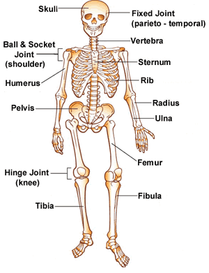
What major bones and joints are labeled in the diagram?
What type of tissue is bone classified as?
What makes bone hard?
What are the two types of bone tissue?
What is compact bone?
What is the structure of compact bone?
What is cancellous bone?
What does the medullary cavity contain?
What is shown in the diagram of a long bone?
What are long bones?
What are short bones?
What are flat bones?
What are irregular bones?
What are pneumatic bones?
What are sesamoid bones?
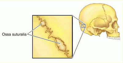
What are sutural bones?
Which bones are considered long bones in the hand and foot?
What are the two main parts of long bones?
What is the shaft of a long bone called?
What does the diaphysis contain?
What lines the shaft of a long bone?
What is the outer covering of the shaft called?
What do osteoblasts in the periosteum do?
What are the ends of long bones called?
What type of bone forms the ends of long bones?
What covers the articular surfaces of the epiphysis?
What separates the diaphysis from the epiphysis during growth?
What is the metaphysis?
What does the metaphysis transmit?
What is depicted in the long bone diagram?
What is the proximal epiphysis of the humerus?
What is the distal epiphysis of the humerus?
What is the diaphysis of the humerus?
What is the metaphysis of the humerus?
What is the function of articular cartilage in the humerus?
What is spongy bone in the humerus?
What does the medullary cavity of the humerus contain?
What is the nutrient artery in the humerus?
What is the epiphyseal line in the humerus?
What is the compact bone in the humerus?
What is the endosteum in the humerus?
What is the function of red bone marrow in the humerus?
What does the diagram of the humerus illustrate?
What is ossification of bone?
When does primary ossification occur?
What do primary ossification centers form?
When do secondary ossification centers appear?
What do secondary ossification centers complete?
What does the diagram illustrate?
What is endochondral ossification?
What is intramembranous ossification?
What type of ossification occurs in long bones?
What type of ossification occurs in flat bones?
What does the diagram of endochondral ossification illustrate?
What does the diagram of intramembranous ossification show?
What are the sources of blood supply for bones?
How do nutrient arteries enter the bone?
What types of nerves supply bones?
What is the function of vasomotor nerves in bones?
What does the diagram of a long bone illustrate?
What is the body of a bone?
What is a capitulum?
What is a condyle?
What is a crest?
What is an epicondyle?
What is a facet?
What is a foramen?
What is a fossa?
What is a groove?
What are the parts of a rib?
What is the spinous process?
What is the greater tubercle?
What is the iliac crest?
What is the greater sciatic notch?
What is the obturator foramen?
What is the lateral malleolus?
What are the articular facets of vertebra?
What is depicted in the skeleton diagram?
What is a head in bone markings?
What is a malleolus?
What does neck refer to in bone markings?
What is a notch in bone terminology?
Define process in the context of bone markings.
What is a protuberance?
What does shaft refer to in bone structure?
What is a spine in bone anatomy?
Define trochanter in bone markings.
What is a tubercle?
What does tuberosity refer to?
What is a trochlea?
What are the components of a rib?
What is the capitulum?
Identify the external occipital protuberance.
What is the greater trochanter?
What is the ischial tuberosity?
What is the radial groove?
What does the spinous process refer to?
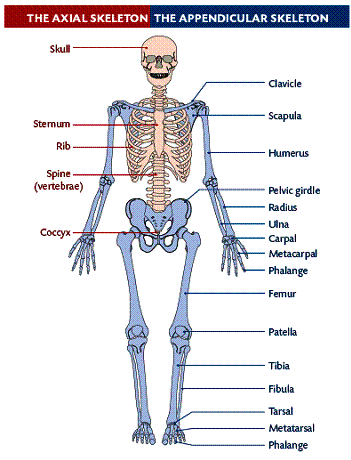
What bones are part of the axial skeleton?
What bones are part of the appendicular skeleton?
What is the function of the axial skeleton?
What is the function of the appendicular skeleton?
What are the major components of the appendicular skeleton?
What is the diagram illustrating the skeleton?
What are the two parts of the skull?
How many bones make up the skull?
What types of joints do the immovable bones of the skull have?
What is the movable bone of the skull?
What does the brain box of the skull consist of?
What does the facial skeleton of the skull consist of?
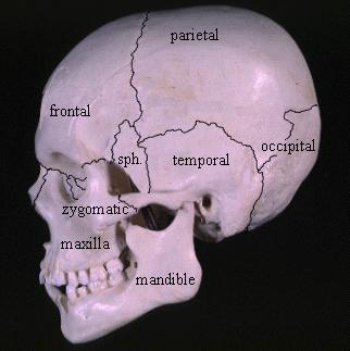
What major bones are labeled in the anatomical illustration of the skull?
What is the source of the anatomical illustration of the skull?
What are the single bones of the skull?
What bone is located at the front of the skull?
Which bone forms the base of the skull?
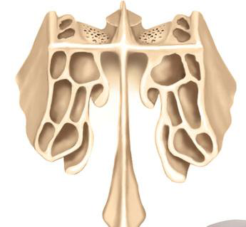
What bone is located between the nasal cavity and the brain?
Which bone is shaped like a butterfly and is located in the middle of the skull?
What is the name of the bone that forms the lower jaw?
What is the name of the bone that separates the nasal cavity from the mouth?
What bones are highlighted in this image? 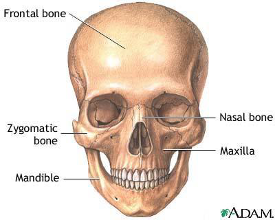
What view shows the major cranial and facial bones? 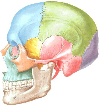
What does this image highlight? 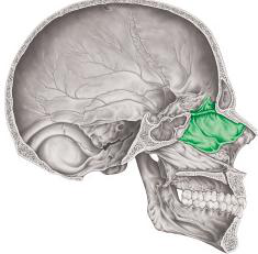
What are the paired bones of the skull?
What is the function of the parietal bone?
What is the location of the temporal bone?
What is the role of the maxilla?
What does the zygomatic bone form?
What is the function of the nasal bone?
What is the role of the lacrimal bone?
What does the palatine bone contribute to?
What is the function of the inferior nasal concha?
What does the inferior view of the skull show?
What does the anterior view of the skull illustrate?
How many cervical vertebrae are in the vertebral column?
How many thoracic vertebrae are in the vertebral column?
How many lumbar vertebrae are in the vertebral column?
How many sacral vertebrae are fused to form the sacrum?
How many coccygeal vertebrae are fused to form the coccyx?
What is the primary function of the vertebral column?
What does the vertebral column protect?
What regions are labeled in the vertebral column diagram?
What is shown in the lateral view of the vertebral column diagram?
What is the shape and position of the vertebra body?
What forms the vertebral arch?
What is the vertebral foramen?
What is formed by the succession of vertebral foramina?
How many processes project from the vertebral arch?
How many transverse processes are there?
What do the superior articular processes articulate with?
What do the inferior articular processes articulate with?
What does the lateral diagram of a vertebra show?
What is the body shape of cervical vertebrae?
What is the spinous process of cervical vertebrae like?
Do cervical vertebrae show a transverse foramen?
What is the vertebral foramen shape of cervical vertebrae?
What is the body shape of thoracic vertebrae?
What is the spinous process of thoracic vertebrae like?
Do thoracic vertebrae have facets for rib articulation?
What is the vertebral foramen shape of thoracic vertebrae?
What is the body shape of lumbar vertebrae?
What is the spinous process of lumbar vertebrae like?
What is the vertebral foramen shape of lumbar vertebrae?
What is shown in the overhead view of lumbar vertebrae?
What is the characteristic feature of cervical vertebrae?
What is the characteristic feature of thoracic vertebrae?
What is the characteristic feature of lumbar vertebrae?
What is depicted in the image comparing cervical, thoracic, and lumbar vertebrae?
What are the three types of vertebrae?
What is the function of the Atlas (C1)?
What is the function of the Axis (C2)?
What are the key features of thoracic vertebrae?
What are the key features of lumbar vertebrae?
What is depicted in the illustration of cervical vertebrae?
What are the features labeled in the thoracic vertebra diagram?
What are the features labeled in the lumbar vertebra diagram?
What is the shape of the sacrum?
How many vertebrae are fused in the sacrum?
What becomes the median sacral crest?
What forms the lateral sacral crest?
What does the vertebral foramen become in the sacrum?
How many vertebrae are fused in the coccyx?
What does the coccyx attach to?
What are intervertebral foramina?
What do adjacent notches in the vertebral arch form?
What is shown in the diagram related to intervertebral discs?
What does the anterior view diagram of the sacrum and coccyx include?
What are the four curves of the vertebral column?
What shape does the vertebral column form at birth?
What are the secondary curves of the vertebral column?
Which curves of the vertebral column are convex forward?
Which curves of the vertebral column are convex backward?
What does the vertebral column look like at three months of gestation?
What is kyphosis?
What is lordosis?
What is scoliosis?
What does this illustration show? 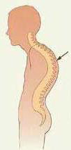
What do the diagrams illustrate? 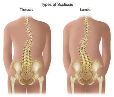
What are the components of the thoracic cage?
What are the three parts of the sternum?
What is the sternal angle?
What does the sternum consist of?
How many pairs of ribs are in the thoracic cage?
What is the function of the thoracic cage?
What is labeled in the diagram of the thoracic cage?
How many pairs of ribs are there?
Where are all the ribs attached at their posterior ends?
What are the true ribs?
How are the false ribs attached?
What are the floating ribs?
What do the true ribs connect to?
What is illustrated in this diagram?
What is the head of a rib?
What is the neck of a rib?
What is the tubercle of a rib?
What is the angle of a rib?
What is the shaft of a rib?
What does the inferior facet of a rib articulate with?
What is the costal groove of a rib?
What is illustrated in the posterior view of a left rib?
What is shown in the lateral view of a vertebra and a single rib?
What is highlighted in the lateral view of a thoracic vertebra?
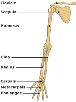
What bones make up the shoulder girdle?
What is the single bone in the arm?
What are the two bones in the forearm?
What bones form the hand?
What bones are included in the carpals?
What is the total number of phalanges in the hand?
What is the diagram of the bones of the upper limb?
What is the coracoid process?
What is the acromion?
What are the greater and lesser tuberosities?
What is the trochlea?
What are the carpal bones?
What are the metacarpal bones?
What are the phalanges?
What is the radius?
What is the ulna?
What is the olecranon process?
What is the trochlear notch?
What is the styloid process?
What is shown in the anterior view of the shoulder joint?
What is depicted in the anterior view of the wrist and hand?
What does the anterior view of the radius and ulna show?
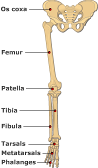
What is the pelvic girdle made of?
How many bones are in the thigh?
What are the bones in the leg?
What forms the foot?
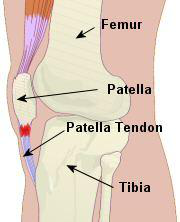
What is the largest bone in the lower limb?
What is the function of the patella?
How many phalanges are in each toe?
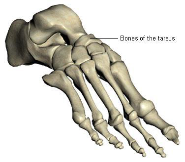
What are the bones of the foot?
What is the image showing?
What does the anterior view of the pelvis label?
What is the structure of the tarsus?
What is the structure of the metatarsus?
What are the three regions of the hip bone?
What are the main parts of the femur?
What bones make up the foot?
What are the main parts of the tibia and fibula?
Karteikarten in diesem Deck (267)
-
anatomy skeletal_system
-
anatomy joints
-
anatomy cartilage
-
What are the functions of bones?
- Form skeleton
- Attachment for muscles & ligaments
- Allow body movements
- Protect vital organs
- Store calcium salts
- Produce blood cells
anatomy bones functions -
anatomy protection
-
anatomy protection
-
What are the connective tissue coverings of bones and cartilage called?
- Bones: Periosteum
- Cartilage: Perichondrium
anatomy connective_tissue -
anatomy skeleton
-
anatomy blood_cells
-
anatomy skeleton diagram
-
biology anatomy
-
biology anatomy
-
biology anatomy
-
What is compact bone?
Compact bone is dense and hard, forming the shaft of long bones and the outer shell of other bones.
biology anatomy -
What is the structure of compact bone?
Compact bone consists of cylindrical units called Haversian systems.
biology anatomy -
What is cancellous bone?
Cancellous (spongy) bone is a delicate bony meshwork filling the inside of bones, except where the medullary cavity exists.
biology anatomy -
biology anatomy
-
What is shown in the diagram of a long bone?
The diagram shows spongy bone, compact bone, and the medullary cavity.
biology anatomy -
What are long bones?
- Tubular and longer than wide
- Found in limbs (e.g. humerus, radius, ulna, femur, tibia, fibula)
anatomy bones -
anatomy bones
-
anatomy bones
-
What are irregular bones?
- Irregular in shape
- Have many processes (e.g. vertebrae, pelvic bones, facial bones)
anatomy bones -
anatomy bones
-
anatomy bones
-
anatomy bones
-
Which bones are considered long bones in the hand and foot?
- Metatarsal bones
- Metacarpal bones
- Phalanges
anatomy bones -
anatomy bones
-
anatomy bones
-
anatomy bones
-
anatomy bones
-
anatomy bones
-
anatomy bones
-
anatomy bones
-
anatomy bones
-
anatomy bones
-
anatomy bones
-
anatomy bones
-
anatomy bones
-
anatomy bones
-
What is the proximal epiphysis of the humerus?
The end part of the humerus closest to the shoulder joint.
anatomy humerus -
What is the distal epiphysis of the humerus?
The end part of the humerus closest to the elbow joint.
anatomy humerus -
anatomy humerus
-
What is the metaphysis of the humerus?
The region between the epiphysis and diaphysis, involved in bone growth.
anatomy humerus -
What is the function of articular cartilage in the humerus?
Cushions and reduces friction at the joint surfaces.
anatomy joints -
What is spongy bone in the humerus?
A porous type of bone found at the ends of the humerus, containing red bone marrow.
anatomy bone_structure -
What does the medullary cavity of the humerus contain?
It contains yellow bone marrow and is involved in fat storage.
anatomy bone_structure -
anatomy circulation
-
What is the epiphyseal line in the humerus?
The remnant of the growth plate, indicating where bone growth occurred.
anatomy growth -
What is the compact bone in the humerus?
The dense outer layer that provides strength and structure.
anatomy bone_structure -
anatomy bone_structure
-
What is the function of red bone marrow in the humerus?
It is responsible for the production of blood cells.
anatomy hematopoiesis -
What does the diagram of the humerus illustrate?
It shows the structure and regional divisions of the humerus.
anatomy illustration -
anatomy ossification
-
When does primary ossification occur?
It starts with the appearance of points where cartilage calcifies and periosteal capillaries grow.
anatomy ossification -
What do primary ossification centers form?
They are responsible for ossification of the central parts of the bone, forming the diaphysis.
anatomy ossification -
When do secondary ossification centers appear?
After birth, they appear in the peripheral parts of the bone.
anatomy ossification -
What do secondary ossification centers complete?
They complete the process of ossification, forming epiphyses.
anatomy ossification -
What does the diagram illustrate?
The process of bone growth and ossification, showing stages from hyaline cartilage model to fully formed bone.
anatomy ossification -
What is endochondral ossification?
Bone formed by replacement of the cartilaginous model by bone tissue (e.g., long bones).
biology ossification -
What is intramembranous ossification?
Bone formed directly from connective tissue membrane (e.g., flat bones, clavicle).
biology ossification -
biology ossification
-
biology ossification
-
What does the diagram of endochondral ossification illustrate?
Stages of bone growth replacing cartilage.
biology ossification -
What does the diagram of intramembranous ossification show?
Bone tissue growing over a membrane, illustrating fetal skull ossification.
biology ossification -
What are the sources of blood supply for bones?
- Nutrient arteries
- Periosteal arteries
- Metaphyseal arteries
- Epiphyseal arteries
anatomy blood_supply -
anatomy blood_supply
-
anatomy nerve_supply
-
anatomy nerve_supply
-
anatomy blood_supply
-
What is the body of a bone?
The principal mass of a bone; the shaft in long bones; anterior weight-bearing portions in vertebrae.
anatomy bones -
anatomy bones
-
What is a condyle?
A rounded, knuckle-like articular area, often occurring in pairs (e.g., lateral and medial femoral condyles).
anatomy bones -
anatomy bones
-
What is an epicondyle?
An eminence superior or adjacent to a condyle (e.g., lateral epicondyle of the humerus).
anatomy bones -
What is a facet?
A smooth flat area, usually covered with cartilage, where a bone articulates with another bone (e.g., superior costal facet on a vertebra).
anatomy bones -
anatomy bones
-
anatomy bones
-
anatomy bones
-
anatomy bones
-
anatomy bones
-
What is the greater tubercle?
A prominent projection on the humerus, important for muscle attachment.
anatomy bones -
anatomy bones
-
What is the greater sciatic notch?
A notch in the ilium that allows passage for nerves and blood vessels.
anatomy bones -
anatomy bones
-
What is the lateral malleolus?
The bony prominence on the outer side of the ankle, part of the fibula.
anatomy bones -
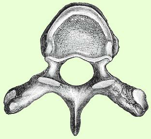
What are the articular facets of vertebra?
Smooth surfaces on vertebrae that articulate with adjacent vertebrae or ribs.
anatomy bones -
What is depicted in the skeleton diagram?
A posterior and anterior view of the human skeleton with labeled bones.
anatomy skeleton -
anatomy bone_markings
-
anatomy bone_markings
-
anatomy bone_markings
-
What is a notch in bone terminology?
An indentation at the edge of a bone (e.g., greater sciatic notch).
anatomy bone_markings -
Define process in the context of bone markings.
An extension or projection serving a particular purpose, having a characteristic shape (e.g., articular process).
anatomy bone_markings -
anatomy bone_markings
-
anatomy bone_markings
-
anatomy bone_markings
-
Define trochanter in bone markings.
A large blunt elevation (e.g., greater trochanter of the femur).
anatomy bone_markings -
anatomy bone_markings
-
anatomy bone_markings
-
What is a trochlea?
A spool-like articular process or process that acts as a pulley (e.g., trochlea of the humerus).
anatomy bone_markings -
anatomy rib
-
anatomy humerus
-
anatomy skull
-
anatomy femur
-
anatomy pelvis
-
anatomy humerus
-
What does the spinous process refer to?
A bony projection on a vertebra, serving as an attachment point for muscles.
anatomy vertebrae -
anatomy skeleton
-
What bones are part of the appendicular skeleton?
- Clavicle
- Scapula
- Humerus
- Radius
- Ulna
- Pelvic girdle
- Femur
- Tibia
- Fibula
anatomy skeleton -
What is the function of the axial skeleton?
Supports the head, neck, and trunk; protects the brain and spinal cord.
anatomy functions -
anatomy functions
-
What are the major components of the appendicular skeleton?
- Bones of upper limb
- Bones of lower limb
- Shoulder girdle
- Pelvic girdle
anatomy components -
anatomy diagram
-
What are the two parts of the skull?
- Brain box (vault): upper & posterior part
- Facial skeleton: anterior part
anatomy skull -
anatomy bones
-
What types of joints do the immovable bones of the skull have?
They articulate by fibrous joints (sutures).
anatomy joints -
anatomy bones
-
anatomy brain_box
-
anatomy facial_skeleton
-
What major bones are labeled in the anatomical illustration of the skull?
- Frontal
- Parietal
- Temporal
- Occipital
- Sphenoid
- Zygomatic
- Maxilla
- Mandible
anatomy illustration -
anatomy illustration
-
What are the single bones of the skull?
- Frontal bone
- Occipital bone
- Ethmoid bone
- Sphenoid bone
- Vomer
- Mandible
anatomy skull -
anatomy skull
-
anatomy skull
-
anatomy skull
-
anatomy skull
-
anatomy skull
-
anatomy skull
-
What bones are highlighted in this image?
- Frontal bone
- Vomer
- Zygomatic bone
- Nasal bone
- Maxilla
- Mandible
anatomy skull -
anatomy skull
-
anatomy skull
-
What are the paired bones of the skull?
- Parietal bone
- Temporal bone
- Maxilla
- Zygomatic bone
- Nasal bone
- Lacrimal bone
- Palatine bone
- Inferior nasal concha
anatomy skull -
anatomy skull bones
-
anatomy skull bones
-
anatomy skull bones
-
anatomy skull bones
-
anatomy skull bones
-
anatomy skull bones
-
What does the palatine bone contribute to?
Forms part of the hard palate and floor of the nasal cavity.
anatomy skull bones -
What is the function of the inferior nasal concha?
Helps to filter and warm the air inhaled through the nose.
anatomy skull bones -
What does the inferior view of the skull show?
Highlights the maxilla, sphenoid bone, palatine bone, and vomer.
anatomy skull -
What does the anterior view of the skull illustrate?
Shows the frontal bone, lacrimal bone, ethmoid bone, and nasal cavity bones.
anatomy skull -
anatomy vertebral_column
-
anatomy vertebral_column
-
anatomy vertebral_column
-
anatomy vertebral_column
-
anatomy vertebral_column
-
anatomy functions
-
anatomy functions
-
What regions are labeled in the vertebral column diagram?
Cervical, Thoracic, Lumbar, Sacrum, Coccyx
anatomy vertebral_column -
What is shown in the lateral view of the vertebral column diagram?
Color-coded regions: Cervical, Thoracic, Lumbar, Sacrum, Coccyx
anatomy vertebral_column -
anatomy vertebra
-
anatomy vertebra
-
anatomy vertebra
-
anatomy vertebra
-
anatomy vertebra
-
anatomy vertebra
-
What do the superior articular processes articulate with?
The inferior articular processes of the vertebra above
anatomy vertebra -
What do the inferior articular processes articulate with?
The superior articular processes of the vertebra below
anatomy vertebra -
anatomy vertebra
-
anatomy vertebrae
-
anatomy vertebrae
-
anatomy vertebrae
-
anatomy vertebrae
-
anatomy vertebrae
-
anatomy vertebrae
-
anatomy vertebrae
-
anatomy vertebrae
-
anatomy vertebrae
-
anatomy vertebrae
-
anatomy vertebrae
-
anatomy vertebrae
-
anatomy vertebrae
-
What is the characteristic feature of thoracic vertebrae?
Have facets for articulation with rib tubercle
anatomy vertebrae -
anatomy vertebrae
-
What is depicted in the image comparing cervical, thoracic, and lumbar vertebrae?
Characteristics of each vertebra type
anatomy vertebrae -
anatomy vertebrae
-
anatomy cervical
-
anatomy cervical
-
anatomy thoracic
-
What are the key features of lumbar vertebrae?
- Body
- Pedicle
- Lamina
- Spinous process
- Transverse process
anatomy lumbar -
What is depicted in the illustration of cervical vertebrae?
Atlas (C1) and Axis (C2) with Dens and bifid spine.
anatomy cervical -
What are the features labeled in the thoracic vertebra diagram?
- Body
- Spinal canal
- Transverse processes
- Facets for ribs
anatomy thoracic -
What are the features labeled in the lumbar vertebra diagram?
- Body
- Pedicle
- Lamina
- Inferior Articular Process
- Spinous Process
anatomy lumbar -
anatomy sacrum
-
anatomy sacrum
-
anatomy sacrum
-
anatomy sacrum
-
anatomy sacrum
-
anatomy coccyx
-
anatomy coccyx
-
anatomy vertebrae
-
anatomy vertebrae
-
anatomy discs
-
What does the anterior view diagram of the sacrum and coccyx include?
Labels for Sacrum, Coccyx, Ala, Spinous tubercles, Sacral canal, Dorsal sacral foramina, and Sacral hiatus
anatomy sacrum -
anatomy vertebral_column
-
What shape does the vertebral column form at birth?
One C-shaped curve (1ry curve) that is convex backward
anatomy development -
What are the secondary curves of the vertebral column?
Curves that appear in the cervical and lumbar regions, convex forward
anatomy curves -
anatomy curves
-
anatomy curves
-
What does the vertebral column look like at three months of gestation?
Developing bones with a C-shaped curve
anatomy development -
anatomy spine
-
anatomy spine
-
anatomy spine
-
What does this illustration show?
It shows abnormal spinal curvatures including kyphosis and scoliosis.
anatomy illustration -
anatomy illustration
-
anatomy thoracic_cage
-
anatomy sternum
-
anatomy sternal_angle
-
anatomy sternum
-
anatomy ribs
-
What is the function of the thoracic cage?
Protects vital organs and supports the upper body structure.
anatomy thoracic_cage -
anatomy thoracic_cage
-
anatomy ribs
-
anatomy ribs
-
anatomy ribs
-
anatomy ribs
-
anatomy ribs
-
anatomy ribs
-
anatomy ribs
-
anatomy ribs
-
anatomy ribs
-
What is the tubercle of a rib?
Articulates with the transverse process of its corresponding vertebra
anatomy ribs -
anatomy ribs
-
What is the shaft of a rib?
Thin and flattened, lower border sharp with a groove for intercostal nerves & vessels
anatomy ribs -
anatomy ribs
-
anatomy ribs
-
What is illustrated in the posterior view of a left rib?
Head, Neck, Superior & inferior facets, Tubercle, Angle, Shaft, Costal groove
anatomy ribs -
What is shown in the lateral view of a vertebra and a single rib?
Articulation points with labels for Head, Neck, Tubercle, Angle, and Shaft
anatomy ribs -
anatomy ribs
-
anatomy skeleton
-
anatomy skeleton
-
anatomy skeleton
-
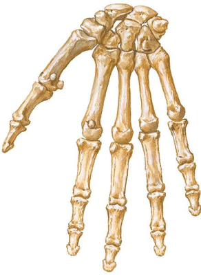
What bones form the hand?
- Carpus (8 bones)
- Metacarpus (5 bones)
- Phalanges (3 in each finger, 2 in thumb)
anatomy skeleton -
anatomy skeleton
-
What is the total number of phalanges in the hand?
- 14 in total: 12 (3 in each finger) + 2 (in thumb)
anatomy skeleton -
anatomy skeleton
-
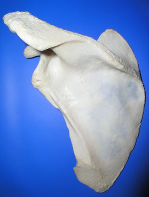
anatomy scapula
-
anatomy scapula
-
What are the greater and lesser tuberosities?
Bony prominences on the humerus for muscle attachment.
anatomy humerus -
anatomy humerus
-
anatomy carpals
-
anatomy metacarpals
-
anatomy phalanges
-
anatomy radius
-
anatomy ulna
-
anatomy ulna
-
What is the trochlear notch?
The concave surface of the ulna that articulates with the trochlea of the humerus.
anatomy ulna -
anatomy styloid
-
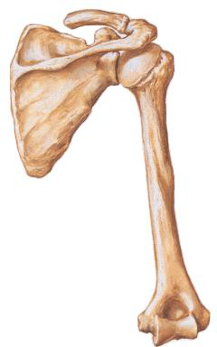
What is shown in the anterior view of the shoulder joint?
The scapula and humerus with articulation points visible.
 anatomy shoulder
 -
What is depicted in the anterior view of the wrist and hand?
The carpals, metacarpals, and phalanges.
 anatomy hand
 -
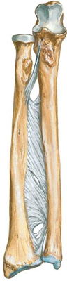
What does the anterior view of the radius and ulna show?
The distal ends with interosseous membrane and ligaments.
 anatomy forearm
 -
anatomy skeleton
-
anatomy skeleton
-
anatomy skeleton
-
anatomy skeleton
-
anatomy skeleton
-
anatomy skeleton
-
anatomy skeleton
-
anatomy skeleton
-
anatomy skeleton
-
What does the anterior view of the pelvis label?
- Hip bone
- Sacrum
- Coccyx
- Pubic symphysis
- Sacroiliac joint
- Sacral promontory
- Pelvic brim
- Acetabulum
anatomy skeleton -
anatomy skeleton
-
anatomy skeleton
-
anatomy hip_bone
-
What are the main parts of the femur?
- Head
- Neck
- Greater Trochanter
- Lesser Trochanter
- Femoral Condyles
anatomy femur -
anatomy foot
-
anatomy tibia fibula