Sign up to unlock more features
- Save this deck to your account
- Study flashcards with spaced repetition
- Export to Anki (.apkg) or PDF
- Process documents up to 100 pages
- Images extracted from PDFs and documents
- Better text extraction from your PDFs and documents
- Better flashcards with our more advanced AI model
What is the first step in the vision pathway?
What follows the retina in the vision pathway?
What is the third component of the vision pathway?
After the optic chiasm, what is the next part in the vision pathway?
What part of the thalamus is involved in the vision pathway?
What comes after the lateral geniculate nucleus in the vision pathway?
Where does the vision pathway end?
What visual information travels from the retina to the visual cortex?
What does the optic chiasm do?
How does information from the temporal side of the visual field travel?
Describe the path of nasal visual information.
What happens to visual fields during processing?
What visual loss occurs with an optic nerve lesion?
What is the result of an optic chiasm lesion?
What vision deficit is associated with lesions in optic radiation or occipital lobe?
What is an ipsilateral monocular scotoma?
What does bitemporal hemianopia indicate?
What does bilateral central scotoma indicate?
When does the development of the eye begin?
What forms the retina during eye development?
What extends from the developing diencephalon?
What does the optic cup form?
What does the optic stalk become?
What induces the formation of the lens in eye development?
From what does the sclera and choroid develop?
What does the choroid give rise to?
What tissues form the eyelids, conjunctiva, and cornea?
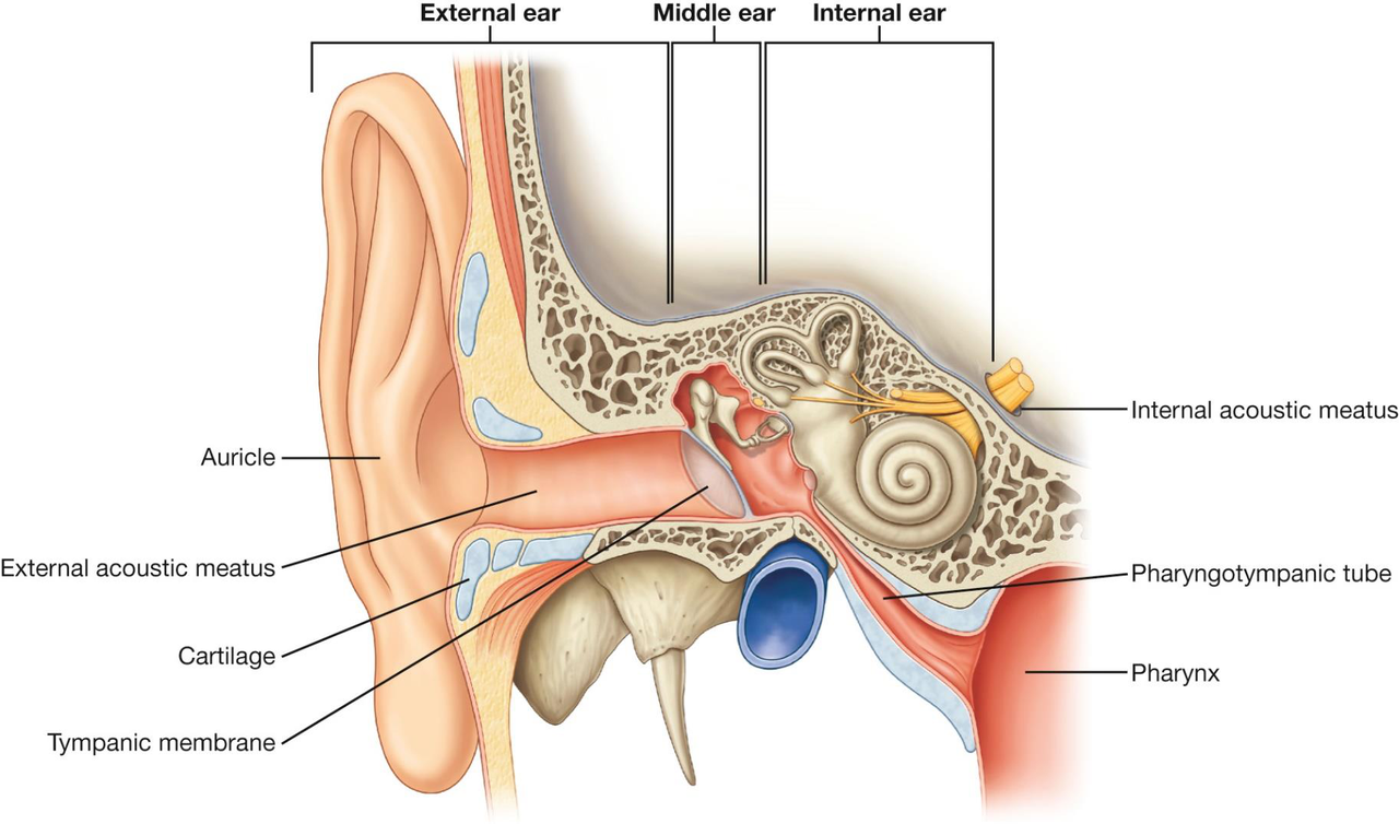
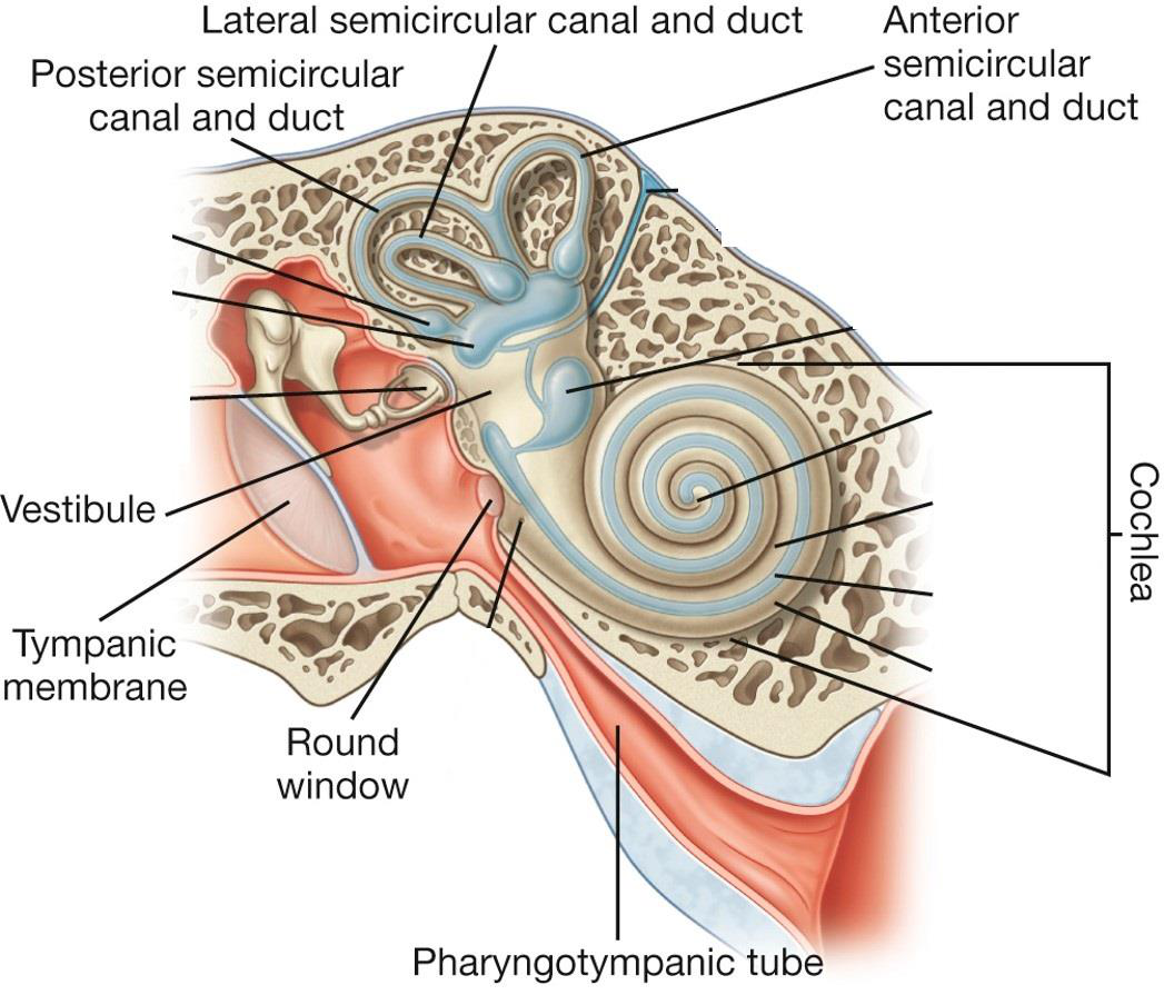
What is the function of the external ear?
What connects the middle ear to the nasopharynx?
What does the internal ear transfer to the CNS?
What sensations are produced by fluid movement in the internal ear?
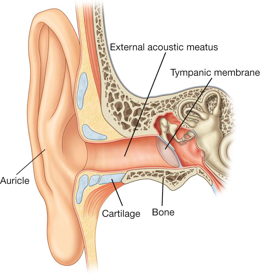
What is the external ear primarily composed of?
What is the function of the tympanic membrane?
What does the external acoustic meatus contain?
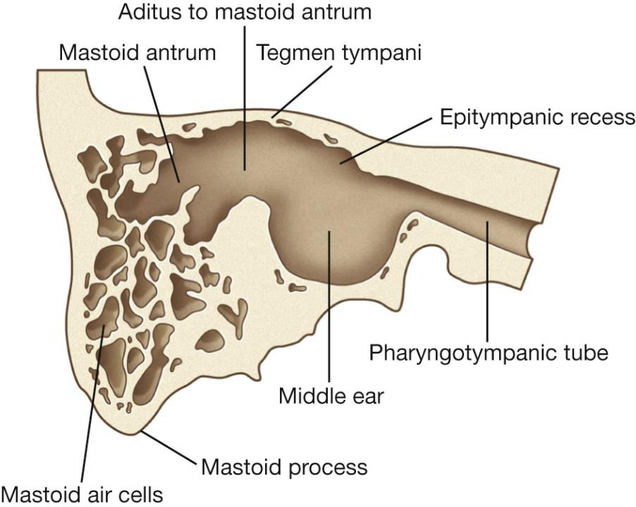
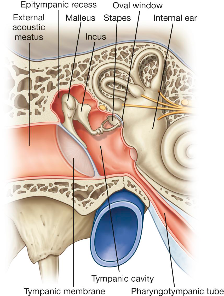
What does the middle ear consist of?
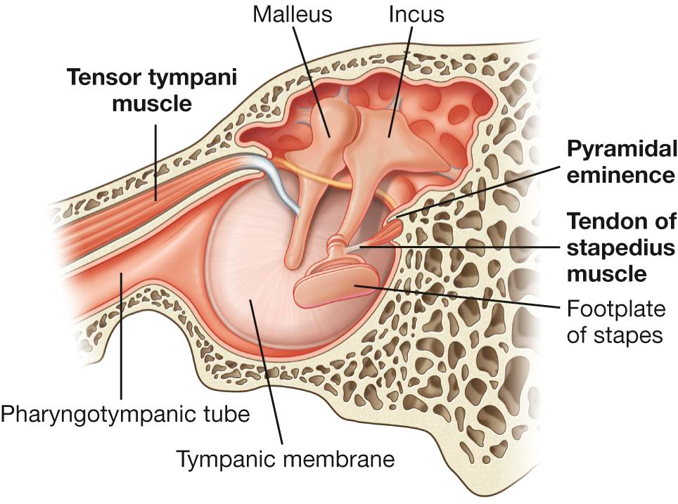
What is the function of the pharyngotympanic tube?
What structure allows the pharyngotympanic tube to open?
What are the openings in the middle ear?
What is the anatomical role of the tympanic membrane?
What connects the middle ear to the nasal cavity?
Which structure in the middle ear is responsible for pressure equalization?
What are the auditory ossicles in the middle ear?
What is the function of the malleus?
What is the function of the stapes?
How does the base of the stapes compare to the tympanic membrane?
What is the function of the Stapedius muscle?
What does the Tensor tympani muscle do?
Which cranial nerve provides the Chorda tympani nerve?
Which cranial nerve is part of the Tympanic plexus?
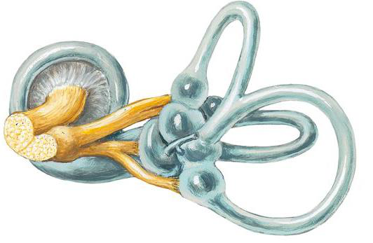
What organ is contained in the inner ear?
What are the functions of the vestibulocochlear organ?
What protects the inner ear?
What fluid fills the bony labyrinth?
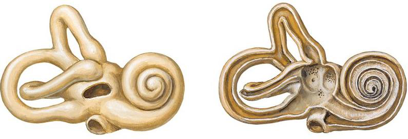
What comprises the bony labyrinth?
What fluid fills the membranous labyrinth?
What are the components of the bony labyrinth in the inner ear?
What are the components of the membranous labyrinth in the inner ear?
What is the primary function of the cochlea?
What fills the cochlea?
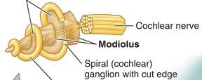
What are the three distinct parts of the cochlea?
What carries the cochlear nerve within the cochlea?
What structure does the cochlea communicate with?
What does the cochlear labyrinth contain?
What is the cochlear labyrinth comprised of?
Where is the spiral organ located?
What is the shape of the cochlear duct?
What is the apex of the cochlear duct called?
What organ sits on the basilar membrane in the cochlear duct?
What types of cells are contained within the spiral organ?
Which nerve synapses with the cells in the spiral organ?
What membranes are associated with the cochlear duct?
What structure is involved in the hearing process found in the cochlear duct?
What are the first steps in sound transmission?
How do vibrations transfer into fluid in the ear?
What leads to hair cells deflection?
What happens to hydraulic waves after the cochlear duct?
What fluid is involved in sound transmission?
How do high frequency sounds affect the basilar membrane?
How do low frequency sounds affect the basilar membrane?
What follows after synapsing on cochlear nerve fibers?
What are the steps in the auditory pathway?
Which nerve is responsible for transmitting auditory signals?
What is the role of the Cochlear nuclei?
What structure helps with auditory acuity and sound localization?
Which part of the brain is responsible for processing auditory information after superior olivary nuclei—> inferior colliculi?
Where does the auditory perception occur in the brain?
What type of hearing loss is linked to the external or middle ear?
What are examples of conductive hearing loss?
Which parts of the ear are affected in sensorineural hearing loss?
What examples are associated with sensorineural hearing loss?
What is the location of conductive hearing loss lesions?
What is the location of sensorineural hearing loss lesions?
What is the function of the vestibule?
What does the vestibule contain?
What is the structure of the vestibular labyrinth?
What are maculae?
How many semicircular canals are there?
What is the orientation of the semicircular canals?
What is the bony ampulla?
What are semicircular ducts?
What is contained in the ampulla of the semicircular ducts?
Where do the semicircular ducts open?
What nerve innervates the vestibular system?
How does the vestibular system sense motion?
What structures are sensitive to gravity and acceleration?
What is the specialized epithelium containing hair cells in the utricle and saccule?
What do hair cells project into?
What are the mineralized structures in the otolithic membrane?
What is the function of the otolithic membrane?
What is the role of hair cells in the vestibular system?
What do the ampullae contain?
What do the ampullae sense?
How do ampullae sense rotations?
What initiates the vestibular sensation pathway?
What represents the primary neurons in the vestibular pathway?
What is the function of the vestibular nerve?
Which cranial nerve is involved in the vestibular sensation pathway?
Where do the secondary neurons synapse in the vestibular pathway?
What is the role of the vestibulospinal tract?
What does the Vestibulo-Ocular Reflex (VOR) coordinate?
Which brain structures receive projections from vestibular signals?
Flashcards in this deck (121)
-
neuroscience vision
-
neuroscience vision
-
neuroscience vision
-
neuroscience vision
-
neuroscience vision
-
neuroscience vision
-
neuroscience vision
-
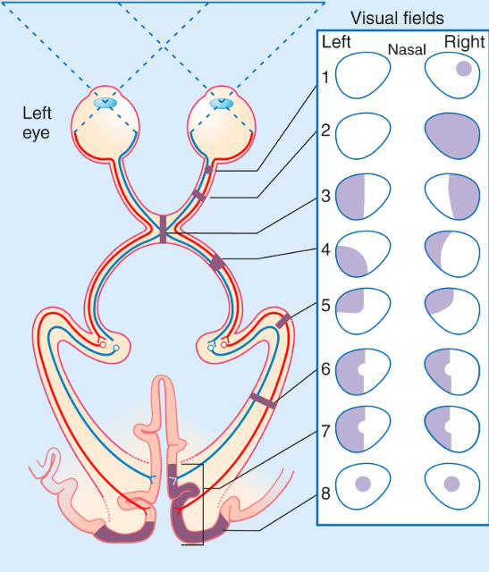
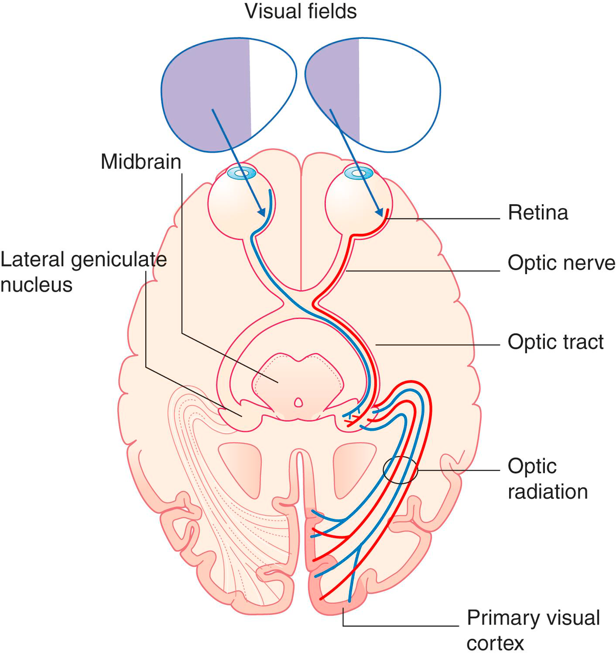
What visual information travels from the retina to the visual cortex?
Information flows from the retina through the optic nerve, chiasm, tract, lateral geniculate nucleus, optic radiation, to the visual cortex.
neuroscience vision -
What does the optic chiasm do?
It allows nasal visual information to cross, entering the contralateral optic tract.
neuroscience vision -
How does information from the temporal side of the visual field travel?
It enters the ipsilateral optic tract.
neuroscience vision -
Describe the path of nasal visual information.
It crosses at the optic chiasm and enters the contralateral optic tract.
neuroscience vision -
What happens to visual fields during processing?
They converge at the optic chiasm, allowing for visual integration.
neuroscience vision -
vision anatomy
-
vision anatomy
-
What vision deficit is associated with lesions in optic radiation or occipital lobe?
Contralateral homonymous hemianopia 6,7
 vision anatomy
 -
vision anatomy
-
vision anatomy
-
vision anatomy
-
development anatomy
-
development retina
-
anatomy diencephalon
-
anatomy optic_cup
-
anatomy optic_nerve
-
What induces the formation of the lens in eye development?
Overlying ectoderm forms lens pit, inducing lens formation.
embryology eye -
embryology eye
-
embryology eye
-
embryology eye
-
anatomy ear senses
-
anatomy ear senses
-
anatomy ear senses
-
anatomy ear senses
-
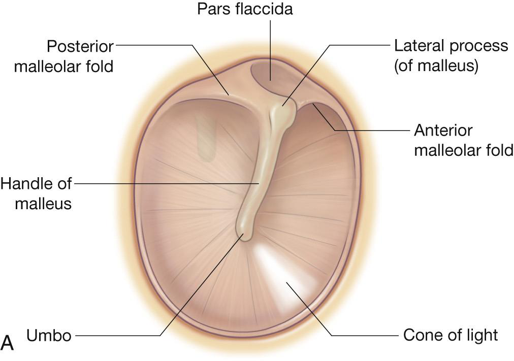
What is the external ear primarily composed of?
- Elastic cartilage
- Temporal bone
- auricle (pinna)
- External acoustic meatus: Outside-ear-hole, Contains ceruminous (produce cerumen/earwax) and sebaceous glands
- Tympanic membrane (ear drum): Oriented like a mini radar or satellite dish to receive sound waves
anatomy ear -
anatomy ear
-
anatomy ear
-
What does the middle ear consist of?
- Tympanic cavity: Lined with mucous membrane
- openings: mastoid air cells, pharyngotympanic tube (Eustachian tube/auditory tube)
- ear bones (malleus, incus, stapes)
anatomy ear -
anatomy ear
-
anatomy ear
-
anatomy ear
-
anatomy ear
-
anatomy ear
-
anatomy ear
-
anatomy middle_ear
-
anatomy physiology
-
anatomy physiology
-
How does the base of the stapes compare to the tympanic membrane?
Its base is smaller than the tympanic membrane, increasing vibratory force by 10x.
anatomy physics -
anatomy middle_ear
-
anatomy middle_ear
-
anatomy nerves
-
anatomy nerves
-
anatomy ear
-
anatomy function
-
anatomy protection
-
anatomy fluid
-
anatomy structure
-
anatomy fluid
-
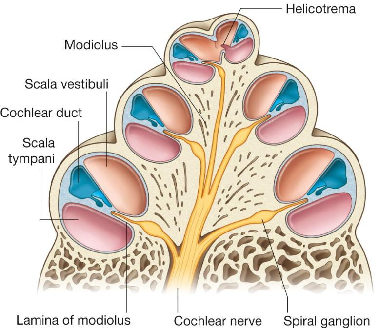
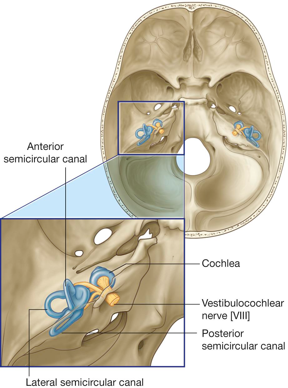
What are the components of the bony labyrinth in the inner ear?
- Cochlea
- Vestibule
- Semicircular canal
anatomy ear inner_ear -
What are the components of the membranous labyrinth in the inner ear?
- Cochlear labyrinth
- Vestibular labyrinth
- Semicircular ducts
anatomy ear inner_ear -
anatomy hearing
-
anatomy cochlea
-
anatomy cochlea
-
anatomy nerves
-
What structure does the cochlea communicate with?
Round window containing a secondary tympanic membrane
anatomy cochlea -
anatomy hearing
-
anatomy hearing
-
anatomy hearing
-
anatomy ear
-
anatomy ear
-
anatomy ear hearing
-
What types of cells are contained within the spiral organ?
- Supporting cells
- Outer hair cells
- Inner hair cells
anatomy ear cells -
anatomy ear nervous_system
-
anatomy ear membranes
-
What structure is involved in the hearing process found in the cochlear duct?
Spiral organ (organ of Corti)
anatomy ear hearing -
What are the first steps in sound transmission?
- Vibrations
- Tympanic membrane
- Ossicles
- Base of stapes at oval window
biology hearing -
How do vibrations transfer into fluid in the ear?
Vibrations transfer into hydraulic waves in perilymph of scala vestibuli
biology hearing -
What leads to hair cells deflection?
Deformation of cochlear duct causes endolymph to deflect hair cells, activating cochlear nerve fibers (CN VIII)
biology hearing -
What happens to hydraulic waves after the cochlear duct?
Hydraulic waves continue through to scala tympani and reach the secondary tympanic membrane in round window
biology hearing -
anatomy audio
-
How do high frequency sounds affect the basilar membrane?
Displace closer to the oval window (shorter distance)
anatomy audio -
How do low frequency sounds affect the basilar membrane?
Displace closer to helicotrema (longer distance)
anatomy audio -
What follows after synapsing on cochlear nerve fibers?
Spiral ganglion → Sound is interpreted by CNS
anatomy audio -
What are the steps in the auditory pathway?
1. Hair cells in spiral organ
2. Spiral ganglion (primary neurons)
3. Cochlear nerve
4. CN VIII
5. Cochlear nuclei in brain stem (synapse- secondary neurons)
6. Superior olivary nuclei (auditory acuity and localizing of sound)
7. Inferior colliculi
8. Thalamus (tertiary neurons)
9. Primary auditory cortex in temporal lobe
anatomy auditory -
Which nerve is responsible for transmitting auditory signals?
Cochlear nerve (branch of CNVIII)—> to CN VIII
anatomy nerves -
anatomy auditory brainstem
-
anatomy auditory brainstem
-
Which part of the brain is responsible for processing auditory information after superior olivary nuclei—> inferior colliculi?
Thalamus (tertiary neurons)
anatomy auditory thalamus -
anatomy auditory cortex
-
hearing anatomy
-
hearing examples
-
hearing anatomy
-
hearing examples
-
hearing anatomy
-
hearing anatomy
-
anatomy equilibrium
-
anatomy vestibule
-
anatomy vestibular_labyrinth
-
anatomy maculae
-
anatomy ear
-
What is the orientation of the semicircular canals?
They are oriented at right angles to occupy three planes in space.
anatomy ear -
anatomy ear
-
What are semicircular ducts?
They are portions of the membranous labyrinth within the semicircular canals.
anatomy ear -
What is contained in the ampulla of the semicircular ducts?
Hair cells for sensation are contained in the ampulla.
anatomy ear sensation -
anatomy ear
-
anatomy nervous_system
-
physiology sensation
-
anatomy vestibular
-
anatomy vestibular
-
anatomy vestibular
-
anatomy vestibular
-
physiology vestibular
-
What is the role of hair cells in the vestibular system?
Transduce mechanical changes into neural signals
physiology neuroscience -
anatomy inner_ear
-
physiology vestibular
-
physiology inner_ear
-
What initiates the vestibular sensation pathway?
- Hair cells in maculae of utricle
- Hair cells in saccule
- Hair cells in ampulla
anatomy vestibular -
anatomy vestibular
-
What is the function of the vestibular nerve?
- Transmits information from vestibular apparatus to brain
neuroanatomy vestibular -
neuroanatomy cranial_nerves
-
anatomy vestibular
-
physiology vestibular
-
What does the Vestibulo-Ocular Reflex (VOR) coordinate?
- Reflexive motor activities
- Coordinates eye (CN III and CN VI), head, and neck movements
physiology vestibular -
Which brain structures receive projections from vestibular signals?
Tertiary neurons
- Cerebellum
- Thalamus
- Cerebral cortex
anatomy vestibular
Vision Pathway
- Retina (nasal or temporal half)
- Optic nerve (through optic canal)
- Optic chiasm
- Optic tract
- Lateral geniculate nucleus of thalamus
- Optic radiation
- Occipital lobe's visual cortex
Optic Chiasm Processing
Visual Information Processing
- Temporal side: ipsilateral optic tract
- Nasal side: contralateral optic tract
- Each optic tract processes contralateral visual fields

Visual Pathway Lesions
Visual Field Deficits
- Optic nerve lesion: visual loss in one eye
- Optic chiasm lesion: bitemporal hemianopia
- Optic radiation/occipital lobe lesion: contralateral homonymous hemianopia
Development of the Eye
Key Stages
- Begins in the 4th week of development
- Retina forms from the developing brain
- Optic vesicles extend from diencephalon
- Forms the optic cup (double layered neural tunic)
- Optic stalk becomes the optic nerve
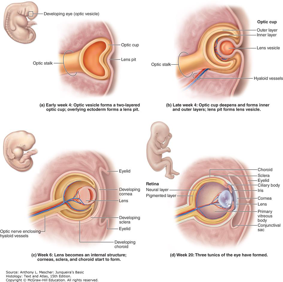
Parts of the Eye Development
Structures Formation
- Lens: overlying ectoderm induces lens formation
- Sclera & Choroid: form from mesenchyme surrounding the optic cup
- Choroid: forms iris, ciliary body, suspensory ligaments
- Eyelids, conjunctiva, cornea: from ectoderm and mesenchyme
Hearing and Balance Overview
Ear Structures
- External Ear: transfers sound, bound internally by tympanic membrane
- Middle Ear: contains ossicles, opens to nasopharynx via pharyngotympanic tube
- Internal Ear: transfers sound to CNS; movement of fluid results in sensations of hearing and balance
External and Middle Ear Components
Key Features
- External Ear Components: auricle, external acoustic meatus, tympanic membrane
- Middle Ear Components: tympanic cavity, lined with mucous membrane
- Openings: mastoid air cells, pharyngotympanic tube (Eustachian tube)
Middle Ear Contents
Auditory Ossicles
- Malleus (hammer): moves with tympanic membrane
- Incus (anvil)
- Stapes (stirrup): interacts with the oval window, amplifying sound
Inner Ear Components
Main Structures
- Bony Labyrinth: perilymph-filled, includes cochlea, vestibule, semicircular canals
- Membranous Labyrinth: filled with endolymph, series of sacs and ducts
Cochlear Structure
Cochlear Labyrinth Functions
- Three Parts: cochlear duct, scala tympani, scala vestibuli
- Modiolus: carries cochlear nerve (CN VIII) and vessels
- Communicates with round window
Sound Transmission Process
- Vibrations → tympanic membrane
- Ossicles → base of stapes at oval window
- Hydraulic waves in perilymph → scala vestibuli
- Hair cell deflection → cochlear nerve (CN VIII)
- Waves reach the round window
Auditory Pathway
- Hair cells in spiral organ
- Spiral ganglion (primary neurons)
- Cochlear nerve
- CN VIII
- Cochlear nuclei in brainstem
- Superior olivary nuclei
- Inferior colliculi
- Thalamus
- Primary auditory cortex
Clinical Connection: Hearing Loss
| Type | Lesion Location | Example |
|---|---|---|
| Conductive | External or middle ear | Otitis media, cerumen impaction |
| Sensorineural | Cochlea or CN VIII | Noise damage, age, drugs |
Vestibular System Overview
Key Components
- Vestibule: contains utricle and saccule
- Maculae: specialized epithelium with hair cells
Semicircular Ducts and Canals
Structure and Function
- Detect head rotation
- Crista Ampullaris: contains hair cells for sensation
Vestibular Sensation Pathway
- Hair cells in maculae
- Vestibular ganglion (primary neurons)
- Vestibular nerve
- CN VIII
- Vestibular nuclei in medulla (secondary neurons)
- Projects to cerebellum