Sign up to unlock more features
- Save this deck to your account
- Study flashcards with spaced repetition
- Export to Anki (.apkg) or PDF
- Process documents up to 100 pages
- Images extracted from PDFs and documents
- Better text extraction from your PDFs and documents
- Better flashcards with our more advanced AI model
What is the purpose of the Acid-Fast Stain?
What is the Endospore Stain used for?
What is the steam heating method used for?
What is the purpose of the Acid-Fast Stain?
Which bacteria are identified using the Acid-Fast Stain?
What diseases are caused by Mycobacterium?
Why doesn't the gram-stain work for certain bacteria?
What are some characteristics of acid-fast organisms?
What are the causative agents in leprosy and tuberculosis?
Staphylococcus
Bacillus
Mycobacterium
Escherichia
The Acid-Fast Stain is used to identify _______.
The Acid-Fast Stain is a differential stain used to detect cells capable of retaining a primary stain when treated with _______.
What is the cytological basis for the Acid-Fast Stain?
What does the Acid-Fast Stain resist?
What are the two procedures for Acid-Fast Staining?
What is unique about the Ziehl-Neelsen method?
What is unique about the Kinyoun method?
What do mycolic acids provide in Acid-Fast Staining?
What components are highlighted in the mycobacterial envelope structure?
What is the first step in the Acid-Fast Stain (ZN) method?
What is the primary stain used in the Acid-Fast Stain (ZN) method?
What is used as a mordant in the Acid-Fast Stain (ZN) method?
What is the decolorizer used in the Acid-Fast Stain (ZN) method?
What is the secondary stain in the Acid-Fast Stain (ZN) method?
What color do acid-fast cells appear after staining?
What color do non acid-fast cells appear after staining?
What is the order of steps in the Acid-Fast Stain (ZN) method?
Carbolfuchsin, Fix cells, Steam heat, Methylene blue, Acid alcohol
Fix cells, Methylene blue, Steam heat, Acid alcohol, Carbolfuchsin
Steam heat, Carbolfuchsin, Acid alcohol, Fix cells, Methylene blue
Fix cells, Carbolfuchsin, Steam heat, Acid alcohol, Methylene blue
The Acid-Fast Stain (ZN) method involves the following steps: 1. _______ 2. _______ 3. _______ 4. _______ 5. _______.
In the Acid-Fast Stain (ZN) method, acid-fast cells appear _______ and non acid-fast cells appear _______.
A diagram illustrating the Acid-Fast ZN staining method shows the appearance of cells as follows: before staining (transparent), after staining with _______ (reddish-purple), after decolorization with _______ (acid-fast cells retain color, others lose it), and after counterstaining with _______ (acid-fast cells are reddish-purple, non-acid-fast cells are blue). 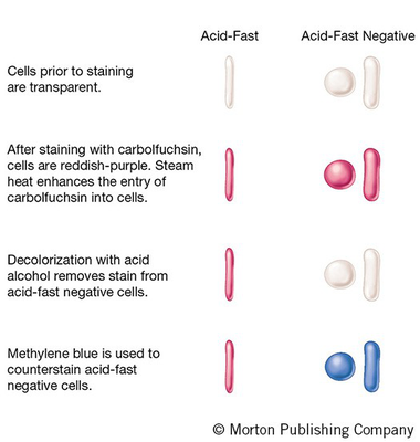
What is the first step in the Acid-Fast Stain (K) method?
What is the primary stain used in Acid-Fast Stain (K)?
How does the concentration of carbolfuchsin in Acid-Fast Stain (K) compare to ZN?
What is the purpose of the incubation time in Acid-Fast Stain (K)?
What is the decolorizer used in Acid-Fast Stain (K)?
What is the secondary stain used in Acid-Fast Stain (K)?
What color do acid-fast cells appear after staining?
What color do non acid-fast cells appear after staining?
Which of the following is used as a secondary stain in Acid-Fast Stain (K)?
Carbolfuchsin
Brilliant green
Methylene blue
Acid alcohol
In the Acid-Fast Stain (K) method, the primary stain is _______ and the decolorizer is _______.
After the Acid-Fast Stain (K), acid-fast cells appear _______ and non acid-fast cells appear _______.
What does the ZN method show in acid-fast staining?
What does the K method show in acid-fast staining?
What is the purpose of the endospore stain?
What are endospores?
Which types of bacteria commonly form endospores?
What are the characteristics of endospores?
How long can spores in soil survive?
Where can endospores form in bacterial cells?
What makes staining endospores difficult?
Low temperature
Small size
Tough outer covering
High moisture content
The endospore stain is a _______ used to detect the presence and location of spores in bacterial cells.
Endospores are _______, non-reproductive structures found in certain gram-positive bacteria.
Endospores are resistant to _______ and _______.
Endospores can survive in soil for _______.
Endospores form in different locations within the cell depending on the _______.
The diagram shows different shapes of bacterial endospores located in different positions within the bacterial cell: 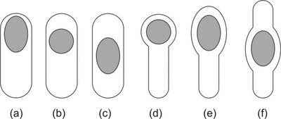
What is the first step in the Schaeffer-Fulton endospore staining method?
What is the primary stain used in the Schaeffer-Fulton method?
What is the purpose of steam heating in the Schaeffer-Fulton method?
What is used to decolorize the cells in the Schaeffer-Fulton method?
What is the counterstain used in the Schaeffer-Fulton method?
What color do vegetative cells appear after counterstaining with safranin?
What color do spores remain after the Schaeffer-Fulton staining process?
What is the purpose of the Schaeffer-Fulton method?
To stain lipids
To stain proteins
To stain endospores
To stain nucleic acids
The Schaeffer-Fulton method involves the following steps: 1. Fix cells, 2. Apply _______ as the primary stain, 3. Use _______ to force the stain into spores, 4. _______ with water, 5. Counterstain with _______.
What does the diagram illustrate in the Schaeffer-Fulton method?
What are the stages of endospore formation?
What color do endospores appear when stained with safranin?
What color do endospores appear when stained with malachite green?
What is the first stage of endospore formation?
Mature stage
Late stage
Early stage
Dormant stage
The early stage of endospore formation shows endospores as _______ structures.
The late stage of endospore formation shows endospores as _______ structures.
What do the images illustrate regarding endospores?
What is the purpose of steam heating in a laboratory?
What equipment is commonly used for steam heating?
What safety equipment is typically worn during steam heating experiments?
What is the main medium used in steam heating?
Steam
Air
Oil
Water
In steam heating, substances are heated using _______ as a medium.
Common equipment for steam heating includes a _______ and a _______.
During steam heating, it is important to wear a _______ and _______ for safety.
What is the visual setup for steam heating?
Flashcards in this deck (80)
-
What is the purpose of the Acid-Fast Stain?
To identify acid-fast bacteria, such as Mycobacterium species, which resist decolorization by acid-alcohol after staining.
microbiology staining -
What is the Endospore Stain used for?
To visualize bacterial endospores, which are resistant structures formed by some bacteria to survive harsh conditions.
microbiology staining -
What is the steam heating method used for?
It is commonly used in sterilization processes, such as autoclaving, to kill microorganisms through high temperature and pressure.
microbiology sterilization -
What is the purpose of the Acid-Fast Stain?
To detect cells that retain a primary stain when treated with acid alcohol.
microbiology staining -
microbiology bacteria
-
microbiology diseases
-
Why doesn't the gram-stain work for certain bacteria?
Because some bacteria are acid-fast and do not retain the gram-stain.
microbiology staining -
What are some characteristics of acid-fast organisms?
They are capable of retaining a primary stain when treated with acid alcohol.
microbiology acid-fast -
What are the causative agents in leprosy and tuberculosis?
Bacillus
Mycobacterium
Staphylococcus
Escherichia
microbiology diseases -
microbiology staining
-
The Acid-Fast Stain is a differential stain used to detect cells capable of retaining a primary stain when treated with acid alcohol.
microbiology staining -
microbiology staining
-
microbiology staining
-
microbiology staining
-
microbiology staining
-
microbiology staining
-
microbiology staining
-
What components are highlighted in the mycobacterial envelope structure?
- Capsule
- Mycolic acids
- Cell wall
microbiology structure -
microbiology staining
-
microbiology staining
-
microbiology staining
-
microbiology staining
-
microbiology staining
-
microbiology staining
-
microbiology staining
-
What is the order of steps in the Acid-Fast Stain (ZN) method?
Carbolfuchsin, Fix cells, Steam heat, Methylene blue, Acid alcohol
Fix cells, Carbolfuchsin, Steam heat, Acid alcohol, Methylene blue
Fix cells, Methylene blue, Steam heat, Acid alcohol, Carbolfuchsin
Steam heat, Carbolfuchsin, Acid alcohol, Fix cells, Methylene blue
microbiology staining -
The Acid-Fast Stain (ZN) method involves the following steps: 1. Fix cells 2. Carbolfuchsin 3. Steam heat 4. Acid alcohol 5. Methylene blue.
microbiology staining -
In the Acid-Fast Stain (ZN) method, acid-fast cells appear reddish purple and non acid-fast cells appear blue.
microbiology staining -
A diagram illustrating the Acid-Fast ZN staining method shows the appearance of cells as follows: before staining (transparent), after staining with carbolfuchsin (reddish-purple), after decolorization with acid alcohol (acid-fast cells retain color, others lose it), and after counterstaining with methylene blue (acid-fast cells are reddish-purple, non-acid-fast cells are blue).
microbiology staining -
microbiology staining
-
microbiology staining
-
microbiology staining
-
microbiology staining
-
microbiology staining
-
microbiology staining
-
microbiology staining
-
microbiology staining
-
Which of the following is used as a secondary stain in Acid-Fast Stain (K)?
Methylene blue
Acid alcohol
Carbolfuchsin
Brilliant green
microbiology staining -
In the Acid-Fast Stain (K) method, the primary stain is carbolfuchsin and the decolorizer is acid alcohol.
microbiology staining -
After the Acid-Fast Stain (K), acid-fast cells appear reddish purple and non acid-fast cells appear blue/green/gray.
microbiology staining -
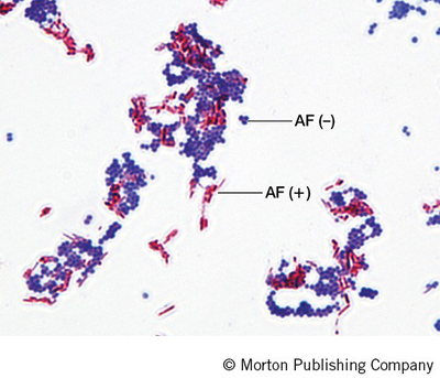
What does the ZN method show in acid-fast staining?
Reddish-purple rod-shaped cells (AF (+)) and blue cells (AF (-)).
 microbiology staining acid-fast
 -
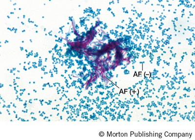
What does the K method show in acid-fast staining?
Reddish-purple clusters of cells (AF (+)) against a blue background of smaller cells (AF (-)).
 microbiology staining acid-fast
 -
What is the purpose of the endospore stain?
To detect the presence and location of spores in bacterial cells.
microbiology staining -
microbiology bacteria
-
Which types of bacteria commonly form endospores?
Certain gram-positive bacteria from the genera Clostridium and Bacillus.
microbiology bacteria -
What are the characteristics of endospores?
Heat and desiccation resistant with a tough outer covering.
microbiology endospores -
microbiology survival
-
Where can endospores form in bacterial cells?
Endospores can form in different locations within the cell, depending on the species.
microbiology endospores -
What makes staining endospores difficult?
Low temperature
Tough outer covering
Small size
High moisture content
microbiology staining -
The endospore stain is a differential stain used to detect the presence and location of spores in bacterial cells.
microbiology staining -
microbiology bacteria
-
microbiology endospores
-
microbiology survival
-
microbiology endospores
-
The diagram shows different shapes of bacterial endospores located in different positions within the bacterial cell:
microbiology diagrams -
microbiology staining
-
microbiology staining
-
What is the purpose of steam heating in the Schaeffer-Fulton method?
To force stain into spores (mordant)
microbiology staining -
microbiology staining
-
microbiology staining
-
microbiology staining
-
microbiology staining
-
What is the purpose of the Schaeffer-Fulton method?
To stain proteins
To stain nucleic acids
To stain endospores
To stain lipids
microbiology staining -
The Schaeffer-Fulton method involves the following steps: 1. Fix cells, 2. Apply malachite green as the primary stain, 3. Use steam heating to force the stain into spores, 4. Decolorize with water, 5. Counterstain with safranin.
microbiology staining -
What does the diagram illustrate in the Schaeffer-Fulton method?
The staining process for spore-producing and non-spore-producing cells.
microbiology staining -
What are the stages of endospore formation?
- Early stage: Small, distinct endospores.
- Late stage: More developed endospores.
biology microbiology -
biology microbiology
-
biology microbiology
-
biology microbiology
-
biology microbiology
-
biology microbiology
-
biology microbiology
-
science heating
-
science equipment
-
science safety
-
science heating
-
science heating
-
science equipment
-
science safety
-
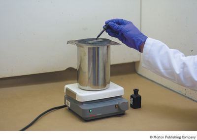
What is the visual setup for steam heating?
A person in a lab coat using a dropper over a metal container on a hot plate.
 science visual