Sign up to unlock more features
- Save this deck to your account
- Study flashcards with spaced repetition
- Export to Anki (.apkg) or PDF
- Process documents up to 100 pages
- Images extracted from PDFs and documents
- Better text extraction from your PDFs and documents
- Better flashcards with our more advanced AI model
Epithelium lines _______ and _______.
The functions of epithelium include _______, _______, and _______.
Histologically, epithelium has one or more _______ of cells that are _______ with one another.
Epithelial cells are _______ and devoid of _______.
Each epithelial cell has a single _______.
The properties of epithelium include being _______ and _______.
The basement membrane consists of two layers: - _______ - _______.
The basement membrane serves to _______ epithelial cells and provides support.
The apical side of epithelial cells _______.
The basal side of epithelial cells _______.
The lateral side of epithelial cells is _______.
In the apical domain, epithelial cells have junctions such as _______ and _______.
The lateral domain of epithelial cells contains _______ and _______ (desmosomes).
The basal domain of epithelial cells includes junctions with the _______, such as _______ and _______.
Most epithelial junctions depend on _______ and _______.
Tight Junctions, also known as _______, are located on the _______ of epithelial cells and seal off lateral spaces to create a _______.
The main proteins involved in tight junctions are _______ and _______, which connect to the cytoskeleton via _______.
Adherens Junctions, also known as _______, are located on the _______ of cells and function as a type of _______.
The main function of Adherens Junctions is to reinforce bonding to deal with _______.
Histological identifiers for Adherens Junctions include SEM and a clear _______ between cells, as well as increased _______ in the cytoplasm.
Key junction proteins in Adherens Junctions include _______, _______, and _______.
Adherens Junctions connect to the cytoskeleton via _______.
Desmosomes, also known as _______, are located on the _______ of cells and function as a type of _______.
The primary function of desmosomes is to reinforce bonding between cells, similar to _______.
Histologically, desmosomes can be identified by a _______ of two dark plates and fibers extending from these plates, known as _______.
Desmosomes contain junction proteins such as _______ which connect to the _______.
Gap junctions are located on the _______ of adjacent cells and are a type of _______.
Gap junctions allow direct passage of _______ between cells.
Histological identifiers for gap junctions include _______ and the _______ between membranes.
Gap junctions are formed by _______ which are composed of _______.
Gap junctions allow small _______ to pass between cells but do not connect the _______.
The hemidesmosome is a type of _______ located on the _______ of cells, connecting them to the _______.
Hemidesmosomes reinforce bonding similar to _______ and are composed of junction proteins which are _______
The hemidesmosome anchors to the basal border via _______.
Focal adhesions are located on the _______ and serve as a type of _______ between the cell and the _______.
Focal adhesions reinforce bonding and are similar to _______ or _______.
The key junction proteins involved in focal adhesions are _______
Focal adhesions anchor to the basal border via _______.
The types of junctions include: - _______: Seals adjacent epithelial cells together; prevents leakage. - _______: Joins actin bundles between cells. - _______: Anchors intermediate filaments between cells. - _______: Allows passage of small water-soluble molecules. - _______: Anchors intermediate filaments to basal lamina.
The junction that binds cells to adjacent cells through _______ is called _______.
The junction that anchors cells to basal lamina through _______ is called _______.
The junction between lateral epithelium cells that forms a _______ is called _______.
The junction that allows the passage of small molecules between adjacent cells is called _______.
The junction that binds cells to adjacent cells through _______ is called _______.
The junction that anchors cells to basal lamina through _______ is called _______.
Apical modifications are present on the _______ and are usually found on _______.
Microvilli are the _______ extension of membrane, while cilia are of _______ length and stereocilia are the _______.
Microvilli function to _______ for absorption and are found on many _______.
The microvilli region can be referred to as _______ or _______.
Histological identifiers for microvilli include a _______ at the apical surface and a _______ at the base of microvilli called the terminal web.
Microvilli contain a core of _______ filaments and are anchored by a _______.
The properties of microvilli include limited _______ and cytoplasmic processes containing a core of _______.
The micrograph shows the apical surfaces of epithelial cells with numerous _______. 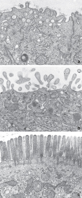
The micrograph of intestinal villi lined by absorptive epithelial cells shows a _______. 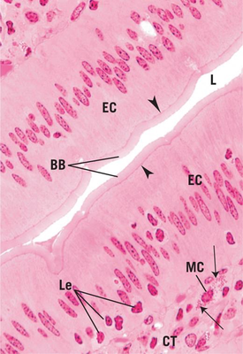
Cilia can be classified into two types: _______ and _______.
The function of motile cilia includes _______ and is commonly found in the _______.
Non-motile cilia serve as _______ receptors, acting as chemical and mechanical sensors on most cells.
Cilia appear as small hairs under a _______ and show a dark line at the base called _______.
The core of cilia is made up of _______ and is anchored to the cell by _______.
Cilia are important for the _______ and _______ functions in various tissues.
Stereocilia function as a _______ in the ear and increase absorption in the _______.
Stereocilia appear as very long hairs under a _______ and have a _______ in scanning electron microscopy (SEM).
The core of stereocilia is made of _______ and is anchored by a _______.
Stereocilia are similar to _______ but not to _______.
The terminal web appears as a _______ at the base of microvilli.
The treadmilling mechanism in actin involves _______ and _______.
Stereocilia have limited _______.
Glands are categorized by how products are released, mechanism of secretion, and type of secretion. The two main types are _______ and _______.
_______ glands secrete a product onto a surface directly or through _______ or tubes.
_______ glands secrete a product into connective tissue to enter the _______.
Endocrine glands are sometimes called '_______' because of their haphazard layering pattern and lack of _______.
The three types of exocrine gland secretion are _______, _______, and _______.
Exocrine glands are characterized by their _______ structure and have a _______ around ducts.
Endocrine glands are distinguished by being _______ and _______.
The three types of exocrine secretion mechanisms are _______, _______, and _______.
In merocrine secretion, the product is secreted via _______.
Apocrine secretion involves the product being secreted with a thin layer of _______ and _______ surrounding it.
An example of apocrine secretion is _______.
Holocrine secretion involves product accumulation within the cell, leading to _______ to release it.
An example of holocrine secretion is _______.
Endocrine secretion is characterized by products being secreted through the basal lamina directly into the _______.
Types of exocrine secretion include: - _______ - _______ - _______ - Others like milk and pancreatic enzymes.
Mucous secretion is characterized by being _______ and slimy with heavily _______ proteins.
Serous secretion is typically _______ and contains proteins that are _______.
Sebaceous secretion consists of _______.
Endocrine glands secrete _______.
Simple glands have a _______ duct, while compound glands have a _______ duct.
The shape of glands can be classified as _______, _______, or _______.
An example of a simple branched alveolar gland is _______.
Examples of compound glands include _______ and _______.
The term _______ describes a tubular shape where the tube is _______ rather than straight.
The _______ are an example of a simple tubular gland that is not found in adults, being a stage in development.
Mucous glands in the mouth and bulbourethral glands in the male reproductive system are examples of _______ glands.
A gland that combines tubular and alveolar shapes is referred to as _______.
Goblet cells are unicellular glands that are specialized for _______ in the _______ and _______ systems.
The structure of a goblet cell includes microvilli, secretory vesicles containing _______, rough ER, Golgi apparatus, and a nucleus.
In histological staining, mucin does not stain well in H&E, giving the goblet cell a _______.
Epithelial tissue can be classified by the number of layers into: - _______ (1 cell layer) - _______ (≥ 2 cell layers).
The unique classifications of epithelial tissue include: - _______ - _______.
Simple squamous epithelium consists of _______ of _______.
Simple squamous epithelium is found in the _______, _______, and _______.
The main functions of simple squamous epithelium include _______ and _______.
In the vascular system, simple squamous epithelium is referred to as _______.
In body cavities, simple squamous epithelium is known as _______.
Simple cuboidal epithelium consists of _______ of _______.
Examples of locations where simple cuboidal epithelium can be found include: - _______ - _______ - _______.
The main functions of simple cuboidal epithelium are _______ and _______.
Simple Columnar Epithelium consists of _______ of _______.
Simple Columnar Epithelium is primarily located in the _______ and the _______.
The main function of Simple Columnar Epithelium is _______ and _______.
Pseudostratified Columnar Epithelium appears _______ but is actually _______.
In Pseudostratified Columnar Epithelium, all cells rest on the _______, but not all reach the _______.
Pseudostratified Columnar Epithelium can be _______ or _______.
Location examples of Pseudostratified Columnar Epithelium include the _______ and _______.
The main functions of Pseudostratified Columnar Epithelium are _______ and _______ as well as _______ (cilia).
Stratified Squamous Epithelium has an apical layer of _______ shaped cells and multiple layers underneath of varying shapes.
Stratified Squamous Epithelium is found in the _______, _______, _______, and _______.
The main function of Stratified Squamous Epithelium is _______.
Stratified cuboidal epithelium has an apical layer of _______ shaped cells and is often _______ layers thick.
Stratified cuboidal epithelium is considered a _______ tissue.
Examples of locations where stratified cuboidal epithelium is found include _______ and _______.
The main functions of stratified cuboidal epithelium are _______ and _______.
Stratified Columnar Epithelium has an apical layer of _______ shaped cells and is considered a _______ tissue.
The main functions of Stratified Columnar Epithelium include _______ and _______.
Stratified Columnar Epithelium is found in the _______ and _______.
Transitional epithelium is characterized by multiple layers of cells with varying shapes, including _______ cells. It is primarily located in the _______, _______, and _______.
The main functions of transitional epithelium include acting as a _______ and being _______.
The top layer of transitional epithelium is referred to as _______ or _______ when not distended.
Transitional epithelium is found in the urinary tract, specifically in the _______, _______, and _______.
Transitional epithelium consists of multiple layers of cells, including the top layer known as _______.
Flashcards in this deck (130)
-
anatomy epithelium
-
anatomy epithelium function
-
histology epithelium
-
histology epithelium
-
histology epithelium
-
anatomy epithelium
-
biology epithelial tissue
-
biology epithelial tissue
-
biology epithelial_cells
-
biology epithelial_cells
-
biology epithelial_cells
-
epithelial junctions apical
-
epithelial junctions lateral
-
The basal domain of epithelial cells includes junctions with the basal lamina, such as hemidesmosomes and focal adhesions.
epithelial junctions basal -
epithelial junctions composition
-
Tight Junctions, also known as Occluding Junctions, are located on the lateral border of epithelial cells and seal off lateral spaces to create a barrier.
biology cell_biology epithelial_cells -
The main proteins involved in tight junctions are occludin and claudin, which connect to the cytoskeleton via actin filaments.
cell_biology tight_junctions proteins -
Adherens Junctions, also known as Zonula Adherens, are located on the lateral border of cells and function as a type of anchoring junction.
cell_biology junctions -
cell_biology junctions
-
Histological identifiers for Adherens Junctions include SEM and a clear space between cells, as well as increased density in the cytoplasm.
histology cell_biology -
cell_biology junctions
-
cell_biology junctions
-
Desmosomes, also known as Macula Adherens, are located on the lateral border of cells and function as a type of anchoring junction.
cell_biology junctions -
cell_biology junctions
-
Histologically, desmosomes can be identified by a sandwich of two dark plates and fibers extending from these plates, known as intermediate filaments.
histology desmosomes -
cell_biology junctions
-
Gap junctions are located on the lateral border of adjacent cells and are a type of communicating junction.
cell_biology gap_junctions -
cell_biology gap_junctions
-
Histological identifiers for gap junctions include SEM and the narrowing of the gap between membranes.
histology gap_junctions -
cell_biology gap_junctions
-
cell_biology gap_junctions
-
The hemidesmosome is a type of anchoring junction located on the basal border of cells, connecting them to the basal lamina.
cell_biology junctions -
Hemidesmosomes reinforce bonding similar to desmosomes and are composed of junction proteins which are integrins
cell_biology junctions -
cell_biology junctions
-
Focal adhesions are located on the basal border and serve as a type of anchoring junction between the cell and the basal lamina.
cell_biology focal_adhesions -
cell_biology junctions
-
cell_biology focal_adhesions proteins
-
cell_biology focal_adhesions actin
-
The types of junctions include: - Tight Junctions: Seals adjacent epithelial cells together; prevents leakage. - Adherens Junctions: Joins actin bundles between cells. - Desmosomes: Anchors intermediate filaments between cells. - Gap Junctions: Allows passage of small water-soluble molecules. - Hemidesmosomes: Anchors intermediate filaments to basal lamina.
cell_biology junctions -
The junction that binds cells to adjacent cells through actin filaments is called Adherens/Zonula Adherens.
cell_biology junctions -
The junction that anchors cells to basal lamina through intermediate filaments is called Hemidesmosomes.
cell_biology junctions -
cell_biology junctions
-
cell_biology junctions
-
The junction that binds cells to adjacent cells through intermediate filaments is called Desmosomes/Macula Adherens.
cell_biology junctions -
cell_biology junctions
-
Apical modifications are present on the apical cell surface and are usually found on columnar shaped cells.
biology epithelial_cells -
Microvilli are the shortest extension of membrane, while cilia are of medium length and stereocilia are the longest.
biology epithelial_cells -
biology microvilli function
-
biology microvilli terminology
-
Histological identifiers for microvilli include a pinkish haze at the apical surface and a dark line at the base of microvilli called the terminal web.
biology histology identifiers -
biology microvilli structure
-
The properties of microvilli include limited movement and cytoplasmic processes containing a core of actin.
biology microvilli properties -
biology microvilli images
-
biology microvilli images
-
biology cilia classification
-
The function of motile cilia includes propulsion of surrounding fluid and is commonly found in the respiratory tract.
biology cilia function -
Non-motile cilia serve as sensory receptors, acting as chemical and mechanical sensors on most cells.
biology cilia function -
Cilia appear as small hairs under a light microscope and show a dark line at the base called basal bodies.
biology cilia histology -
biology cilia properties
-
biology cilia function
-
Stereocilia function as a mechanoreceptor in the ear and increase absorption in the male reproductive system.
biology function stereocilia -
Stereocilia appear as very long hairs under a light microscope and have a tiered appearance in scanning electron microscopy (SEM).
biology histology stereocilia -
biology properties stereocilia
-
biology comparison stereocilia
-
biology histology microvilli
-
biology actin treadmilling
-
biology properties stereocilia
-
Glands are categorized by how products are released, mechanism of secretion, and type of secretion. The two main types are exocrine and endocrine.
glands secretion biology -
glands exocrine secretion
-
glands endocrine secretion
-
Endocrine glands are sometimes called 'epitheloid tissues' because of their haphazard layering pattern and lack of basal lamina.
glands endocrine tissues -
glands exocrine secretion
-
Exocrine glands are characterized by their duct structure and have a layered organization around ducts.
histology glands exocrine -
histology glands endocrine
-
anatomy physiology glands
-
anatomy physiology merocrine
-
Apocrine secretion involves the product being secreted with a thin layer of cytoplasm and plasma membrane surrounding it.
anatomy physiology apocrine -
anatomy physiology apocrine
-
Holocrine secretion involves product accumulation within the cell, leading to cell lysis to release it.
anatomy physiology holocrine -
anatomy physiology holocrine
-
Endocrine secretion is characterized by products being secreted through the basal lamina directly into the blood stream.
anatomy physiology endocrine -
Types of exocrine secretion include: - Mucous - Serous - Sebaceous - Others like milk and pancreatic enzymes.
biology histology secretion -
biology histology mucous
-
biology histology serous
-
biology histology sebaceous
-
biology endocrine hormones
-
anatomy glands
-
anatomy glands
-
anatomy glands
-
anatomy glands
-
anatomy glands
-
The intestinal glands are an example of a simple tubular gland that is not found in adults, being a stage in development.
anatomy glands -
Mucous glands in the mouth and bulbourethral glands in the male reproductive system are examples of compound glands.
anatomy glands -
anatomy glands
-
Goblet cells are unicellular glands that are specialized for mucus secretion in the gastrointestinal and respiratory systems.
anatomy histology glands -
The structure of a goblet cell includes microvilli, secretory vesicles containing mucin, rough ER, Golgi apparatus, and a nucleus.
anatomy histology glands -
In histological staining, mucin does not stain well in H&E, giving the goblet cell a goblet appearance.
anatomy histology glands -
Epithelial tissue can be classified by the number of layers into: - simple (1 cell layer) - stratified (≥ 2 cell layers).
epithelium classification anatomy -
anatomy epithelium classification
-
anatomy epithelium
-
anatomy locations epithelium
-
The main functions of simple squamous epithelium include secretion/lubrication and gas exchange/diffusion.
anatomy functions epithelium -
anatomy vascular epithelium
-
anatomy body_cavities epithelium
-
biology histology epithelium
-
Examples of locations where simple cuboidal epithelium can be found include: - small ducts - surface of ovaries - kidney tubules.
biology locations epithelium -
biology functions epithelium
-
biology epithelium
-
biology epithelium location
-
biology epithelium function
-
histology epithelium
-
In Pseudostratified Columnar Epithelium, all cells rest on the basement membrane, but not all reach the apical surface.
histology epithelium -
histology epithelium
-
histology epithelium location
-
The main functions of Pseudostratified Columnar Epithelium are secretion and absorption as well as propulsion (cilia).
histology epithelium function -
Stratified Squamous Epithelium has an apical layer of squamous shaped cells and multiple layers underneath of varying shapes.
anatomy epithelium -
anatomy location epithelium
-
anatomy function epithelium
-
Stratified cuboidal epithelium has an apical layer of cuboidal shaped cells and is often two layers thick.
histology epithelium -
histology epithelium
-
Examples of locations where stratified cuboidal epithelium is found include sweat gland ducts and large ducts of exocrine glands.
histology locations epithelium -
histology function epithelium
-
Stratified Columnar Epithelium has an apical layer of columnar shaped cells and is considered a rare tissue.
anatomy epithelium -
anatomy epithelium function
-
anatomy location
-
Transitional epithelium is characterized by multiple layers of cells with varying shapes, including dome-shaped cells. It is primarily located in the ureters, bladder, and urethra.
anatomy histology epithelium -
anatomy histology epithelium
-
The top layer of transitional epithelium is referred to as umbrella cells or dome-shaped cells when not distended.
anatomy histology epithelium -
Transitional epithelium is found in the urinary tract, specifically in the ureters, bladder, and urethra.
anatomy urinary epithelium -
Transitional epithelium consists of multiple layers of cells, including the top layer known as umbrella cells.
anatomy histology epithelium