Sign up to unlock more features
- Save this deck to your account
- Study flashcards with spaced repetition
- Export to Anki (.apkg) or PDF
- Process documents up to 100 pages
- Images extracted from PDFs and documents
- Better text extraction from your PDFs and documents
- Better flashcards with our more advanced AI model
Flashcards in this deck (54)
-
anatomy meninges
-
anatomy meninges
-
terminology meninges
-
anatomy meninges
-
Where do dural folds typically occur?
In the fissures of the brain, such as between the cerebral hemispheres.
anatomy meninges -
anatomy circulation
-
anatomy csf
-
anatomy csf
-
anatomy meninges
-
anatomy meninges
-
anatomy csf
-
anatomy meninges
-
anatomy csf
-
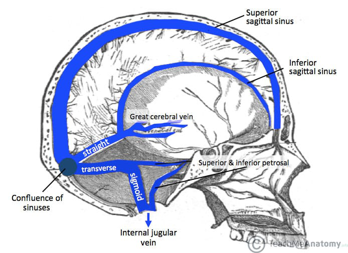
What are the labeled structures in the venous sinuses diagram?
- Superior sagittal sinus
- Inferior sagittal sinus
- Transverse sinus
- Sigmoid sinus
anatomy venous_sinuses -
List the main contents of the cranial meninges diagram.
- Dura mater
- Arachnoid mater
- Pia mater
- Cerebral cortex
anatomy meninges -
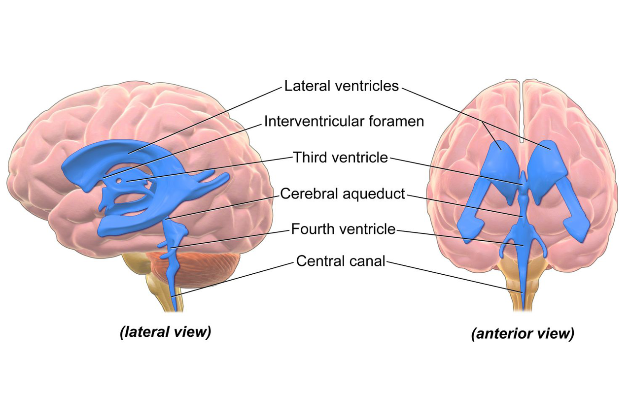
What are the key labeled parts of the ventricles?
- Lateral ventricles
- Third ventricle
- Fourth ventricle
anatomy ventricles -
What is the superior venous sinus?
A venous sinus between the left and right hemisphere formed where the dura mater splits at the top.
anatomy sinus -
What is the arachnoid mater?
The middle layer of the meninges, meaning 'spider mother', with fibrous attachments to the pia mater called trabeculae.
anatomy meninges -
What does the pia mater mean?
'Tender mother'; it is the innermost layer that attaches to the brain and spinal cord.
anatomy meninges -
What is the subarachnoid space?
The space between the arachnoid and pia mater that contains trabeculae and CSF.
anatomy meninges -
What are arachnoid granulations?
Outcroppings of the arachnoid mater into a venous sinus, allowing waste products and old CSF to flow into the venous system.
anatomy meninges -
anatomy meninges
-
anatomy ventricles
-
anatomy meninges
-
How does the subarachnoid space contribute to the CNS?
It facilitates the flow of CSF and contains trabeculae for support.
anatomy csf -
anatomy csf
-
anatomy csf
-
What are the main functions of CSF?
- Protects the brain
- Provides buoyancy
- Acts as a marker of nervous system health
anatomy csf functions -
anatomy protection
-
Why is buoyancy important for the brain?
It prevents the brain from squashing the cerebellum and brainstem.
anatomy buoyancy -
medical health
-
anatomy ventricles
-
anatomy ventricles
-
anatomy ventricles
-
anatomy ventricles
-
anatomy ventricles
-
anatomy ventricles
-
anatomy ventricles
-
anatomy cns
-
anatomy meninges
-
medical health
-
What is the fourth ventricle responsible for?
Last point for cerebrospinal fluid (CSF) before being absorbed.
anatomy ventricles -
What composes the choroid plexus?
- Blood capillaries
- Specialized cuboid epithelial cells (ependymal cells)
anatomy choroid -
physiology csf
-
anatomy csf_pathway
-
What follows the lateral ventricles in the CSF flow?
CSF travels through the intraventricular foramen into the third ventricle.
anatomy csf_pathway -
After the third ventricle, how does CSF reach the fourth ventricle?
It passes through the cerebral aqueduct.
anatomy csf_pathway -
After the fourth ventricle, what are the CSF exit points?
- Two lateral apertures
- One median aperture
- Central canal
anatomy csf_pathway -
What is the function of the apertures in CSF movement?
Allow CSF to exit into the subarachnoid space.
physiology csf -
anatomy central_canal
-
Can CSF skip ventricles during its pathway?
Yes, if produced in the fourth ventricle, it can bypass others.
physiology csf_pathway -
anatomy meninges
-
physiology meninges
-
What are the core functions of cerebrospinal fluid (CSF)?
Protects the CNS and provides buoyancy, nourishment, and waste removal.
physiology csf
Meninges
Meninges
The meninges are protective layers surrounding the brain and spinal cord, consisting of three layers:
- Dura mater: The outermost layer, known as the "tough mother". It splits in some areas to create dural folds, enhancing protection and facilitating cerebrospinal fluid (CSF) flow.
- Arachnoid mater: The middle layer, or "spider mother", characterized by trabeculae that connect it to the pia mater.
- Pia mater: The innermost layer, meaning "tender mother", that adheres directly to the brain and spinal cord.
Dural Fold & Venous Sinuses
- Dural folds occur when the dura mater splits, creating spaces that facilitate drainage of deoxygenated blood.
- Venous sinuses: Spaces formed by layered dura mater, acting as veins for blood drainage.
Subarachnoid Space
- The space between the arachnoid mater and the pia mater that contains CSF and trabeculae.
Arachnoid Granulations
- Projections of the arachnoid mater into the venous sinuses that allow waste transfer and CSF reabsorption.
Ventricles
Ventricles
The brain contains four ventricles that produce and circulate cerebrospinal fluid (CSF):
- Lateral ventricles: Two ventricles that shape closely around the basal nuclei, connected by the intraventricular foramen.
- Third ventricle: Located near the thalamus and connected to the fourth ventricle by the cerebral aqueduct.
- Fourth ventricle: The last ventricle before CSF enters subarachnoid space.
CSF Functions
- Protection: Cushions the brain, preventing impact damage.
- Buoyancy: Aids in support, reducing the effective weight on the spinal column.
- Health Indicator: CSF analysis can reveal nervous system issues.
CSF Pathway
- Produced in the lateral ventricles.
- Travels through the intraventricular foramen to the third ventricle.
- Passes through the cerebral aqueduct to the fourth ventricle.
- Finally, it can:
- Exit to the subarachnoid space via apertures.
- Travel down the central canal.