Sign up to unlock more features
- Save this deck to your account
- Study flashcards with spaced repetition
- Export to Anki (.apkg) or PDF
- Process documents up to 100 pages
- Images extracted from PDFs and documents
- Better text extraction from your PDFs and documents
- Better flashcards with our more advanced AI model
What is the main function of the nervous system?
What are the two main parts of the nervous system?
What does the PNS stand for?
What is the role of reflex pathways?
What does the CNS consist of?
What is depicted in this diagram?

What are the major components of the nervous system?
What does the nervous system transmit?
What is the function of the brain in the nervous system?
What does the nervous system interpret?
What role does the brain play in homeostasis?
What are the two major control centers of the human body?
What is depicted in the image related to the nervous system?
What is the function of the nervous system related to sensing?
What does CNS stand for in the nervous system?
What are the main components of the CNS?
What are the components of the PNS?
What is the role of the integrating function of the nervous system?
How does the nervous system control body responses?
What is depicted in the diagram of the human nervous system?
What are the two main structural divisions of the nervous system?
What does the Central Nervous System (CNS) consist of?
What does the Peripheral Nervous System (PNS) consist of?
What is the function of the Peripheral Nervous System (PNS)?
What is the function of the Central Nervous System (CNS)?
What are the components of the Peripheral Nervous System (PNS)?
What are the major organs of the central nervous system?
What does the CNS stand for?
What are the components of the peripheral nervous system?
What is the primary role of the spinal cord?
What type of processing occurs primarily in the brain?
What basic processing does the spinal cord perform?
What are the major organs of the peripheral nervous system?
What do nerves consist of?
What does the PNS do?
What are the components of the central nervous system (CNS)?
What are the components of the peripheral nervous system (PNS)?
What is the function of the enteric nervous system?
How does the PNS function like a network?
What information can each neuron within a nerve send?
What is a nerve composed of?
What are the groups of axons in a nerve called?
What types of neurons can be found in nerves?
What surrounds the axons in a nerve?
What is the function of the myelin sheath?
What does the illustration of a nerve show?
What is the function of sensory neurons?
What do motor neurons do?
What is another name for sensory neurons?
What is another name for motor neurons?
Do neurons send signals in multiple directions?
What is the direction of signal transmission for sensory neurons?
What is the direction of signal transmission for motor neurons?
What do sensory neurons carry impulses from?
What do motor neurons carry impulses to?
What does the diagram compare?
What are the two main divisions of the nervous system?
What does the PNS consist of?
What is the function of the sensory division?
What is the function of the motor division?
What does the autonomic nervous system do?
What does the somatic nervous system do?
What is the function of the sympathetic division?
What is the function of the parasympathetic division?
What type of information does the PNS neurons send?
What are the two types of motor divisions in the PNS?
What is illustrated in the flowchart of the nervous system?
What is the responsibility of the sensory division?
What does CNS stand for?
What are the components of the CNS?
What does the sensory division carry messages from?
What is the function of the motor division?
What is the role of the somatic nervous system?
What does the sympathetic division prepare the body for?
What does the parasympathetic division control?
How does the CNS process stimulus information?
What is the function of the sensory division in the brain?
Where are visual stimuli processed in the brain?
What happens to stimuli that are not consciously perceived?
What is the optic chiasm?
What are the left and right visual fields?
What do the green and purple lines in the diagram represent?
What is the role of the left visual cortex?
What is the role of the right visual cortex?
What is depicted in the diagram related to visual pathways?
How do sensory signals from the body reach the brain?
Where do sensory signals from specialized organs go?
What is the role of the spinal cord in sensory processing?
What are examples of specialized sensory organs?
What do sensory signals from the skin and visceral organs do?
What does the diagram illustrate about sensory pathways?
What is the function of sensory receptors in the skin?
What does the motor division do?
What are the effectors in the motor division?
What is the central nervous system (CNS)?
What does the sensory division do?
What is the function of the autonomic nervous system?
What does the somatic nervous system control?
What is the role of the sympathetic division?
What does the parasympathetic division do?
What is the origin of a motor signal in the pathway?
What does the motor signal descend through?
What does the motor signal stimulate in the pathway?
What part of the brain is involved in the motor pathway?
What is the function of the motor division?
What structure exits to stimulate a response in muscle fibers?
What is the role of the medulla oblongata in the motor pathway?
What is illustrated in the diagram related to the motor pathway?
What is the function of the sensory division?
What is the role of the somatic nervous system?
What does the sympathetic division prepare the body for?
What does the parasympathetic division control?
What type of muscles does the somatic nervous system control?
What is an example of a voluntary muscle?
What does the somatic nervous system innervate?
What does the autonomic nervous system innervate?
What is the primary difference between the somatic and autonomic nervous systems?
What are the two types of neurons in the autonomic nervous system?
What type of muscle does the autonomic nervous system control?
What is the role of the ganglion in the autonomic nervous system?
What is the effect of the somatic nervous system on muscle?
What is the effect of the autonomic nervous system on muscle?
What type of muscle is controlled by the somatic nervous system?
What type of glands are controlled by the autonomic nervous system?
What is depicted in the diagram comparing somatic and autonomic nervous systems?
What are the two branches of the Autonomic Nervous System?
What does the Sympathetic Division do?
What does the Parasympathetic Division control?
What are the two main divisions of the Nervous System?
What does the Sensory Division do?
What does the Motor Division do?
What does the Somatic Nervous System control?
How do the Sympathetic and Parasympathetic Divisions interact?
What is the primary function of the parasympathetic nervous system?
What is the primary function of the sympathetic nervous system?
What happens to pupils under sympathetic activation?
What happens to salivation under parasympathetic activation?
What is the effect on heartbeat during sympathetic activation?
What is the effect on airways during sympathetic activation?
What happens to glucose release under sympathetic activation?
What happens to bladder contraction under parasympathetic activation?
What is the effect on intestines during parasympathetic activation?
What effect does the sympathetic nervous system have on the gallbladder?
What is secreted by the adrenal gland during sympathetic activation?
What is the effect of parasympathetic activation on the heart rate?
What happens to bladder relaxation under sympathetic activation?
What is the effect on genital function under parasympathetic activation?
What is the effect on ejaculation under sympathetic activation?
Which nerves are involved in the parasympathetic system?
Which nerves are involved in the sympathetic system?
What is the effect of sympathetic activation on salivation?
What is the effect of parasympathetic activation on stomach activity?
What happens to stomach activity under sympathetic activation?
What is the effect of sympathetic activation on pupils?
What is the diagram comparing in the autonomic nervous system?
What does the sympathetic nervous system prepare the body for?
What are the effectors of the sympathetic nervous system?
What happens to pupils during a sympathetic response?
What is the effect of the sympathetic nervous system on salivation?
How does the sympathetic nervous system affect heartbeat?
What happens to the airways during a sympathetic response?
What effect does the sympathetic nervous system have on stomach activity?
What does the sympathetic nervous system stimulate the release of?
What is the traditional term for the body's response to stressors?
What type of threats were stressors primarily in our evolutionary past?
What is the effect of the sympathetic response on the digestive system?
How does the sympathetic response affect cardiac output?
What happens to the pupils during a sympathetic response?
What is the effect of the sympathetic response on airways?
What cranial nerve effects are associated with the sympathetic response?
What sacral nerve effects are associated with the sympathetic response?
What hormones are secreted during the sympathetic response?
What happens to the bladder during a sympathetic response?
What reproductive effects occur during the sympathetic response?
What is stimulated in the body during a sympathetic response?
What is a stressor?
How do individuals perceive stress?
What activates the sympathetic nervous system?
What are some types of stressors?
What is a common academic stressor?
What kind of stress can social media cause?
What is a physical stressor example?
What does the parasympathetic nervous system coordinate?
What is one effect of the parasympathetic nervous system on pupils?
What effect does the parasympathetic nervous system have on heart rate?
What is the effect of the parasympathetic nervous system on digestive activity?
What happens to cardiac output under the parasympathetic nervous system?
What is one effect of the parasympathetic nervous system on airways?
What does the parasympathetic nervous system stimulate in the bladder?
What is one effect of the parasympathetic nervous system on the stomach?
What does the parasympathetic nervous system promote in the genitals?
What does the parasympathetic nervous system inhibit regarding glucose?
What is one effect of the parasympathetic nervous system on the intestines?
What is the general association of the parasympathetic nervous system?
What type of autonomic response is associated with the parasympathetic nervous system?
What is the effect of the parasympathetic nervous system on vasodilation?
What diagram represents the effects of the parasympathetic nervous system?

What is a reflex?
What triggers a reflex response?
What are spinal reflexes?
What are cranial reflexes?
What is the role of the afferent neuron?
What is the role of the efferent neuron?
What is the integration center in a reflex arc?
What is the purpose of reflexes?
What is the pathway of a reflex arc?
What does the diagram of a reflex arc illustrate?
What are the components of a spinal reflex pathway?
What is the role of the sensory neuron in a spinal reflex pathway?
What is the function of the motor neuron in a spinal reflex pathway?
What is the function of an interneuron?
Where are interneurons located?
What do sensory receptors do in a spinal reflex pathway?
What is the neuromuscular junction?
What is the structure shown in the diagram?
Where do motor neurons exit the spinal cord?
Where do sensory neurons enter the spinal cord?
What is the spinal nerve?
What connects skin and muscles in the spinal reflex pathway?
What is the role of neuromuscular junction?
What is found in the dorsal root?
What is the ventral root responsible for?
What type of receptors are found in the skin?
What triggers the nerve impulse in a withdrawal reflex?
What carries the nerve impulse to the spinal cord?
What does the motor neuron stimulate in a withdrawal reflex?
What is the purpose of the withdrawal reflex?
Does the reflex pathway generate a feeling of pain?
What is the stimulus in the simple spinal reflex example?
What is the effector in a withdrawal reflex?
What type of diagram illustrates the withdrawal reflex?
What causes a nerve impulse to be generated in a sensory neuron?
What is the role of the sensory neuron in the reflex pathway?
What is the effector in the withdrawal reflex?
What type of receptor detects skin damage from a cactus needle?
What is the integration center for the withdrawal reflex?
What is the pathway of a simple spinal reflex?
What is the first structure in the withdrawal reflex pathway?
What is the role of the dorsal root ganglion in the reflex pathway?
What structure do sensory neurons enter after the dorsal root ganglion?
What type of neuron connects sensory and motor neurons in the spinal cord?
What is the next step after the relay neuron in the reflex pathway?
What structure do motor neurons exit through?
What is the final effector in the withdrawal reflex pathway?
What are the two types of matter in the spinal cord involved in the reflex pathway?
What type of nerve is the spinal nerve classified as?
What is the stimulus in the withdrawal reflex example?
What does the diagram of the reflex arc illustrate?
What is the first step in the reflex pathway?
What type of nerve is the spinal nerve?
Where does the sensory axon enter the spinal cord?
What do interneurons do in the reflex pathway?
Where does the motor neuron exit the spinal cord?
What is the role of the effector in the reflex pathway?
What structures are highlighted in the withdrawal reflex pathway diagram?
What does the dorsal root contain?
What is the function of the spinal cord grey matter?
What are the components of a sensory neuron?
What type of neurons are sensory neurons classified as?
Where are the cell bodies of sensory neurons located?
What is a ganglion in the PNS?
What is the dorsal root ganglion?
What structures are part of the spinal nerve?
What do the arrows in the spinal cord diagram indicate?
What are the two pathways mentioned in the spinal nerve?
What is the function of somatosensory neurons?
What is the role of motor nerve fibers?
What does the posterior root ganglion contain?
What is shown in the diagram of a sensory neuron?
What is illustrated in the cross-section of the spinal cord diagram?
What is the spinal cord?
What is the vertebral column?
Where does the spinal cord end?
What are the components of spinal cord pathways?
What is shown in the image provided?
What is the function of the spinal cord?
What are the protective layers of the spinal cord?
What is found in the epidural space?
What does the subarachnoid space contain?
What is the function of PNS nerves?
What is shown in the cross-section image of the spinal cord?
What initiates the knee-jerk reflex?
What travels to the spinal cord during the knee-jerk reflex?
What does the afferent neuron do in the knee-jerk reflex?
What is the effect of efferent signals in the knee-jerk reflex?
What happens to the hamstrings during the knee-jerk reflex?
What is the purpose of the knee-jerk reflex?
What does the inhibitory motor neuron do in the knee-jerk reflex?
What is illustrated in the diagram of the knee-jerk reflex pathway?
What does a neurological assessment typically include?
What do reflex tests provide insight into?
What may an abnormal reflex response suggest?
What are possible causes of abnormal reflex responses?
What does the biceps tendon reflex assess?
What is the receptor involved in the biceps reflex?
What is the effector in the biceps reflex?
What is illustrated in the biceps reflex diagram?
What are the common deep tendon reflexes tested?
What does a reflex scale indicate?
What is a normal reflex rating?
What are signs of abnormal reflexes?
What does clinical shorthand summarize?
What is the link to reflex test demonstration?
What is the function of reflex tests in neuro assessments?
What is the scale for grading reflex responses?
What spinal nerves are associated with the Biceps reflex?
What spinal nerves are associated with the Patellar reflex?
What spinal nerves are associated with the Achilles tendon reflex?
What is a cranial reflex?
Where is the integration of cranial reflexes located?
What is the pupillary light reflex?
Is the pupillary light reflex processed in the cerebral cortex?
What nerves are involved in the pupillary light reflex pathway?
What does the pupillary light reflex respond to?
What type of reaction does the pupillary light reflex include?
What is the role of the midbrain in cranial reflexes?
What is depicted in the diagram of the pupillary light reflex?
What is the stimulus for the Pupillary Light Reflex?
What are the receptors involved in the Pupillary Light Reflex?
Where does integration occur in the Pupillary Light Reflex?
What is the effector in the Pupillary Light Reflex?
What is the response of the Pupillary Light Reflex?
What is the purpose of the Pupillary Light Reflex?
What pathway does the light reaction take to the iris?
What is the direct reaction in the Pupillary Light Reflex?
What is the consensual reaction in the Pupillary Light Reflex?
What is the location of the midbrain?
What is a key function of the midbrain?
Are you consciously aware of activities in the midbrain?
What reflex is associated with the midbrain?
What does the sagittal view of the brain illustrate?
What is the role of sensory receptors?
What is the function of the spinal cord in reflexes?
What differentiates reflex pathways from conscious perception pathways?
Where does a conscious sensation, like pain, reach in the brain?
What is the function of the thalamus?
What is the role of interneurons in reflex actions?
What happens to the muscle during a reflex action?
What is illustrated in the diagram comparing reflex pathways?
What do ascending pathways do?
What is the role of motor neurons in reflexes?
What is required for you to consciously feel a stimulus?
How does a sensory signal travel to the brain?
What triggers a voluntary muscle contraction?
What happens to the signal after it descends the spinal cord?
What does the sensory pathway diagram illustrate?
What does the motor pathway diagram illustrate?
Flashcards in this deck (347)
-
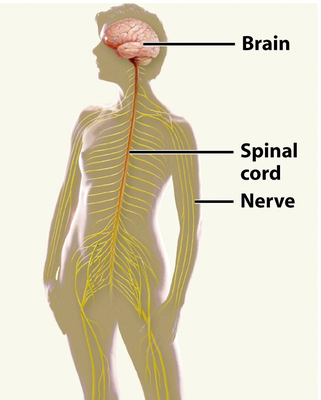
What is the main function of the nervous system?
To transmit signals between different parts of the body and coordinate actions.
biology nervous_system -
What are the two main parts of the nervous system?
- Central Nervous System (CNS)
- Peripheral Nervous System (PNS)
biology nervous_system -
biology nervous_system
-
What is the role of reflex pathways?
To allow quick responses to stimuli without direct involvement of the brain.
biology nervous_system -
biology nervous_system
-
biology nervous_system
-
biology nervous_system
-
biology nervous_system
-
What is the function of the brain in the nervous system?
Coordinates all of your actions and reactions
biology nervous_system -
biology nervous_system
-
biology nervous_system
-
biology nervous_system
-
biology nervous_system
-
What is the function of the nervous system related to sensing?
Receptors detect stimuli and send sensory information to the CNS.
nervous_system sensing -
nervous_system cns
-
nervous_system cns
-
nervous_system pns
-
What is the role of the integrating function of the nervous system?
Sensory information is processed, connecting it with memories and emotions.
nervous_system integration -
How does the nervous system control body responses?
Motor neurons send information to effectors (muscles or glands) to produce responses.
nervous_system control -
nervous_system diagram
-
What are the two main structural divisions of the nervous system?
- Central Nervous System (CNS)
- Peripheral Nervous System (PNS)
anatomy nervous_system -
anatomy cns
-
What does the Peripheral Nervous System (PNS) consist of?
- Cranial nerves
- Spinal nerves
- Ganglia
- Sensory receptors
anatomy pns -
What is the function of the Peripheral Nervous System (PNS)?
Consists of all the nerves, ganglia, and sensory receptors.
function pns -
What is the function of the Central Nervous System (CNS)?
Processes information and coordinates activities of the body.
function cns -
anatomy pns
-
anatomy nervous_system
-
acronyms nervous_system
-
anatomy nervous_system
-
anatomy nervous_system
-
anatomy nervous_system
-
anatomy nervous_system
-
What are the major organs of the peripheral nervous system?
- Nerves (cranial and spinal)
- Ganglia (swellings of neuron cell bodies)
anatomy nervous_system -
anatomy nervous_system
-
function nervous_system
-
anatomy cns
-
What are the components of the peripheral nervous system (PNS)?
- Cranial nerves
- Spinal nerves
- Ganglia
anatomy pns -
What is the function of the enteric nervous system?
It is involved in the gut, but the PNS does not process information or make decisions.
function nervous_system -
How does the PNS function like a network?
It acts like electrical highways leading to and from the organs of the CNS.
function nervous_system -
What information can each neuron within a nerve send?
- From a sensory receptor to the CNS
- From the CNS to an effector (muscle or gland)
function nervous_system -
anatomy nervous_system
-
anatomy nervous_system
-
What types of neurons can be found in nerves?
- All motor neurons
- All sensory neurons
- A mixture of motor and sensory neurons
anatomy nervous_system -
anatomy nervous_system
-
anatomy nervous_system
-
What does the illustration of a nerve show?
A cross-section of a nerve with axons bundled together and labeled components.
anatomy nervous_system -
What is the function of sensory neurons?
They carry sensory impulses from sensory organs to the central nervous system (CNS).
biology neuroscience -
What do motor neurons do?
They carry motor impulses from the central nervous system to specific effectors.
biology neuroscience -
biology neuroscience
-
biology neuroscience
-
Do neurons send signals in multiple directions?
No, each neuron sends its signal in only one direction.
biology neuroscience -
biology neuroscience
-
What is the direction of signal transmission for motor neurons?
From the CNS to the effectors (muscles or glands).
biology neuroscience -
biology neuroscience
-
biology neuroscience
-
What does the diagram compare?
It compares sensory and motor neurons and illustrates their structure.
biology neuroscience -
What are the two main divisions of the nervous system?
- Central Nervous System (CNS)
- Peripheral Nervous System (PNS)
nervous_system cns pns -
nervous_system pns
-
What is the function of the sensory division?
Carries messages from sense organs and internal organs to CNS
nervous_system sensory_division -
What is the function of the motor division?
Carries messages from CNS to internal organs, glands, and muscles
nervous_system motor_division -
nervous_system autonomic
-
nervous_system somatic
-
nervous_system sympathetic
-
nervous_system parasympathetic
-
nervous_system pns neurons
-
nervous_system motor_divisions
-
What is illustrated in the flowchart of the nervous system?
Divisions of the nervous system including CNS, PNS, autonomic, and somatic systems
nervous_system flowchart -
nervous_system sensory_division
-
nervous_system abbreviations
-
nervous_system cns
-
nervous_system sensory_division
-
What is the function of the motor division?
Carries messages from CNS to internal organs, glands, and muscles
nervous_system motor_division -
nervous_system somatic
-
nervous_system sympathetic
-
nervous_system parasympathetic
-
nervous_system cns_processing
-
What is the function of the sensory division in the brain?
Processes various types of stimuli, including visual input from the retinas.
neuroscience sensory -
Where are visual stimuli processed in the brain?
In the visual cortex located in the dorsal portion of the brain.
neuroscience vision -
What happens to stimuli that are not consciously perceived?
They are processed for homeostatic responses, like blood pressure regulation.
neuroscience homeostasis -
What is the optic chiasm?
A structure where optic nerves cross, allowing visual information from both fields to be processed.
neuroscience anatomy -
What are the left and right visual fields?
Areas that detect visual stimuli, processed in the left and right visual cortex, respectively.
neuroscience vision -
What do the green and purple lines in the diagram represent?
Green lines represent signals from the left visual field; purple lines represent signals from the right visual field.
neuroscience visual_pathways -
What is the role of the left visual cortex?
Processes visual information from the right visual field.
neuroscience vision -
What is the role of the right visual cortex?
Processes visual information from the left visual field.
neuroscience vision -
What is depicted in the diagram related to visual pathways?
It illustrates the visual pathways, including the eyes, optic nerves, and processing in the visual cortex.
neuroscience visual_pathways -
How do sensory signals from the body reach the brain?
Sensory signals from the body are sent to the spinal cord before ascending to the brain for processing.
nervous_system sensory_signals -
Where do sensory signals from specialized organs go?
Sensory signals from specialized organs (like eyes, nose, ears) go directly to the brain.
nervous_system sensory_organs -
What is the role of the spinal cord in sensory processing?
The spinal cord acts as a relay for sensory information from the body to the brain.
nervous_system spinal_cord -
anatomy sensory_organs
-
What do sensory signals from the skin and visceral organs do?
They are brought into the spinal cord before reaching the brain.
nervous_system sensory_signals -
What does the diagram illustrate about sensory pathways?
It shows the pathway from skin receptors to the spinal cord and then to the brain.
anatomy diagrams -
What is the function of sensory receptors in the skin?
They detect stimuli and send signals to the spinal cord.
anatomy sensory_receptors -
nervous_system motor_division
-
What are the effectors in the motor division?
- Skeletal muscles
- Smooth muscles
- Cardiac muscles
- Glands (e.g. sweat, endocrine)
nervous_system effectors -
nervous_system cns
-
What does the sensory division do?
Carries messages from sense organs and internal organs to the CNS.
nervous_system sensory_division -
nervous_system autonomic
-
nervous_system somatic
-
nervous_system sympathetic
-
nervous_system parasympathetic
-
anatomy nervous_system
-
anatomy nervous_system
-
anatomy nervous_system
-
anatomy nervous_system
-
anatomy nervous_system
-
anatomy nervous_system
-
anatomy nervous_system
-
What is illustrated in the diagram related to the motor pathway?
Signal transmission from brain to muscles
anatomy nervous_system -
What is the function of the sensory division?
Carries messages from sense organs and internal organs to CNS
sensory_division function -
somatic_nervous_system function
-
sympathetic_division function
-
parasympathetic_division function
-
somatic_nervous_system muscles
-
muscles examples
-
nervous_system somatic
-
What does the autonomic nervous system innervate?
- Smooth muscles (involuntary)
- Cardiac muscles (involuntary)
- Glands (e.g. sweat glands)
nervous_system autonomic -
What is the primary difference between the somatic and autonomic nervous systems?
The type of effector each innervates.
nervous_system difference -
What are the two types of neurons in the autonomic nervous system?
- Preganglionic neurons
- Postganglionic neurons
nervous_system neurons -
nervous_system muscles
-
What is the role of the ganglion in the autonomic nervous system?
It acts as a relay point for postganglionic neurons.
nervous_system ganglion -
What is the effect of the somatic nervous system on muscle?
It causes voluntary muscle contractions.
nervous_system voluntary -
What is the effect of the autonomic nervous system on muscle?
It causes involuntary muscle contractions.
nervous_system involuntary -
nervous_system skeletal
-
nervous_system glands
-
What is depicted in the diagram comparing somatic and autonomic nervous systems?
Connections to different effectors: skeletal muscle, smooth muscle, cardiac muscle, and glands.
nervous_system diagram -
What are the two branches of the Autonomic Nervous System?
- Sympathetic Division
- Parasympathetic Division
nervous_system autonomic -
nervous_system sympathetic
-
nervous_system parasympathetic
-
What are the two main divisions of the Nervous System?
- Central Nervous System (CNS)
- Peripheral Nervous System (PNS)
nervous_system divisions -
nervous_system sensory
-
nervous_system motor
-
nervous_system somatic
-
How do the Sympathetic and Parasympathetic Divisions interact?
Typically, sympathetic excites an effector, while parasympathetic relaxes it.
nervous_system interactions -
nervous_system physiology
-
nervous_system physiology
-
nervous_system physiology
-
nervous_system physiology
-
nervous_system physiology
-
nervous_system physiology
-
nervous_system physiology
-
nervous_system physiology
-
What is the effect on intestines during parasympathetic activation?
Stimulate activity of intestines
nervous_system physiology -
What effect does the sympathetic nervous system have on the gallbladder?
Inhibit gallbladder activity
nervous_system physiology -
nervous_system physiology
-
nervous_system physiology
-
nervous_system physiology
-
What is the effect on genital function under parasympathetic activation?
Promote erection of genitals
nervous_system physiology -
What is the effect on ejaculation under sympathetic activation?
Promote ejaculation and vaginal contraction
nervous_system physiology -
nervous_system anatomy
-
nervous_system anatomy
-
nervous_system physiology
-
nervous_system physiology
-
nervous_system physiology
-
nervous_system physiology
-
What is the diagram comparing in the autonomic nervous system?
Effects of parasympathetic and sympathetic systems
nervous_system anatomy -
What does the sympathetic nervous system prepare the body for?
Stressful situations (Fight or Flight response)
nervous_system physiology -
What are the effectors of the sympathetic nervous system?
- Smooth muscle
- Cardiac muscle
- Various glands
nervous_system effectors -
nervous_system fight_or_flight
-
nervous_system fight_or_flight
-
nervous_system fight_or_flight
-
nervous_system fight_or_flight
-
What effect does the sympathetic nervous system have on stomach activity?
It inhibits activity of the stomach.
nervous_system fight_or_flight -
What does the sympathetic nervous system stimulate the release of?
- Glucose
- Epinephrine
- Norepinephrine
nervous_system hormones -
nervous_system fight_or_flight
-
What type of threats were stressors primarily in our evolutionary past?
Physical threats (e.g., dangerous animals).
evolution nervous_system -
What is the effect of the sympathetic response on the digestive system?
Inhibition of the digestive system: smooth muscle relaxes, decreasing peristaltic waves.
physiology nervous_system -
How does the sympathetic response affect cardiac output?
Increases cardiac output: cardiac muscle contracts at a higher frequency, causing vasoconstriction.
physiology nervous_system -
What happens to the pupils during a sympathetic response?
Pupil dilation: radial smooth muscle of the iris contracts.
physiology nervous_system -
What is the effect of the sympathetic response on airways?
Bronchodilation: smooth muscle in bronchiolar walls relaxes, decreasing airway resistance.
physiology nervous_system -
What cranial nerve effects are associated with the sympathetic response?
Dilate pupils, inhibit salivation, increase heartbeat, relax airways.
physiology nervous_system -
What sacral nerve effects are associated with the sympathetic response?
Inhibit activity of stomach, stimulate release of glucose, inhibit gallbladder, inhibit intestines.
physiology nervous_system -
What hormones are secreted during the sympathetic response?
Secretion of epinephrine and norepinephrine into the blood.
physiology nervous_system -
What happens to the bladder during a sympathetic response?
Inhibition of urination: smooth muscle in bladder is relaxed.
physiology nervous_system -
What reproductive effects occur during the sympathetic response?
Promotes ejaculation and vaginal contraction.
physiology nervous_system -
What is stimulated in the body during a sympathetic response?
Stimulates secretion of adrenaline (epinephrine) into the blood.
physiology nervous_system -
What is a stressor?
A stressor is anything that causes stress, activating the sympathetic nervous system for a 'fight' or 'flight' response.
psychology stress -
How do individuals perceive stress?
Stress is subjective; what stresses one person may relax another.
psychology stress -
What activates the sympathetic nervous system?
The perception of a stressor activates the sympathetic nervous system.
biology nervous_system -
psychology stress
-
What is a common academic stressor?
Pressure from exams and studying can cause significant stress for students.
education stress -
What kind of stress can social media cause?
Constant notifications and communication can lead to overwhelming stress for individuals.
psychology social_media -
What is a physical stressor example?
An encounter with a dangerous animal, such as a bear, can be a physical stressor.
biology stress -
nervous_system physiology
-
nervous_system physiology
-
nervous_system physiology
-
What is the effect of the parasympathetic nervous system on digestive activity?
Increase in digestive activity (more peristalsis)
nervous_system physiology -
nervous_system physiology
-
nervous_system physiology
-
nervous_system physiology
-
What is one effect of the parasympathetic nervous system on the stomach?
Stimulate activity of stomach
nervous_system physiology -
nervous_system physiology
-
nervous_system physiology
-
What is one effect of the parasympathetic nervous system on the intestines?
Stimulate activity of intestines
nervous_system physiology -
nervous_system physiology
-
What type of autonomic response is associated with the parasympathetic nervous system?
Rest and relaxation
nervous_system physiology -
What is the effect of the parasympathetic nervous system on vasodilation?
Decreased heart rate and vasodilation
nervous_system physiology -
nervous_system physiology
-
What is a reflex?
A simple neural pathway producing a quick motor reaction in response to a specific stimulus.
neuroscience reflexes -
What triggers a reflex response?
Stimuli that are potentially damaging, such as excess heat or light.
neuroscience stimuli -
What are spinal reflexes?
Reflexes integrated in the spinal cord, producing reactions below the neck (e.g. patellar reflex).
neuroscience spinal_reflexes -
What are cranial reflexes?
Reflexes integrated in the brain, producing responses like the pupillary reflex.
neuroscience cranial_reflexes -
What is the role of the afferent neuron?
It carries sensory information from the receptor to the spinal cord.
neuroscience neurons -
What is the role of the efferent neuron?
It transmits signals from the spinal cord to the effector (muscle).
neuroscience neurons -
What is the integration center in a reflex arc?
It is typically an interneuron in the spinal cord that processes the sensory input.
neuroscience integration -
neuroscience survival
-
What is the pathway of a reflex arc?
- Stimulus
- Receptor
- Afferent neuron
- Integration center
- Efferent neuron
- Effector
neuroscience reflex_arc -
What does the diagram of a reflex arc illustrate?
It shows the pathway from stimulus to response, including receptor, sensory neuron, spinal cord, motor neuron, and effector.
neuroscience diagram -
neuroscience reflexes
-
What is the role of the sensory neuron in a spinal reflex pathway?
It transmits signals from pain receptors in the skin to the spinal cord.
neuroscience reflexes -
What is the function of the motor neuron in a spinal reflex pathway?
It carries signals from the spinal cord to the skeletal muscle to initiate contraction.
neuroscience reflexes -
What is the function of an interneuron?
It integrates information between the sensory and motor neurons in the CNS.
neuroscience reflexes -
neuroscience reflexes
-
What do sensory receptors do in a spinal reflex pathway?
They detect stimuli like pain and initiate the reflex action.
neuroscience reflexes -
neuroscience reflexes
-
neuroscience reflexes
-
anatomy nervous_system
-
anatomy nervous_system
-
anatomy nervous_system
-
anatomy nervous_system
-
anatomy nervous_system
-
anatomy nervous_system
-
anatomy nervous_system
-
anatomy nervous_system
-
What triggers the nerve impulse in a withdrawal reflex?
A strong pain stimulus, like touching a cactus, activates a sensory neuron.
neuroscience reflexes -
What carries the nerve impulse to the spinal cord?
The sensory neuron carries the nerve impulse to the spinal cord.
neuroscience reflexes -
What does the motor neuron stimulate in a withdrawal reflex?
The motor neuron stimulates an effector (muscle) causing contraction.
neuroscience reflexes -
What is the purpose of the withdrawal reflex?
To remove the body part in potential danger as quickly as possible.
neuroscience reflexes -
Does the reflex pathway generate a feeling of pain?
No, the pain signal is sent to the conscious brain along a different pathway.
neuroscience reflexes -
neuroscience reflexes
-
What is the effector in a withdrawal reflex?
The effector is the muscle that contracts to withdraw the hand.
neuroscience reflexes -
What type of diagram illustrates the withdrawal reflex?
A four-step diagram showing the pathway from stimulus to response.
neuroscience reflexes -
What causes a nerve impulse to be generated in a sensory neuron?
A strong pain stimulus (e.g., skin damage from cactus needle).
biology neuroscience -
What is the role of the sensory neuron in the reflex pathway?
Carries the nerve impulse to the spinal cord.
biology neuroscience -
biology muscles
-
What type of receptor detects skin damage from a cactus needle?
Nociceptor (detects damage molecules binding to dendrites).
biology sensory -
biology neuroscience
-
What is the pathway of a simple spinal reflex?
- Stimulus (pain)
- Receptor (nociceptor)
- Integration (spinal cord)
- Effector (muscle contraction).
biology reflexes -
neuroanatomy reflex
-
What is the role of the dorsal root ganglion in the reflex pathway?
Contains cell bodies of sensory neurons
neuroanatomy reflex -
What structure do sensory neurons enter after the dorsal root ganglion?
Dorsal root of the spinal cord
neuroanatomy reflex -
What type of neuron connects sensory and motor neurons in the spinal cord?
Relay neuron (interneuron)
neuroanatomy reflex -
neuroanatomy reflex
-
neuroanatomy reflex
-
neuroanatomy reflex
-
What are the two types of matter in the spinal cord involved in the reflex pathway?
- White matter
- Grey matter
neuroanatomy reflex -
neuroanatomy reflex
-
neuroanatomy reflex
-
neuroanatomy reflex
-
What is the first step in the reflex pathway?
The axon of the sensory neuron passes through a series of nerves to reach the spinal nerve.
neurobiology reflexes -
What type of nerve is the spinal nerve?
The spinal nerve is a mixed nerve that includes motor neurons.
neurobiology anatomy -
Where does the sensory axon enter the spinal cord?
The sensory axon enters the spinal cord at the dorsal root.
neurobiology anatomy -
What do interneurons do in the reflex pathway?
Interneurons receive the signal from sensory neurons in the spinal cord grey matter.
neurobiology reflexes -
Where does the motor neuron exit the spinal cord?
The motor neuron exits the spinal cord by the ventral root.
neurobiology anatomy -
What is the role of the effector in the reflex pathway?
The effector, such as a muscle, responds to the signal from the motor neuron.
neurobiology reflexes -
What structures are highlighted in the withdrawal reflex pathway diagram?
- Dorsal root ganglion
- Dorsal root
- Ventral root
- White matter
- Grey matter
neurobiology anatomy -
neurobiology anatomy
-
What is the function of the spinal cord grey matter?
The spinal cord grey matter processes signals from sensory neurons.
neurobiology reflexes -
nervous_system neurons
-
nervous_system neurons
-
nervous_system neurons
-
nervous_system ganglia
-
nervous_system ganglia
-
nervous_system spinal_nerves
-
nervous_system pathways
-
nervous_system pathways
-
What is the function of somatosensory neurons?
To transmit sensory information from receptors to the spinal cord
nervous_system somatosensory -
nervous_system motor_neurons
-
nervous_system ganglia
-
nervous_system neurons
-
What is illustrated in the cross-section of the spinal cord diagram?
The posterior and anterior roots, and the dorsal root ganglion
nervous_system spinal_cord -
What is the spinal cord?
The spinal cord is the central nervous system organ that begins at the base of the brain and ends at the L2 vertebral bone.
anatomy nervous_system -
What is the vertebral column?
The vertebral column, also called the spine, consists of all the vertebrae that surround and protect the spinal cord.
anatomy spine -
anatomy nervous_system
-
anatomy nervous_system
-
What is shown in the image provided?
The image shows an overview of the spinal cord and its pathways, including the brain, brainstem, and vertebral levels.
anatomy nervous_system -
What is the function of the spinal cord?
It protects and transmits information between the brain and body through PNS nerves.
anatomy nervous_system -
anatomy meninges
-
anatomy epidural_space
-
anatomy subarachnoid_space
-
nervous_system pns
-
What is shown in the cross-section image of the spinal cord?
It illustrates the spinal cord and its protective layers.
anatomy spinal_cord -
What initiates the knee-jerk reflex?
A tap on the patellar ligament excites nerve endings of muscle spindle in quadriceps femoris.
anatomy reflex -
What travels to the spinal cord during the knee-jerk reflex?
Stretch signals travel via afferent nerve fiber and posterior root.
anatomy reflex -
What does the afferent neuron do in the knee-jerk reflex?
It excites the motor neuron in the spinal cord.
anatomy reflex -
What is the effect of efferent signals in the knee-jerk reflex?
They stimulate quadriceps to contract, producing knee jerk.
anatomy reflex -
What happens to the hamstrings during the knee-jerk reflex?
Hamstring contraction is inhibited so they do not antagonize quadriceps.
anatomy reflex -
anatomy reflex
-
What does the inhibitory motor neuron do in the knee-jerk reflex?
It inhibits the motor neuron that supplies hamstring muscles.
anatomy reflex -
What is illustrated in the diagram of the knee-jerk reflex pathway?
It shows the steps of the reflex and the effects on quadriceps and hamstrings.
 anatomy reflex
 -
neurology assessment
-
neurophysiology reflexes
-
neurology reflexes
-
What are possible causes of abnormal reflex responses?
- Sensory receptor damage
- Peripheral nerve damage
- Spinal cord lesion
- Muscle or neuromuscular junction issues
neurology reflexes -
What does the biceps tendon reflex assess?
The spinal cord and spinal nerves at the C5 and C6 level.
neurology reflexes -
anatomy reflexes
-
anatomy reflexes
-
What is illustrated in the biceps reflex diagram?
The pathway of sensory and motor neurons involved in the reflex.
anatomy reflexes -
What are the common deep tendon reflexes tested?
- Biceps reflex C5/C6
- Brachioradialis reflex C6
- Triceps reflex C7
- Patellar reflex L4
- Achilles tendon S1
neurology reflexes -
What does a reflex scale indicate?
0 = absent 1+ = hypoactive 2+ = normal 3+ = hyperactive 4+ = hyperactive with clonus 5+ = sustained clonus
neurology reflexes -
neurology reflexes
-
neurology reflexes
-
What does clinical shorthand summarize?
It summarizes reflex findings during neurological assessments.
neurology reflexes -
What is the link to reflex test demonstration?
<a href="https://www.youtube.com/watch?v=0sqCIzuotWo">Reflex Test Demonstration</a>
neurology reflexes -
What is the function of reflex tests in neuro assessments?
To identify affected spinal cord levels based on reflex responses.
neurology reflexes -
What is the scale for grading reflex responses?
Reflex responses are graded from 0 to 5 based on their activity level.
neurology reflexes -
neurology reflexes
-
neurology reflexes
-
neurology reflexes
-
What is a cranial reflex?
A neural pathway designed to minimize bodily damage to harmful stimuli, integrated in the brain rather than the spinal cord.
biology reflexes -
biology anatomy
-
What is the pupillary light reflex?
An automatic decrease in pupil diameter in response to increased light intensity.
biology reflexes -
Is the pupillary light reflex processed in the cerebral cortex?
No, it is not processed in the cerebral cortex.
biology nervous_system -
What nerves are involved in the pupillary light reflex pathway?
- Optic nerve (sensory)
- Oculomotor nerve (motor)
biology nervous_system -
biology reflexes
-
What type of reaction does the pupillary light reflex include?
Both direct and consensual reactions of the iris.
biology nervous_system -
What is the role of the midbrain in cranial reflexes?
It serves as a center for integration of cranial reflexes.
biology anatomy -
What is depicted in the diagram of the pupillary light reflex?
Neural pathways involved in light detection and pupil constriction.
biology anatomy -
reflex physiology
-
reflex physiology
-
reflex physiology
-
reflex physiology
-
reflex physiology
-
What is the purpose of the Pupillary Light Reflex?
Minimize potential damage to the retinas from bright light
reflex physiology -
reflex physiology
-
reflex physiology
-
reflex physiology
-
anatomy brain
-
What is a key function of the midbrain?
The midbrain serves as an integrating center for eye reflexes like the pupillary light reflex.
anatomy functions -
Are you consciously aware of activities in the midbrain?
No, activities in the midbrain occur without your conscious awareness.
anatomy brain -
What reflex is associated with the midbrain?
The pupillary light reflex is associated with the midbrain.
anatomy reflexes -
What does the sagittal view of the brain illustrate?
It illustrates the major divisions: Forebrain (orange), Midbrain (green), and Hindbrain (pink).
 anatomy brain
 -
biology nervous_system
-
What is the function of the spinal cord in reflexes?
It relays signals between sensory neurons and motor neurons, enabling quick reflex actions.
biology nervous_system -
What differentiates reflex pathways from conscious perception pathways?
Reflex pathways are shorter and quicker than pathways to the cortex for conscious perception.
biology nervous_system -
Where does a conscious sensation, like pain, reach in the brain?
It reaches the cerebral cortex after traveling through the spinal cord and thalamus.
biology nervous_system -
What is the function of the thalamus?
It relays sensory information to the cerebral cortex for conscious perception.
biology nervous_system -
What is the role of interneurons in reflex actions?
They relay signals between sensory neurons and motor neurons in the spinal cord.
biology nervous_system -
What happens to the muscle during a reflex action?
The muscle contracts before the sensation reaches the cortex.
biology nervous_system -
What is illustrated in the diagram comparing reflex pathways?
It shows the speed and processing difference between reflex pathways (red) and ascending pathways (blue).
 biology nervous_system
 -
biology nervous_system
-
What is the role of motor neurons in reflexes?
They carry signals from the spinal cord to muscles to produce a response.
biology nervous_system -
What is required for you to consciously feel a stimulus?
The signal from the sensory neuron must reach the brain.
neuroscience sensation -
How does a sensory signal travel to the brain?
Through 3 neurons from skin receptors, ascending the spinal cord.
neuroscience sensation -
What triggers a voluntary muscle contraction?
A signal initiated in the brain that descends the spinal cord.
neuroscience muscle -
What happens to the signal after it descends the spinal cord?
It is passed to a second neuron, which transmits it to muscle fibers.
neuroscience muscle -
anatomy diagrams
-
What does the motor pathway diagram illustrate?
The pathway from the brain to muscles for voluntary movement.
anatomy diagrams