Sign up to unlock more features
- Save this deck to your account
- Study flashcards with spaced repetition
- Export to Anki (.apkg) or PDF
- Process documents up to 100 pages
- Images extracted from PDFs and documents
- Better text extraction from your PDFs and documents
- Better flashcards with our more advanced AI model
Where is the pancreas located in the human body?
Transversely across the posterior abdominal wall at the level of L1 & L2 vertebrae
Near the liver and gallbladder
In the upper right quadrant of the abdomen
Along the left side of the ribcage
What structures does the pancreas extend between?
The liver and the small intestine
The concavity of the duodenum and the spleen
The stomach and the kidneys
The gallbladder and the esophagus
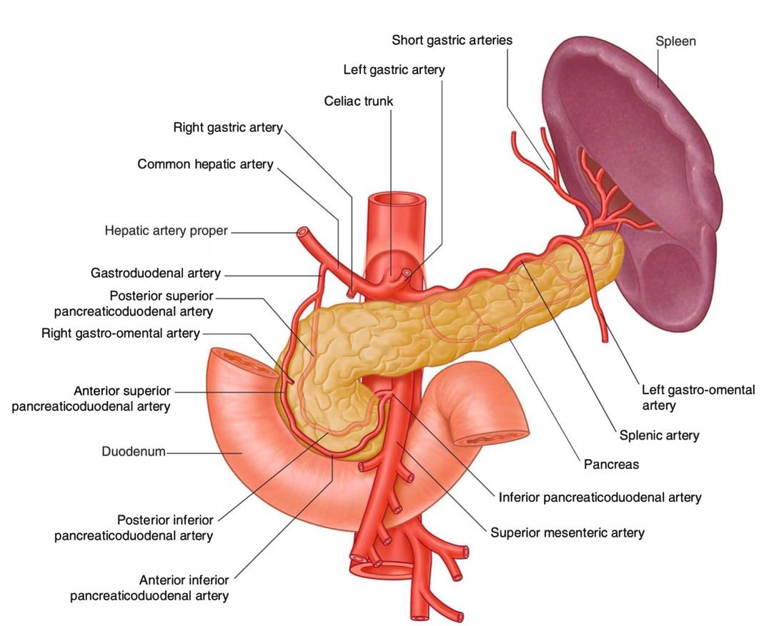
Which image shows the pancreas and spleen's anatomical relationship?


Where does the head of the pancreas occupy?
The concavity of the duodenum
In the liver
In the spleen
In the stomach
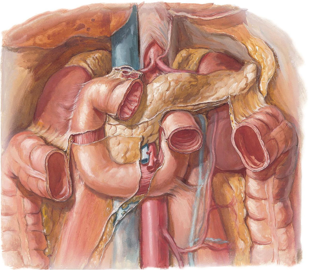
What is the projection behind the superior mesenteric vessels called?
Uncinate process
Celiac trunk
Common bile duct
Pancreatic duct
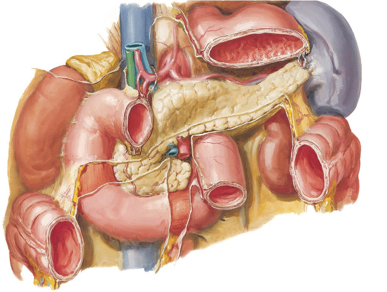
Which organ's anatomical relations are highlighted in the given illustration?
Kidney
Liver
Spleen
Pancreas
What major vessels are related to the head of the pancreas?
Celiac trunk
Hepatic veins
Renal arteries
Superior mesenteric vessels
Identify the source of the anatomical illustration provided.
Cardiac vessels
Major vessels of abdominal organs
Cervical vessels
Pulmonary vessels
What structure is located anteriorly to the head of the pancreas?
Aorta
IVC
Common bile duct
Transverse colon
Which vessel is located posteriorly to the head of the pancreas?
Common bile duct
Transverse colon
Superior mesenteric vessels
Aorta
What is the anterior relation of the uncinate process of the pancreas?
Transverse colon
IVC
Superior mesenteric vessels
Common bile duct
Which structure is behind the uncinate process of the pancreas?
Superior mesenteric vessels
Transverse colon
Aorta
IVC
Identify the major vessel located posteriorly to the head of the pancreas.
Aorta
Transverse colon
Common bile duct
IVC
What is anteriorly related to the neck of the pancreas?
Gastro-duodenal junction
Superior mesenteric vein
Portal vein
Splenic vein
Which veins form the portal vein posteriorly to the neck of the pancreas?
Hepatic veins and splenic veins
Inferior mesenteric and superior mesenteric veins
Splenic and superior mesenteric veins
Gastro-duodenal and splenic veins
Which artery originates from the abdominal aorta and supplies the pancreas?
Splenic artery
Superior mesenteric artery
Renal artery
Celiac trunk
Which vein is associated with the pancreas?
Inferior vena cava
Hepatic portal vein
Renal vein
Splenic vein
Which muscle is located in relation to the pancreas?
Quadratus lumborum
Rectus abdominis
Left psoas major
Transverse abdominis
What gland is found in close proximity to the pancreas?
Thyroid gland
Adrenal gland
Left suprarenal gland
Pituitary gland
What anatomical feature runs along the upper border of the pancreas?
Splenic artery
Inferior mesenteric artery
Hepatic artery
Celiac trunk
What passes between the layers of the lieno-renal ligament with splenic vessels?
Spleen
Tail of the pancreas
Body of the pancreas
Head of the pancreas
Which vessel is closely associated with the tail of the pancreas?
Superior mesenteric artery
Hepatic artery
Splenic vessels
Celiac trunk
What structure is located adjacent to the tail of the pancreas?
Kidney
Duodenum
Liver
Spleen
What is the main pancreatic duct also known as?
Duct of Santorini
Duct of Wirsung
Accessory pancreatic duct
Common bile duct
Where does the main pancreatic duct emerge and unite with the common bile duct?
Inside the wall of the 2nd part of the duodenum
In the stomach
In the tail of the pancreas
At the head of the pancreas
What structure surrounds the hepatopancreatic ampulla?
Sphincter of Hoffmann
Ileocecal valve
Pyloric sphincter
Sphincter of Oddi
Where does the accessory pancreatic duct (of Santorini) open?
Into the first part of the duodenum
Into the 2nd part of the duodenum above the main duct
Into the bile duct
Into the stomach
Which artery supplies the body and tail of the pancreas?
Splenic artery
Superior mesenteric artery
Inferior mesenteric artery
Celiac trunk
What arteries supply the head of the pancreas?
Splenic artery
Celiac trunk
Common hepatic artery
Superior and inferior pancreaticoduodenal arteries
Where can you find the anatomical illustration of the pancreas and spleen?
In surgical manuals
In histology textbooks
In physiology lectures
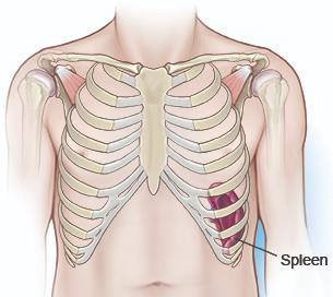
Where is the spleen located?
Lower abdomen
Left hypochondrium near ribs 9, 10, 11
Right hypochondrium
Thoracic cavity
What does the anterior end of the spleen not extend beyond?
Rib 11
Rib 10
Midaxillary line
Median plane
How far is the posterior end of the spleen from the median plane?
1 inch
3 inches
2 inches
1.5 inches
What is the shape of the spleen?
Cube
Triangle
Sphere
Cupped hand
Which borders does the spleen have?
Left and right
Upper and lower
Medial and lateral
Anterior and posterior
What separates the spleen from the 9th, 10th, and 11th ribs?
Diaphragm
Kidneys
Pancreas
Liver
Which ribs are related to the diaphragmatic surface of the spleen?
7th, 8th, 9th
6th, 7th, 8th
10th, 11th, 12th
9th, 10th, 11th
What feature is described as smooth and convex regarding the diaphragm?
Spleen surface
Diaphragmatic surface
Liver edges
Stomach wall
What is the purpose of the hilum on the visceral surface of the spleen?
It allows for renal function.
It gives passage to splenic vessels and lymphatic vessels.
It connects to the stomach.
It supports the pancreas.
What area is located above the hilum on the visceral surface of the spleen?
Gastric impression
Renal impression
Colic impression
Pancreatic impression
Which impression is related to the left kidney on the spleen's visceral surface?
Gastric impression
Colic impression
Renal impression
Pancreatic impression
What is related to the tail of the pancreas on the visceral surface of the spleen?
Colic impression
Pancreatic impression
Gastric impression
Renal impression
Which impression is related to the left colic flexure?
Renal impression
Colic impression
Pancreatic impression
Gastric impression
What structure is illustrated in this image?
Pancreas
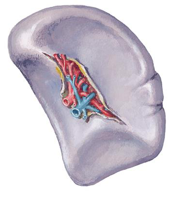
Visceral surface of the spleen
Kidney
Liver
Which ligament connects the splenic hilum to the fundus of the stomach?
Short gastric ligament
Gastrosplenic ligament
Lienorenal ligament
Left gastroepiploic ligament
What vessels does the gastrosplenic ligament carry?
Splenic vessels and tail of the pancreas
Hepatic arteries
Celiac trunk
Short gastric and left gastroepiploic vessels
Where does the lienorenal ligament extend to?
The front of the left kidney
The liver
The right kidney
The diaphragm
What is the position of the spleen in relation to the peritoneum?
It is not covered
It is posterior to the peritoneum
It is completely covered by peritoneum
It is only partially covered
Flashcards in this deck (48)
-
Where is the pancreas located in the human body?
Along the left side of the ribcage
Transversely across the posterior abdominal wall at the level of L1 & L2 vertebrae
In the upper right quadrant of the abdomen
Near the liver and gallbladder
anatomy pancreas -
What structures does the pancreas extend between?
The concavity of the duodenum and the spleen
The gallbladder and the esophagus
The liver and the small intestine
The stomach and the kidneys
anatomy relations -
anatomy illustration
-
Where does the head of the pancreas occupy?
The concavity of the duodenum
In the liver
In the spleen
In the stomach
anatomy pancreas -
What is the projection behind the superior mesenteric vessels called?
Common bile duct
Celiac trunk
Uncinate process
Pancreatic duct
anatomy pancreas -
Which organ's anatomical relations are highlighted in the given illustration?
Pancreas
Kidney
Spleen
Liver
anatomy organs -
What major vessels are related to the head of the pancreas?
Hepatic veins
Renal arteries
Celiac trunk
Superior mesenteric vessels
anatomy blood_supply -
Identify the source of the anatomical illustration provided.
Pulmonary vessels
Cardiac vessels
Cervical vessels
Major vessels of abdominal organs
anatomy illustration -
What structure is located anteriorly to the head of the pancreas?
Aorta
IVC
Common bile duct
Transverse colon
anatomy pancreas -
Which vessel is located posteriorly to the head of the pancreas?
Common bile duct
Superior mesenteric vessels
Aorta
Transverse colon
anatomy pancreas -
What is the anterior relation of the uncinate process of the pancreas?
Common bile duct
Transverse colon
Superior mesenteric vessels
IVC
anatomy pancreas -
Which structure is behind the uncinate process of the pancreas?
Superior mesenteric vessels
Transverse colon
IVC
Aorta
anatomy pancreas -
Identify the major vessel located posteriorly to the head of the pancreas.
Transverse colon
Aorta
IVC
Common bile duct
anatomy pancreas -
What is anteriorly related to the neck of the pancreas?
Gastro-duodenal junction
Superior mesenteric vein
Portal vein
Splenic vein
anatomy pancreas relationships -
Which veins form the portal vein posteriorly to the neck of the pancreas?
Splenic and superior mesenteric veins
Hepatic veins and splenic veins
Gastro-duodenal and splenic veins
Inferior mesenteric and superior mesenteric veins
anatomy veins pancreas -
Which artery originates from the abdominal aorta and supplies the pancreas?
Renal artery
Superior mesenteric artery
Splenic artery
Celiac trunk
anatomy blood_vessels -
Which vein is associated with the pancreas?
Splenic vein
Renal vein
Inferior vena cava
Hepatic portal vein
anatomy veins -
Which muscle is located in relation to the pancreas?
Quadratus lumborum
Transverse abdominis
Left psoas major
Rectus abdominis
anatomy muscles -
What gland is found in close proximity to the pancreas?
Adrenal gland
Left suprarenal gland
Pituitary gland
Thyroid gland
anatomy glands -
What anatomical feature runs along the upper border of the pancreas?
Splenic artery
Inferior mesenteric artery
Celiac trunk
Hepatic artery
anatomy vascular -
What passes between the layers of the lieno-renal ligament with splenic vessels?
Head of the pancreas
Tail of the pancreas
Body of the pancreas
Spleen
anatomy pancreas -
Which vessel is closely associated with the tail of the pancreas?
Superior mesenteric artery
Celiac trunk
Hepatic artery
Splenic vessels
anatomy vascular -
anatomy spleen
-
What is the main pancreatic duct also known as?
Common bile duct
Duct of Santorini
Accessory pancreatic duct
Duct of Wirsung
anatomy pancreas -
Where does the main pancreatic duct emerge and unite with the common bile duct?
In the tail of the pancreas
Inside the wall of the 2nd part of the duodenum
In the stomach
At the head of the pancreas
anatomy duodenum -
What structure surrounds the hepatopancreatic ampulla?
Pyloric sphincter
Sphincter of Oddi
Sphincter of Hoffmann
Ileocecal valve
anatomy sphincters -
Where does the accessory pancreatic duct (of Santorini) open?
Into the first part of the duodenum
Into the bile duct
Into the 2nd part of the duodenum above the main duct
Into the stomach
anatomy pancreas -
Which artery supplies the body and tail of the pancreas?
Inferior mesenteric artery
Superior mesenteric artery
Celiac trunk
Splenic artery
anatomy pancreas -
What arteries supply the head of the pancreas?
Superior and inferior pancreaticoduodenal arteries
Common hepatic artery
Celiac trunk
Splenic artery
anatomy pancreas -
Where can you find the anatomical illustration of the pancreas and spleen?
In physiology lectures
In surgical manuals
In histology textbooks
anatomy illustration -
Where is the spleen located?
Left hypochondrium near ribs 9, 10, 11
Right hypochondrium
Lower abdomen
Thoracic cavity
anatomy spleen -
What does the anterior end of the spleen not extend beyond?
Rib 11
Rib 10
Median plane
Midaxillary line
anatomy spleen -
How far is the posterior end of the spleen from the median plane?
3 inches
2 inches
1 inch
1.5 inches
anatomy measurements -
anatomy spleen
-
Which borders does the spleen have?
Medial and lateral
Left and right
Upper and lower
Anterior and posterior
anatomy spleen -
anatomy spleen
-
Which ribs are related to the diaphragmatic surface of the spleen?
10th, 11th, 12th
7th, 8th, 9th
6th, 7th, 8th
9th, 10th, 11th
anatomy ribs -
What feature is described as smooth and convex regarding the diaphragm?
Stomach wall
Diaphragmatic surface
Spleen surface
Liver edges
anatomy diaphragm -
What is the purpose of the hilum on the visceral surface of the spleen?
It allows for renal function.
It gives passage to splenic vessels and lymphatic vessels.
It supports the pancreas.
It connects to the stomach.
anatomy spleen -
What area is located above the hilum on the visceral surface of the spleen?
Colic impression
Renal impression
Pancreatic impression
Gastric impression
anatomy spleen -
Which impression is related to the left kidney on the spleen's visceral surface?
Renal impression
Gastric impression
Colic impression
Pancreatic impression
anatomy spleen -
What is related to the tail of the pancreas on the visceral surface of the spleen?
Renal impression
Pancreatic impression
Gastric impression
Colic impression
anatomy spleen -
Which impression is related to the left colic flexure?
Gastric impression
Renal impression
Colic impression
Pancreatic impression
anatomy spleen -
anatomy illustration
-
Which ligament connects the splenic hilum to the fundus of the stomach?
Lienorenal ligament
Gastrosplenic ligament
Short gastric ligament
Left gastroepiploic ligament
anatomy spleen ligaments -
What vessels does the gastrosplenic ligament carry?
Short gastric and left gastroepiploic vessels
Splenic vessels and tail of the pancreas
Hepatic arteries
Celiac trunk
anatomy spleen vascular -
Where does the lienorenal ligament extend to?
The front of the left kidney
The right kidney
The diaphragm
The liver
anatomy spleen ligaments -
What is the position of the spleen in relation to the peritoneum?
It is only partially covered
It is completely covered by peritoneum
It is posterior to the peritoneum
It is not covered
anatomy spleen peritoneum
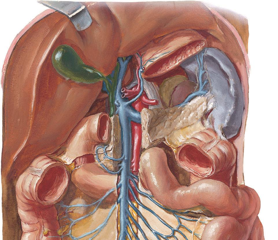
Pancreas: Overview
Pancreas
Site: Elongated gland located transversely across the posterior abdominal wall at the level of L1 & L2 vertebrae.
- Extends from the duodenum on the right to the spleen on the left.
- Located in the lower epigastric region.
Parts of the Pancreas
1. Head
- Occupies the concavity of the duodenum.
- Lower left part forms the uncinate process behind the superior mesenteric vessels.
Relations of the Head
Anterior Relations
- Transverse colon
- Superior mesenteric vessels (in front of uncinate process)
Posterior Relations
- Common bile duct
- I.V.C (Inferior Vena Cava)
- Aorta (behind uncinate process)
Neck Relations
2. Neck
- Narrow part connecting head and body.
- Anteriorly: Related to gastro-duodenal junction.
- Posteriorly: Junction of splenic and superior mesenteric vein forming the portal vein.
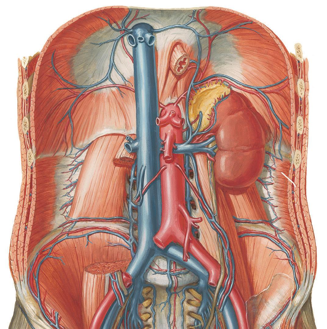
Body Relations
3. Body
Posterior Relations:
1. Abdominal aorta
2. Superior mesenteric artery (origin)
3. Veins: Splenic vein, Left renal vein
4. Muscles: Left crus of diaphragm, Left psoas major
5. Glands: Left kidney, Left suprarenal gland
Tail Relations
4. Tail
- Passes between layers of lieno-renal ligament with splenic vessels.
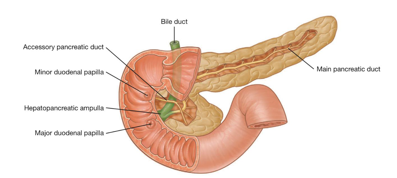
Ducts of the Pancreas
Main Pancreatic Duct (Wirsung)
- Begins at the tail, extends to the head.
- Merges with the common bile duct to form the hepatopancreatic ampulla, opening to the duodenum.
- Sphincter of Oddi controls the duct.
Accessory Pancreatic Duct (Santorini)
- Opens above the main duct into the duodenum at the minor duodenal papilla.

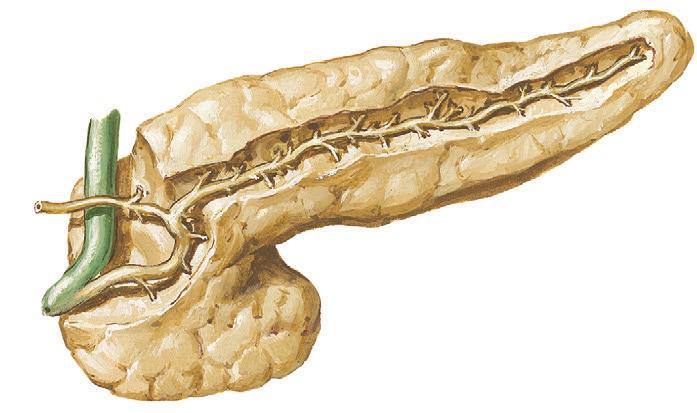
Blood Supply of the Pancreas
Blood Supply
- Splenic artery: Supplies the body & tail.
- Superior and inferior pancreaticoduodenal arteries: Supply the head.
Spleen: Overview
Spleen (Lien)
Site: Located in the left hypochondrium, opposite ribs 9, 10, 11, with the long axis parallel to the 10th rib.
- Posterior end lies 1.5 inches from the median plane; anterior end stays within midaxillary line.
Shape of the Spleen
Shape
- Resembles a cupped hand.
- Surfaces: Visceral and diaphragmatic.
- Borders: Upper (notches) and lower.
- Ends: Medial (tapering) and lateral (broad).
Relations of the Spleen
Diaphragmatic Surface
- Smooth and convex, related to the diaphragm, separating it from ribs 9, 10, and 11.
Visceral Surface Impressions
- Hilum: Passage for splenic vessels, lymphatics.
- Impressions: Gastric, renal, pancreatic, and colic.
Peritoneal Relations of the Spleen
Peritoneal Relations
- Completely covered by peritoneum.
- Forms the left lateral extremity of the lesser sac.
- Ligaments:
- Gastrosplenic ligament: Connects hilum to stomach, carries vessels.
- Lieno-renal ligament: Connects hilum to left kidney, contains splenic vessels.