Sign up to unlock more features
- Save this deck to your account
- Study flashcards with spaced repetition
- Export to Anki (.apkg) or PDF
- Process documents up to 100 pages
- Images extracted from PDFs and documents
- Better text extraction from your PDFs and documents
- Better flashcards with our more advanced AI model
What are the three longitudinal divisions of the brainstem?
What are the three transverse divisions of the brainstem?
Which brainstem division is the most rostral?
What is the midbrain position in the brainstem?
Which brainstem division lies between the midbrain and the medulla?
Where is the pons located?

Which brainstem division is most caudal?
What is the medulla position and its landmark relation?
What landmark marks the spinomedullary junction?
What is the landmark for the spinomedullary junction?
What structure lies dorsal to the pons?
Which view best shows the transverse divisions/segments of the brainstem?
In midsagittal view, which divisions are best appreciated as transverse segments?
Name four landmarks seen in the midsagittal view of the brainstem.
What is another name for the cerebral aqueduct?
What is the cerebral aqueduct?
What ventricular space is posterior to the pons?
Where is the fourth ventricle located?
Name a midsagittal landmark listed near the midbrain.
What is the apex of the fourth ventricle called?
What are the labeled structures commonly shown with the brainstem in sagittal diagrams?
What is the floor or base of the fourth ventricle called?
What sulci are associated with the pons on midsagittal diagrams?
What marks the ponto-midbrain junction ventrally?
What is the role of transverse divisions in brainstem anatomy?
What marks the pontomedullary junction ventrally?
Which structure connects the third and fourth ventricles?
What are the three longitudinal divisions of the brainstem from dorsal to ventral?
What is the anatomical position of the pons relative to the cerebellum?
Where is the tectum located and what is its role?
Which structure lies just above the spinal cord?
What is the superior medullary velum?
Which bony opening is shown in the brainstem overview?
Where is the tegmentum located relative to the neural cavity and basis?
List the main brainstem parts as labeled in introductory diagrams.
What is the rhomboid fossa in relation to the pontine tegmentum?
What is the common name for the fourth ventricle as shown in posterior diagrams?
Name a feature mentioned as part of the pontine region.
What term refers to the transverse divisions of the brainstem?
What is the tegmentum of the brainstem?
Which diagram label indicates the location of the pons between other brainstem parts?
How is gray and white matter organized in the pontine tegmentum?
Why is the midsagittal view useful in brainstem anatomy?
Where is the basis of the pons located relative to the tegmentum?
What are the two major neural cavities inside the brainstem?
What is the trapezoid body and what does it mark?
What is the cerebral aqueduct?
Which cranial nerve nuclei are located in the pontine tegmentum?
What are alternative names for the cerebral aqueduct?
Which major types of tracts pass through the pontine tegmentum?
Where is the fourth ventricle located?
Give examples of specific tracts or fibers found in the tegmentum.
What is the shape of the fourth ventricle?
Which descending motor tracts are located in the basis of the pons?
What is the peak or apex of the fourth ventricle called?
What is the difference between descending cortical and descending subcortical motor tracts?
What is the floor or base of the fourth ventricle called?
Outline the corticospinal tract pathway and termination.
What structure houses the superior pontine sulcus?
Where does most of the corticospinal tract decussate and what happens afterward?
What does the superior pontine sulcus demarcate?
What do the lateral motor neurons in the pons supply?
What imaginary line marks the ponto-midbrain junction?
What does the ventral corticospinal tract (remaining 10%) synapse on and what do those neurons supply?
What anatomical points does the superior pontine sulcus line pass through?
How do corticospinal tracts control body movement relative to the pyramidal decussation?
What structure houses the inferior pontine sulcus?
What is the pathway of the corticobulbar (corticonuclear) tract?
What does the inferior pontine sulcus demarcate?
What is the function of the corticobulbar tract?
What imaginary line marks the ponto-medullary junction?
What are the corticospinal and corticobulbar tracts collectively called?
What anatomical point does the ponto-medullary junction line pass through?
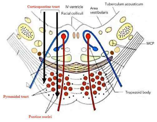
What is the pathway and termination of the corticopontine tract?
What are the three longitudinal divisions of the brainstem from dorsal to ventral?
Where does the pontocerebellar tract originate and how does it reach the cerebellum?
What is the tectum?
Why is the pontocerebellar tract not considered ascending or descending?
How does the tectum relate to the neural cavities?
Which major tracts are found in the basis of the midbrain, pons, and medulla?
What is the superior medullary velum in relation to the tectum?
Why is the medullary basis called the 'medullary pyramids'?
What is notable about the superior medullary velum's physical properties?
What are the three sulci of the pons?
What is the tegmentum and where is it located?
What does the superior pontine sulcus mark?
What is the rhomboid fossa?
What does the inferior pontine sulcus mark?
What is the trapezoid body mentioned in relation to the pons?
What is the basilar sulcus?
What is the pontine tegmentum?
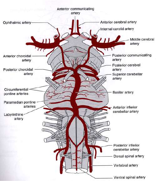
Where do the vertebral arteries course on the brainstem?
How does the tegmentum differ from the spinal cord?
How is the basilar artery formed?
Which cranial nerve nuclei are located in the pontine tegmentum?
What occurs at the inferior pontine sulcus regarding arteries?
What major fiber tracts pass through the pontine tegmentum?
What is the general role of the pons sulci?
Which tracts do NOT pass through the tegmentum but through the basis?
Where does the basilar artery run on the pons?
What is the basis of the pons?
Where does the basilar artery terminate and what does it form?
What are pontine nuclei and where are they located?
Where does the basilar artery commence?
What is the trapezoid body and where is it found?
What is the middle cerebellar peduncle (MCP) also called?
What is the medial longitudinal fasciculus (MLF)?
Where is the MCP located and what is its main connection?
What is the central tegmental tract (CTT)?
Which cranial nerve emerges at the junction of the pons with the MCP?
What autonomic fibers run through the tegmentum?
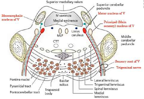
What are the roots of the trigeminal nerve and their functions?
What types of nuclei are found in the pontine tegmentum?
Where does the abducens nerve (VI) emerge and what is its root type?
Which major descending cortical motor tracts are in the pons basis?
What are the main roots of the facial nerve (VII)?
What is the corticospinal tract pathway in brief?
What fibers does the nervus intermedius contain?
Where does the corticospinal tract decussate?
What are the roots of the vestibulocochlear nerve (VIII) and their lamination?
What percentage of corticospinal fibers decussate at the pyramids?
Which three cerebellar peduncles are most prominent on the lateral surface of the pons?
What happens to corticospinal fibers after decussation?
What does the superior pontine sulcus mark ventrally?
What are descending subcortical (extrapyramidal) tracts?
Which cranial nerves emerge along the inferior pontine sulcus/pontomedullary junction ventrally?
What is the function of pontocerebellar tracts?
What are the three pairs of cerebellar peduncles?
Where do corticobulbar fibers project?
What is the main anatomical role of the cerebellar peduncles?
What role does the pontine basis play in motor pathways?
What is a cerebellar afferent and a cerebellar efferent?
How are fiber tracts oriented in the trapezoid body?
What is the primary fiber direction and connections of the SCP?
What is the general developmental note about the tegmentum?
What does the MCP contain and connect?
Which tracts pass through the pons basis illustrated in diagrams?
What are the main inputs carried by the ICP?
What is meant by 'pyramidal tract' in the pons context?
What is the pontocerebellar tract and where does it travel?
What is the main function of corticospinal (CS) tracts in the pons?
Which tract carries nonconscious proprioception from the dorsal nucleus of Clarke and where does it enter the cerebellum?
What does 'above the pyramidal decussation' imply for CS tract control?
Which medullary nucleus sends the cuneocerebellar tract and through which peduncle does it enter?
What does 'below the pyramidal decussation' imply for CS tract control?
What is the main function of the inferior cerebellar peduncle (ICP)?
Which motor neurons do lateral corticospinal fibers synapse onto and what do they supply?
Which cranial nerves emerge from the lateral aspect of the pons?
What percentage of corticospinal fibers remain ventral and what do they synapse on?
Where does the trigeminal nerve (CN V) emerge from the pons?
What muscles are supplied by the medial motor neurons targeted by ventral corticospinal fibers?
Where do CN VI, VII, and VIII emerge relative to the pons?
What is the corticobulbar (corticonuclear) tract pathway end point?
Which nerve emerges ventro-medially from the pons?
What head movements does the corticobulbar tract help control?
Which nerves emerge posterolaterally from the cerebello-pontine angle?
What collective name is given to the corticospinal and corticobulbar tracts?
What is the cerebello-pontine angle?
What is the corticopontine tract pathway?
What are the main fiber types in the superior cerebellar peduncle (SCP)?
Where do corticopontine fibers terminate and where do they not terminate?
What are the main fiber types in the middle cerebellar peduncle (MCP)?
What structure gives rise to the pontocerebellar tract?
What are the main fiber types in the inferior cerebellar peduncle (ICP)?
How do pontocerebellar fibers run through the pons?
How is the dorsal surface of the pons related to the cerebellum and how is it exposed?
Do pontocerebellar fibers pass through the tegmentum to reach the cerebellum?
What is characteristic of the superior medullary velum?
Why is the pontocerebellar tract not considered strictly ascending or descending?
What is the rhomboid fossa and what are its rostral and caudal limits?
Which tracts are listed in the basis of the midbrain?
How is the pontomedullary junction identified on the rhomboid fossa?
Which tracts are listed in the basis of the pons?
Along which plane does the pontomedullary junction lie dorsally?
Which tracts are listed in the basis of the medulla?
What is the locus coeruleus and what does it contain?
Why is the medullary basis called the 'medullary pyramids'?
What is the substantia ferruginea and where is it located?
What surface of the pons is referred to as the 'basis'?
How can the substantia ferruginea be exposed anatomically?
What is the anatomical relation of corticobulbar fibers in the brainstem?
What are the roles of the stria medullaris of the 4th ventricle?
Which cranial nerve motor nuclei are explicitly mentioned as corticobulbar targets?
What does the median sulcus do in the rhomboid fossa?
What role do pontine nuclei play in cortico-cerebellar communication?
Where is the sulcus limitans located and what is its significance?
Do corticopontine fibers reach the medulla?
How is the medial sulcus formed?
What is the primary functional division between lateral and ventral corticospinal systems?
Name key landmarks visible on the posterior view of the 4th ventricle.
Where is the pyramidal decussation located?
Where are the vagal and hypoglossal triangles located?
How do corticospinal fibers travel through the brainstem basis layers?
What causes the dark pigment in catecholamine-producing cells like the locus coeruleus?
What distinguishes basis fibers from tegmental structures in the brainstem?
Close-up: which structures are shown around the substantia ferruginea in the posterior 4th ventricle?
Why are transverse pontine fibers clinically important?
Where are the prominent bulges/enlargements located on the pons?
What is one functional consequence of lesions in the pontine basis affecting pontocerebellar fibers?
Which bulge is present in the upper (rostral) part of the dorsal pons?
What does the superior pontine sulcus mark?
What are the prominences in the lower dorsal pons from medial to lateral?
What junction is indicated by the superior pontine sulcus?
Where are the prominences more prominent on the pons?
Which structure lies immediately above the superior pontine sulcus?
What causes the bulge of the medial eminence internally?
What does the inferior pontine sulcus mark?
Where does the medial eminence lie relative to the median sulcus and sulcus limitans?
What junction is indicated by the inferior pontine sulcus?
Where is the area vestibularis located relative to the sulcus limitans and facial colliculus?
Which structure lies immediately below the inferior pontine sulcus?
What are the roof, lateral and floor boundaries near these dorsal pons prominences?
What is the basilar sulcus?
What are the transverse fibers coursing on the dorsal pons called?
What important vessel lies in the basilar sulcus?
Where is the tuberculum acousticum located?
How is the basilar artery formed?
How does the prominence of the tuberculum acousticum compare to the facial colliculus?
Where do the vertebral arteries course prior to forming the basilar artery?
Which cranial nerve is contained in the upper half of the pons?
At which anatomical level do the vertebral arteries fuse to form the basilar artery?
Which cranial nerves are contained in the lower half of the pons?
What does the term MCP stand for in brainstem anatomy?
What anatomical level corresponds to the upper half of the pons?
What is the main connection function of the middle cerebellar peduncle (MCP)?
What anatomical level corresponds to the lower half of the pons?
What is the general role of the sulci of the pons?
How does the fourth ventricle differ between upper and lower pons?
Which cranial nerves are commonly labeled at the lateral base of the pons in anatomical views?
In cross-sectional orientation, which surface faces up in anatomical sections?
Relative to the pons, where is the cerebellum located?
In cross-sectional orientation, which surface faces down in clinical sections?
What ventral feature indicates the midline position of the basilar artery on the pons?
Name the main cross-sectional regions of the pons.
What anatomical view highlights the pons and medulla beneath the forebrain?
Which cranial nerve uniquely exits from the dorsal surface of the brainstem?
Why is the inferior pontine sulcus clinically important regarding arterial anatomy?
Give one dorsal limit of the pons.
Name a visible landmark that helps identify the rostral and caudal borders of the pons on the ventral surface.
Give another dorsal landmark marking the pons.
Which arteries form the basilar artery and where are they located before fusion?
In anatomical orientation when viewing slides, which direction is dorsal?
Where does the basilar artery run on the pons?
When viewing MRI or CT scans of a patient in the supine position, which surface faces down and which faces up?
Where does the basilar artery commence?
If ventral surfaces face down (anatomical orientation for slides), what structures are found above and below the pons?
Where does the basilar artery terminate ventrally?
If the patient is supine for MRI/CT (dorsal faces down), what structures are placed above and below the pons?
What are the terminal branches of the basilar artery?
What are the three prominent bulgings on the lower half of the floor of the 4th ventricle?
What structure does the basilar sulcus relate to?
What are key features of the IV ventricle at the level of the lower pons?
What is the Middle Cerebellar Peduncle also called?
What defines the pontine tegmentum in the lower pons?
Where is the Middle Cerebellar Peduncle located relative to the pons?
Where is the pontine basis located and what is its composition?
Is the Middle Cerebellar Peduncle paired or unpaired?
Which major descending motor tracts and related structures are contained in the pontine basis?
What does the Middle Cerebellar Peduncle connect?
What is the orientation and function of the facial colliculus on the 4th ventricle floor?
Where does the trigeminal nerve (V) emerge on the pons?
Name common sensory nuclei and roots found lateral to the pontine tegmentum in the lower pons.
What roots does the trigeminal nerve have?
Where is the tegmentum of the pons located?
What is the main function of the trigeminal sensory root?
What is the basilar sulcus relation to pontine structures?
What does the trigeminal motor root supply?
How many middle cerebellar peduncles are there and where are they located?
Which nerves emerge along the inferior pontine sulcus/pontomedullary junction ventrally?
Where do corticopontine fibers terminate?
Where does the abducens nerve (VI) emerge?
Where does the corticospinal tract originate?
What roots compose the facial nerve (VII)?
What is the effect of a corticospinal lesion above the decussation?
What is the main function of the facial nerve large motor root?
What percent of corticospinal fibers cross at the pyramidal decussation?
Where does the nervus intermedius emerge?
What are the two corticospinal divisions after the caudal medulla and their general targets?
What fiber types are contained in the nervus intermedius?
Describe the cortico-ponto-cerebellar pathway from the right cerebral hemisphere.
Is it correct to call the facial smaller root purely sensory?
What tract connects the cerebral cortex with the pontine nuclei?
Where does the vestibulocochlear nerve (VIII) emerge on the pons?
What tract connects the pons and the cerebellum and where does it decussate?
What roots compose the vestibulocochlear nerve?
How do pontine nuclei axons reach the opposite cerebellar hemisphere?
According to the Law of Lamination, which vestibulocochlear root is more medial and primitive?
What is the primary role of the cerebellum in movement?
Which vestibulocochlear root is more lateral and developed later?
Which main descending tracts pass through the pons basis?
What are the three cerebellar peduncles prominent on the lateral surface of the pons?
What does the pontine tegmentum (lower half) contain?
What is the Superior Cerebellar Peduncle also called?
What is the trapezoid body and where is it located?
Which sulcus marks the ponto-midbrain junction ventrally?
What sensory modalities travel in the medial lemniscus?
Which sulcus corresponds to the ponto-medullary junction?
Where are the second-order neurons for the medial lemniscus located?
What are the three pairs of cerebellar peduncles?
Where does the medial lemniscus first form and what is its relation to the trapezoid body?
Anatomically, what is the role of the cerebellar peduncles?
Through which cerebellar peduncle do pontocerebellar fibers enter the cerebellum?
What would happen if you sliced across the cerebellar peduncles?
What sensory deficit occurs with a lesion in the pons?
Define cerebellar afferents.
Why is dorsal column sensation ipsilateral in the spinal cord?
Define cerebellar efferents.
After the medullary sensory decussation, the medial lemniscus conveys sensation to which side?
What is the primary fiber direction of the SCP?
How does the medial lemniscus appearance change between medulla and pons?
Which spinocerebellar tract enters the cerebellum via the SCP?
Which dorsal column fibers enter the spinal cord first?
Which spinocerebellar tract enters the cerebellum via the ICP?
How is the dorsal column divided at cervical and upper thoracic levels?
Functionally, what does the SCP connect?
Where do dorsal column fibers terminate and where do they decussate?
What is the main functional content of the MCP?
What tracts form the spinal lemniscus?
Where does the pontocerebellar tract originate?
What modalities does the spinal lemniscus convey?
Describe the path of the pontocerebellar tract.
Why do the spinothalamic tracts convey contralateral sensation?
Which cerebellar peduncle is the largest?
Where is the medial longitudinal fasciculus (MLF) located and what is its role?
What are the components of the ICP as named in the text?
Where is the inferior cerebellar peduncle (ICP) located in the pons?
What does the ICP primarily contain?
Where is the central tegmental tract (CTT) located relative to the medial lemniscus?
Which nucleus mediates nonconscious proprioception feeding the dorsal spinocerebellar tract?
What modalities are carried by the descending nucleus and tract of CN V?
What is the role of the accessory (lateral) cuneate nucleus in the medulla?
Where is the descending nucleus and tract of CN V located?
Where does the cuneocerebellar tract enter the cerebellum?
Which muscle is innervated by CN VI (abducens nerve) and what is its action?
What alternate name is given for the SCP in the text?
Where does the abducens nucleus lie in relation to the facial colliculus?
What alternate name is given for the MCP in the text?
How does the abducens nerve exit the pons?
Summarize the overall function of the three cerebellar peduncles.
Does CN VI decussate?
Which peduncle anatomically and functionally connects the pons with the cerebellum?
What is the clinical sign of a left abducens nerve lesion?
Name a small group of cerebellar afferents that come from the spinal cord and enter via the SCP.
What two structures make the facial colliculus prominent?
Does the ICP contain any cerebellar efferents?
Describe the course of the facial motor root relative to the abducens nucleus.
What is the basis of the pons important component mentioned in the text?
Where is the facial motor nucleus located in the pontine tegmentum relative to the abducens nuclei?
What is the function of the Inferior Cerebellar Peduncle (ICP)?
What is the facial genu in the pons?
Which cranial nerve emerges at the junction of the ventral pons and the middle cerebellar peduncle?
What is the modality and main targets of the facial motor nucleus?
Which cranial nerves emerge along the inferior pontine sulcus at the pontomedullary junction?
Which muscles/nerves close and open the eyelids?
Where does the Abducens nerve (CN VI) emerge?
Which half of the face does the facial motor root supply?
Where do the Facial (CN VII) and Vestibulocochlear (CN VIII) nerves emerge?
What are the main signs of a peripheral (Bell's) facial nerve lesion?
What is the cerebello-pontine angle?
How do corticobulbar projections innervate the facial nucleus for upper vs lower face?
What covers the dorsal surface of the pons?
What facial deficit results from a unilateral central (corticobulbar) lesion?
How must the cerebellum be removed to view the dorsal surface of the pons?
Give an example of a facial muscle and its function mentioned in the text.
What structures are exposed after removing the cerebellum?
Which parasympathetic nuclei are associated with CN VII?
Describe the superior medullary velum.
What is the 'nervus intermedius' (intermediate nerve)?
Why is the superior medullary velum often not seen in dissections?
Where is the intermediate nerve located relative to the facial motor nucleus?
What is the rhomboid fossa?
What efferent modality does the intermediate nerve carry?
How is the rhomboid fossa bounded laterally?
Which nuclei provide the parasympathetic fibers in the intermediate nerve?
What is the rostral limit of the rhomboid fossa?
What are the parasympathetic targets and functions of the intermediate nerve?
What is the caudal limit of the rhomboid fossa?
Which ganglion contains sensory cell bodies for the intermediate nerve?
What feature bisects the rhomboid fossa marking the pontomedullary junction?
Which taste region is carried by the intermediate nerve?
What are the three cerebellar peduncles abbreviations?
Where do taste fibers from the intermediate nerve terminate centrally?
What fibers does the Superior Cerebellar Peduncle (SCP) mainly carry?
What GVA sensations does the intermediate nerve carry?
What fibers does the Middle Cerebellar Peduncle (MCP) carry?
What GSA fibers are carried by the intermediate nerve?
What fibers does the Inferior Cerebellar Peduncle (ICP) carry?
After the geniculate ganglion, which two central pathways do sensory fibers take?
Which cranial nerve exits dorsally at the ponto-midbrain junction?
Which other cranial nerves contribute to taste sensation alongside CN VII?
Which cranial nerves are considered the lateral cranial nerves of the pons?
Pons cross-section: which structures are near the facial colliculus and facial motor nucleus?
Why must you slice across all three cerebellar peduncles to remove the cerebellum?
What is the visceral afferent center in the brainstem?
What does the pontomedullary junction divide the 4th ventricle and rhomboid fossa into?
Which nerve is CN VIII and what are its two roots?
Dorsally, along which plane does the pontomedullary junction lie?
What sensory modality does the vestibular root mediate?
Which transversely crossing fibers mark the pontomedullary junction dorsally?
What are the peripheral receptors for the vestibular root?
What posterior border does the stria medullaris of the 4th ventricle mark?
Name the four vestibular nuclei.
What dorsal limits does the stria medullaris indicate?
Where are the vestibular nuclei located relative to the cochlear nuclei?
Which deeply pigmented cell group is overlain by the substantia ferruginea?
Which vestibular nuclei are found in the pons versus the medulla?
Which neurotransmitter is found in the locus coeruleus?
What is Scarpa's ganglion and what is unique about its neurons?
What pigment causes the dark color in locus coeruleus cells?
What are the roles of the dorsal and ventral cochlear nuclei?
Where is neuromelanin typically found?
Where do the dorsal cochlear nuclei lie anatomically?
What is the substantia ferruginea in the rhomboid fossa?
Which central connection of vestibular nuclei coordinates eye and head movement?
How can the substantia ferruginea be exposed during dissection?
Which vestibular connection maintains upright posture and balance?
What forms the floor of the 4th ventricle (rhomboid fossa)?
How does the cerebellum connect to the vestibular nuclei and what is its role?
What structure divides the rhomboid fossa into two symmetrical halves?
What is the role of the cerebral motor cortex in vestibular function?
What sulcus runs lateral and parallel to the median sulcus?
What type of neurons make up the spiral ganglion in the auditory pathway?
What is the rostral extension of the sulcus limitans that becomes discolored?
What is the first cell station of the auditory pathway?
What is the medial eminence?
Which nerve root carries auditory impulses from the organs of Corti to the brainstem?
Where is the facial colliculus located?
Upon entering the pons, where do cochlear root fibers first synapse?
What are the vagal and hypoglossal triangles?
Which nucleus in the pons serves as an early relay nucleus in the auditory pathway?
What does SCP, MCP, and ICP stand for?
What is the trapezoid body also known as?
Name key landmarks visible on the posterior view of the 4th ventricle floor.
Name an acoustic stria that contributes to the formation of the lateral lemniscus.
Why is the substantia ferruginea visible in living and dead tissue?
Name another acoustic stria involved in lateral lemniscus formation.
What structure lies immediately lateral to the median sulcus?
Where is the trapezoid body located within the brainstem?
Which structure should be specified when referring to the 'stria medullaris' in this region?
What is the principal ascending auditory pathway tract that originates in the pons?
Where are the prominent bulges (enlargements) of the pons located?
Give the main ascending relay sequence from cochlear nuclei to auditory cortex.
Which enlargement is present in the upper part of the pons?
Where are cochlear nuclei and the main auditory structures in the pons located?
Which three prominences appear in the lower part of the dorsal pons from medial to lateral?
Which auditory nucleus is described as lateral to the spinal lemniscus?
Are the dorsal prominences of the pons more prominent rostrally or caudally?
What is the main projection of the dorsal cochlear nucleus?
What lies between the median sulcus and the sulcus limitans on each side?
How does the ventral cochlear nucleus send crossed fibers?
What is the median sulcus on the dorsal pons?
What uncrossed projection arises from the ventral cochlear nucleus?
Where is the facial colliculus located relative to the median sulcus and sulcus limitans?
What fibers form the trapezoid body (ventral acoustic stria)?
What internal structures form the bulge of the facial colliculus?
What is the main function of the lateral lemniscus?
What is the facial genu?
Which nuclei provide the crossed auditory fibers in the lateral lemniscus?
What is the abducens nucleus function relevant to the facial colliculus?
Which nucleus provides the uncrossed auditory fibers in the lateral lemniscus?
Where is the area vestibularis located relative to the sulcus limitans?
What hearing deficit occurs with a central lesion affecting the lateral lemniscus?
What is the tuberculum acousticum?
Why does a central lesion cause bilateral partial deafness more on the contralateral side?
What forms the roof of the IV ventricle in the region of the dorsal pons?
What is the effect of a peripheral lesion destroying the cochlear nerve?
What structures form the lateral boundaries near the dorsal pons?
Where is the paramedian pontine reticular formation (PPRF) located relative to the abducens nucleus?
What forms the floor in the region of the dorsal pons and IV ventricle?
Where are the main inspiratory and expiratory respiratory centers located?
What are the stria medullaris on the floor of the IV ventricle?
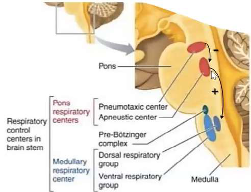
Which pons region contains the pneumotaxic center?
Where is the stria medullaris of the IV ventricle relative to the facial colliculi?
Which pons region contains the apneustic center?
What is the sulcus limitans and its functional significance?
What is the primary action of the apneustic center?
Which prominence is noted as the only enlargement at the upper portion of the pons?
What happens if the apneustic center is unchecked?
What marks the pontomedullary junction?
Which structures inhibit the apneustic center?
What anatomical landmarks are labeled on the dorsal pons dissection described in the lecture?
What is the effect of stretch-receptor impulses at maximal inspiration?
How do facial nerve fibers relate to the facial colliculus externally?
What is the effect of inhibiting the inspiratory center?
Which cerebellar peduncle abbreviations appear on the dorsal pons diagrams?
Name the main respiratory control components in the brain stem. 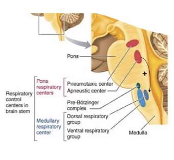
What is the practical surgical relevance of recognizing dorsal pontine prominences?
What pons function is related to horizontal eye movements?
What is the tuberculum acousticum?
Which cranial nerves are associated with the pons region?
Where is the tuberculum acousticum located relative to the area vestibularis?
Name key nuclei related to the facial and lacrimation functions in the pons.
How does the tuberculum acousticum compare to the facial colliculus in prominence?
List important ascending and descending tracts found in the upper pons. 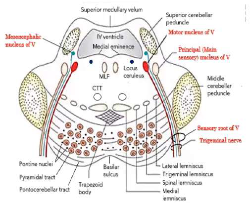
What does area vestibularis refer to?
Name cerebellar-related structures visible in the upper pons cross-section.
Name the three anatomical views used to divide the pons in the lecture.
Which pontine nuclei relay information to the cerebellum?
Which cranial nerve corresponds to the upper half level of the pons?
Which pathways in the pons are involved in auditory and vestibular inputs?
Which cranial nerves are present at the lower half level of the pons?
How does the IV ventricle in the upper pons compare to the lower half?
What is the median sulcus in the floor of the 4th ventricle?
What structure roofs the IV ventricle?
What is the substantia ferruginea?
What forms the lateral walls of the IV ventricle at this level?
What creates the facial colliculus clinically?
What structure lies ventral to the 4th ventricle?
How does the 4th ventricle width differ between pons halves?
What forms the floor of the IV ventricle?
What do the abbreviations SCP and MCP stand for?
What prominent bulge is displayed on the rhomboid fossa?
What is the pontine basis?
Which eminence is the only one in the upper half of the rhomboid fossa?
What is the pontine tegmentum?
Where is the trapezoid body located in the pons?
What is the superior medullary velum?
Why is the trapezoid body still seen in the upper half of the pons?
What structure lies between the pons and medulla at the junction?
Where is the bulk of the trapezoid body located?
Which cranial nerve is the only one to exit the brainstem dorsally?
Where does the basilar sulcus lie?
What is the stria medullaris of the 4th ventricle?
Where is the middle cerebellar peduncle located relative to the basis?
List the main dorsal surface bulges of the pons mentioned in the lecture.
Which descending tracts appear as rounded fascicles in the basis?
Which major cerebellar connection is visible ventrolaterally on the pons?
Where do descending corticopontine tract fibers terminate?
Which major cerebellar connection runs from the cerebellum toward the midbrain above the pons?
How do pontocerebellar fibers reach the cerebellum?
What are the functional contents of the pontine basis?
What is the substantia ferruginea and what does it overlie?
What types of structures does the pontine tegmentum contain?
Name key structures/components of the basis in the upper half of the pons.
What is the clinical orientation used in cross-sections and how does it differ from anatomical orientation?
Which main structures are labeled in the pons diagram?
In anatomical orientation for slides, where is 'dorsal' and where is 'ventral'?
What is the trapezoid body?
How is orientation different for MRI/CT images of supine patients?
What is the medial eminence in the pons?
In slide anatomical view of the pons, which structures are above and which are below?
What does the medial lemniscus convey?
In MRI/CT supine patient view, which pons structures are above and which are below?
Where is the medial lemniscus located relative to the trapezoid body?
At what level is the 'lower half of the pons' described in this lesson?
What do the MLF and CTT share in the pons?
Name three prominent bulgings in the lower half of the pontine tegmentum.
What does the spinal lemniscus convey?
Where is the IV (fourth) ventricle located relative to the pons?
What is the trigeminal lemniscus and what does it convey?
What is the rhomboid fossa in relation to the pons?
What is the lateral lemniscus?
What forms the pontine basis and where is it located?
Where is the locus coeruleus located?
Which major descending motor tracts run in the pontine basis?
What neurotransmitter system is the locus coeruleus a major source of?
Which cerebellar-related tracts and nuclei are found in the pontine basis?
What key functional roles does the locus coeruleus play?
What are the contents of the pontine tegmentum shown in the lower pons?
What pigment and surface change are associated with the locus coeruleus?
What is the facial colliculus anatomically?
What is the dorsolateral pontine reticular formation and its role?
Which nuclei form the tuberculum acousticum and area vestibularis?
Which ascending tracts lie lateral to the medial lemniscus in the pons?
What structures are labeled as cochlear and vestibular roots in the lower pons?
Where is the pneumotaxic center located?
What is the trapezoid body and where is it located?
What is the primary function of the pneumotaxic center?
Which cranial nerve nuclei or roots are prominent in the lower pons section?
How does the pneumotaxic center directly end inspiration?
What sensory ascending tracts are present in the pontine tegmentum?
How does the pneumotaxic center limit inspiration via the apneustic center?
What are the superior salivatory nuclei and lacrimal nuclei associated with in the pons?
How does the pneumotaxic center affect respiratory rate when its signals change?
What is the medial longitudinal fasciculus (MLF) role in the pons?
What is the respiratory effect of opioids on the pneumotaxic center?
Describe the pontine cavity appearance in cross section at the dorsal surface.
Which respiratory centers are located in the brainstem?
What neural elements demarcate the boundary between the pontine tegmentum and basis?
What does the motor root (portio minor) of CN V innervate?
Which motor nucleus is associated with the facial genu in the lower pons?
Where does the motor root of CN V emerge?
What is the relationship of the superior medullary velum to the dorsal pons?
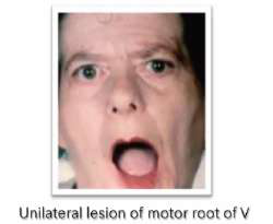
What are signs of a unilateral lesion of the motor root of CN V?
Which structure forms the basilar sulcus and where is it seen?
Where is the motor root of V located within the pons?
Name a compact list of key internal structures found in the lower half of the pons.
Where do sensory afferents of CN V synapse and what is the ganglion type?
What is the pontine nuclei's main projection target?
What does the ophthalmic division (V1) of CN V supply?
Which tract carries cortical input from the pons to the cerebellum?
What does the maxillary division (V2) of CN V supply?
What fibers make up the most ventral part of the pontine basis?
What does the mandibular division (V3) of CN V supply?
Where is the tegmentum of the pons located?
Which sensory ganglia are exceptions to being pseudounipolar?
What is the basilar sulcus on the pons?
Which nucleus of CN V conveys conscious proprioception, fine touch, and vibration from the face?
What are the middle cerebellar peduncles (MCP)?
Where is the principal nucleus of V located within the pons?
Where are the pontine nuclei located?
What peripheral receptors send input to the principal nucleus of V?
What is the role of the pontine nuclei?
Where are the sensory cell bodies located for fibers entering the principal nucleus of V?
Outline the corticopontine tract pathway
Which nucleus of V is considered the homologue of the nucleus cuneatus and nucleus gracilis?
What happens after cortical fibers terminate in pontine nuclei?
What modalities does the descending (spinal) nucleus of V convey?
Describe the corticoponto-cerebellar route from right cortex to cerebellum
Where is the descending (spinal) nucleus of V located?
Why does each cerebral hemisphere communicate with the opposite cerebellar hemisphere?
Briefly describe the pathway for facial pain and temperature fibers via CN V.
Do corticopontine fibers form fascicles in the basis like corticospinal fibers?
What is unique about the mesencephalic nucleus of V among CNS neurons?
Where do ponto-cerebellar fibers enter the cerebellum?
Which nerve and fiber type carry taste from the anterior 2/3 of the tongue?
What structures are part of the pontine dorsal surface (floor of 4th ventricle)?
Name the three branches of the trigeminal nerve (CN V). Include the diagram.
What forms the facial colliculus?
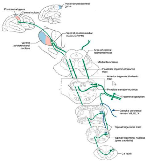
What are the two trigeminothalamic tracts?
What is the trapezoid body in the pons?
Describe the origin and crossing of the anterior (ventral) trigeminothalamic tract.
What is the function of the area vestibularis?
Describe the origin of the posterior (dorsal) trigeminothalamic tract.
What is the clinical effect of a lesion in the corticospinal tract?
What happens when some trigeminal fibers descend?
At the caudal medulla, what percentage of corticospinal fibers cross at the pyramidal decussation?
Which sensory modalities are conveyed by the ventral trigeminothalamic tract?
What are the two main divisions of the corticospinal tract after the medulla?
Which sensory modalities are conveyed by the dorsal trigeminothalamic tract?
Where does the lateral corticospinal tract descend in the spinal cord?
How do bilateral and crossed sensory patterns apply to trigeminal modalities?
What is the primary role of the ventral (anterior) corticospinal tract?
Where is the mesencephalic nucleus of V located?
Summarize the cortico-ponto-cerebellar system in one line
What are the function and unique features of the mesencephalic nucleus of V?
What is the pontine basis composed of?
Which nuclei are homologues for non-conscious proprioception and their body regions?
What tract connects the cerebral cortex with the pontine nuclei?
Which cerebellar tracts arise from the non-conscious proprioception nuclei?
Which tract connects the pons to the cerebellum and decussates in the basis of the pons?
Refer to the diagram: which nucleus sends uncrossed fibers ascending as the posterior trigeminothalamic tract?
How do cerebral hemispheres share motor plans via the pons?
What is a reflex?
What is the role of the cerebellum in movement?
What are the functional components of a basic reflex arc?
Where do axons of pontine nuclei run to reach the cerebellum?
At what brain level are these reflexes elicited?
Name the main descending tracts summarized in the pons section.
What is the jaw jerk reflex and how is it elicited?
What does the pontine tegmentum contain?
What type of synapse is the jaw jerk reflex?
List key internal structures found in the pontine tegmentum (lower half).
What is the stimulus for the jaw jerk reflex?
What structure marks the most ventral part of the pontine tegmentum?
What is the receptor for the jaw jerk reflex?
What type of fibers does the trapezoid body contain?
What is the afferent limb and its cell body for the jaw jerk reflex?
What sensory modalities travel in the medial lemniscus?
What special feature does the mesencephalic nucleus of CN V have?
Where do the medial lemniscus fibers first form?
Where is the central integrator for the jaw jerk reflex?
Which nuclei serve as the second-order neurons for dorsal column modalities?
What is the efferent limb and motor cell body for the jaw jerk reflex?
Where is the medial lemniscus located relative to the trapezoid body?
What is the effector and the response in the jaw jerk reflex?
From which primary afferents do the fasciculus cuneatus and gracilis receive input?
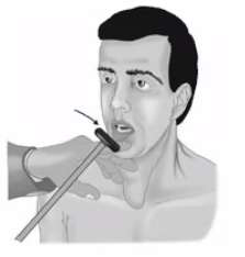
What is the corneal reflex and its pathway type?
What is the pathway from DRG to cortex for fine touch and proprioception?
What happens to the corneal reflex with an afferent limb lesion?
What is the sensory decussation related to the medial lemniscus?
What happens to the corneal reflex with an efferent limb lesion?
What is the role of the pontine nuclei?
What is the afferent limb of the corneal reflex?
Where do pontocerebellar fibers enter the cerebellum?
Where is the afferent neuron cell body for the corneal reflex located?
What marks the boundary between the tegmentum and basis of the pons?
Describe the central pathway for the corneal reflex afferent signal.
Which tract is frequently referred to as the pyramidal tract?
What is the efferent limb of the corneal reflex?
What is the cortico-ponto-cerebellar pathway?
Where are the efferent cell bodies for the corneal reflex?
What is contained in the basis of the pons relevant to decussation?
What are the effector organs in the corneal reflex?
What sensory modalities are lost with medial lemniscus lesions?
What is the normal response of the corneal reflex to corneal stimulation?
Why is transverse orientation of pontine fibers important?
Through which foramina do the divisions of CN V exit the skull?
What structure is labeled as 'Area vestibularis' in the pontine tegmentum?
Where does CN VII exit the skull to reach facial muscles?
What is the facial colliculus?
Which cranial nerves exit via the internal auditory meatus?
Why does dorsal column sensation appear ipsilateral in the spinal cord but contralateral at the level of the pons?
Which arteries from the basilar artery supply the pons?
What is the shape of the medial lemniscus as it appears in the brainstem?
What deficits can result from obstruction of paramedian pontine branches?
How are fibers from lower trunk and limbs organized when entering the dorsal column?
What artery lies in the basilar groove on the ventral surface of the pons?
How is the dorsal column divided at the cervical and upper thoracic levels?
What are the three main branches of the basilar artery supplying the pons?
Why is there no cuneatus division in lower thoracic and lumbosacral regions?
Which basilar branch supplies the ventrolateral areas of the pontine basis?
Where do dorsal column first-order fibers terminate?
Which basilar branch supplies the pontine tegmentum and wedge of the middle cerebellar peduncle?
What happens to axons of the dorsal column nuclei in the medulla?
Which artery reinforces the long circumferential supply at the caudal pontine level?
After decussation, how do dorsal column second-order axons ascend?
Which artery reinforces the long circumferential supply at the rostral pontine level?
Which modalities are conveyed by the lateral spinothalamic tract?
Which four cranial nerves exit at the ventral surface / pontine basis?
Which modality does the ventral spinothalamic tract primarily convey?
Where does CN VI (abducens) emerge from the pons?
Where do the spinothalamic second-order neurons decussate?
Where do CN VII and CN VIII emerge from the pons?
Why do spinothalamic tracts convey contralateral sensation?
What are the three cerebellar peduncles visible on the lateral surface of the pons?
What is the spinal lemniscus?
What does the dorsal surface of the pons consist of?
What sensations does the spinal lemniscus convey?
How is the pons ventrally and dorsally demarcated from midbrain and medulla?
Where is the spinal lemniscus located relative to the medial lemniscus?
What ocular sign indicates lateral rectus weakness?
What is the clinical effect of a lesion in the pons affecting the medial lemniscus?
Which nerve innervates the lateral rectus muscle?
Summarize the dorsal column-medial lemniscus pathway sequence from periphery to cortex.
Name major arteries surrounding the brainstem included in the summary
Summarize the spinothalamic pathway sequence from periphery to cortex.
What are the three enlargements of the rhomboid fossa of the pons (medial to lateral)?
Where is the medial longitudinal fasciculus (MLF) located and what is its function?
What does the facial colliculus overlie?
Where is the central tegmental tract (CTT) located relative to the medial lemniscus?
What does the area vestibularis overlie?
Where is the inferior cerebellar peduncle (ICP) in the pons and what does it contain?
What does the tuberculum acousticum overlie?
Which tract conveys discriminative touch and proprioception to cortex?
What is the substancia ferruginea associated with in the pons?
Where is the descending nucleus and tract of the trigeminal nerve located in the pons?
What are the constant contents of the pontine basis?
What modalities are carried by the descending nucleus and tract of V?
Which descending cortical motor tracts terminate in the pons?
What muscle does the abducens nerve (CN VI) innervate?
Where do the corticopontine fibers terminate and what do they give rise to?
What is the primary action of the lateral rectus muscle?
What is the role of the cortico-ponto-cerebellar pathway?
What happens to the eye in a lesion of the abducens nerve?
Name three key auditory-related structures in the pontine tegmentum and their roles.
Why does a unilateral abducens nerve lesion cause medial deviation rather than lateral?
What is the function of the medial longitudinal fasciculus (MLF) and the PPRF in the pons?
Where is the abducens nucleus located relative to the facial colliculus?
What respiratory centers are located in the pons and their functions?
Where does the abducens nerve emerge from the brainstem?
What sensory pathways in the pons carry somatosensory information from the body?
Does the abducens nerve decussate?
What does the trigeminal lemniscus convey?
Which cranial nerve is the only one that decussates among the ocular motor nerves?
Which cranial reflexes are elicited at the level of the pons?
What is the functional modality of CN VI?
What are the afferent and efferent limbs of the jaw jerk reflex?
Which muscle is the antagonist to lateral rectus and which nerve supplies it?
What are the afferent and efferent limbs of the corneal reflex?
What two structures make the facial colliculus prominent?
What is the main blood supply of the pons?
Where is the facial motor nucleus located in the pons?
What is the main muscle innervated by the Abducens (VI) nerve?
Describe the initial course of the facial motor root in the pons.
What is the fiber type of the Abducens (VI) nerve motor supply?
Why does the facial motor root form a 'genu' over the abducens nucleus?
Through which foramen does the Abducens (VI) nerve exit?
How does the facial genu relate to the facial colliculus?
What is the motor function of the Facial (VII) motor root?
What is a clinical sign of a left abducens nerve lesion specifically affecting the nerve (not the nucleus)?
What is the branch of facial nerve that carries autonomic fibers for lacrimation and salivation?
What additional function do abducens nucleus neurons have besides innervating lateral rectus?
Which taste region is served by the Facial (VII) / intermediate nerve?
What major pontine structures are near the facial motor nucleus and genu (as labeled in cross-sections)?
What cutaneous sensation does the Facial (VII) nerve contribute to?
How does a lesion of the abducens nucleus differ clinically from a lesion of the abducens nerve?
What visceral sensation is carried by the Facial (VII) nerve?
What does 'GSE' stand for in cranial nerve modalities?
Through which foramina does the Facial (VII) nerve exit?
What is the facial genu?
What are the two roots of the Vestibulocochlear nerve and their functions?
Where does the facial motor root emerge from the pons?
What is the fiber type for both roots of the Vestibulocochlear nerve?
Why does the facial nerve lie beneath the facial colliculus?
Through which foramen does the Vestibulocochlear nerve exit?
What is the modality of the facial nerve motor component?
What is the motor function of the Trigeminal (V) motor root (Portio minor)?
Which muscles does CN VII innervate?
Through which foramen does the mandibular division (V3) that carries trigeminal motor fibers exit?
Give an example of a muscle innervated by CN VII and its function.
What sensory modalities are carried by the Trigeminal (V) sensory root (Portio major)?
Which nerve closes the eyes and which opens them?
Which trigeminal division carries non-conscious proprioception from muscles of mastication?
List common signs of a peripheral facial nerve lesion.
Where does the Ophthalmic division (V1) of trigeminal exit?
What is Bell's palsy in simple terms?
Where does the Maxillary division (V2) of trigeminal exit?
Why does a peripheral CN VII lesion risk a dry cornea?
Where does the Mandibular division (V3) of trigeminal exit?
How does a CN III lesion differ from a peripheral CN VII lesion at the eyelid?
What cortical input does the facial motor nucleus receive?
How are corticobulbar projections to the upper face organized?
How are corticobulbar projections to the lower face organized?
What is the clinical result of a unilateral lesion of corticobulbar fibers?
Which half of the face does the facial motor root supply?
What additional nuclei are associated with CN VII?
What is the embryological origin of most facial muscles innervated by CN VII?
Name two facial signs mentioned for a right facial nerve lesion.
What facial sign indicates loss of orbicularis oculi function?
What facial feature becomes shallow with facial weakness?
What facial expression becomes asymmetric in lower facial weakness from corticobulbar lesion?
What simple mnemonic describes CN VII and CN III roles at the eye?
Where do corticobulbar fibers synapse in relation to facial control?
Summarize the two main patterns of facial weakness and their lesion locations.
What is the nervus intermedius (intermediate nerve of Wrisberg)?
Where is the nervus intermedius located relative to the facial motor nucleus?
Which modalities does the nervus intermedius carry?
What is the GVE component of the intermediate nerve?
Where do the GVE fibers of CN VII synapse?
What is the SVA component of the intermediate nerve?
Where do taste fibers from CN VII terminate centrally?
What is the function of the rostral NTS?
What is the GVA component of the intermediate nerve?
Where do GVA fibers of CN VII project centrally?
What is the GSA component of the intermediate nerve?
Where do GSA fibers from the intermediate nerve project centrally?
Which nuclei provide parasympathetic preganglionic fibers for lacrimation and salivation?
Which glands are targeted by parasympathetic fibers of CN VII?
What is the role of the geniculate ganglion for CN VII?
Which central nucleus is the visceral afferent center of the brain?
Is the Nucleus of Tractus Solitarius found in the pons?
What are the two main central pathways after the geniculate ganglion?
Which cranial nerves contribute to taste sensation?
Which cranial nerves supply somatosensory input from the external ear?
What is the distinction between the nervus intermedius and the facial motor root?
Which ganglion relays taste from the anterior 2/3 of the tongue?
Name the parasympathetic ganglia associated with CN VII targets.
Which nucleus processes general visceral sensation for CN VII GVA fibers?
What is the role of the Nucleus of Tractus Solitarius?
Which cranial nerve is the Vestibulocochlear nerve?
What are the vestibular nuclei?
Which vestibular nucleus is the largest and where is it found?
Which vestibular nuclei are located in the area vestibuli of the pons?
Which vestibular nuclei are present in the area vestibularis of the medulla?
Relative position: where are vestibular nuclei compared to cochlear nuclei?
What is the primary function of the vestibular nuclei?
What are the dorsal and ventral cochlear nuclei for?
Where do the dorsal cochlear nuclei lie relative to the tuberculum acousticum?
Why are cochlear nuclei lateral to vestibular nuclei?
Where does the vestibular root of CN VIII synapse?
Where does the cochlear root of CN VIII synapse?
What sensory modality classification applies to the vestibular root?
What receptors provide input to the vestibular root?
What are the otolith organs?
What is Scarpa's ganglion notable for?
Which central connections link vestibular nuclei to eye movements?
How do vestibular nuclei connect to the spinal cord and why?
How do vestibular nuclei interact with the cerebellum?
What is the vestibular nuclei connection to the cerebral cortex for?
List the central connections of vestibular nuclei.
What root lies medial relative to the cochlear root?
Summarize the functions of CN VIII roots in one line each.
What modality do cochlear root neurons carry?
Where are the cell bodies of cochlear nerve first-order neurons located?
After receptors in the organ of Corti, through which structure do auditory impulses travel first?
Upon entering the pons, to which nuclei do cochlear roots synapse?
Where are the cochlear nuclei located within the brainstem?
What is the superior olivary nucleus role in the auditory system?
Name the three main acoustic striae from the cochlear nuclei.
What is the trapezoid body?
What structure forms the principal ascending auditory tract?
Where does the lateral lemniscus originate and where is it found?
List the main ascending stations of the central auditory pathway.
Which midbrain structure is a major auditory center before the thalamus?
Which thalamic nucleus relays auditory information to cortex?
What is indicated by solid lines versus dotted lines in the lateral lemniscus formation diagram?
Which cranial nerve carries the cochlear and vestibular roots?
What are the receptor organs for auditory impulses?
Do cochlear roots have central connections?
Which structures in the pons are specifically associated with the auditory pathway?
How is the trapezoid body positioned relative to tegmentum and basis?
What nuclei are grouped as cochlear nuclei?
Name one label found on the pons cross-sectional diagram related to facial nerve anatomy.
Which tract connects pontine nuclei to the cerebellum?
Which long ascending sensory tract is listed near auditory structures in the pons?
Which descending motor tract is labeled in the pontine cross-section?
What are the two roots of the VIII nerve named in the text?
Which lemniscus is lateral and involved in auditory transmission?
Give an atomic definition of the lateral lemniscus formation.
What is the relationship between superior olivary nucleus and lateral lemniscus in the pons?
What is the main projection pattern of the dorsal cochlear nucleus?
Name the two main projection options of the ventral cochlear nucleus.
If ventral cochlear nucleus axons synapse with the superior olivary nucleus ipsilaterally, what are the two possible subsequent routes?
What is the trapezoid body (ventral acoustic stria)?
What is the primary function of the lateral lemniscus?
Which sources provide crossed auditory fibers to the lateral lemniscus?
Which structure provides uncrossed auditory fibers to the lateral lemniscus?
What is the auditory deficit pattern in a central lesion of the lateral lemniscus?
Why does a central lesion cause bilateral but predominantly contralateral hearing loss?
What is the auditory deficit pattern in a peripheral lesion that destroys the cochlear nerve?
Why does cochlear nerve damage produce ipsilateral complete deafness?
List the main acoustic striae mentioned in the pathway.
What role does the superior olivary nucleus play in the auditory pathway?
Which nucleus is labeled as NII in the notes?
What structures form the ascending auditory pathway from the inner ear to the midbrain in this text?
What is the paramedian pontine reticular formation (PPRF)?
Where is the PPRF located relative to the abducens nucleus?
What is the MLF and its general role in brainstem gaze systems?
How does damage to the PPRF typically affect eye movements?
What is the coordinating center for horizontal conjugate eye movements in the pons?
Which cranial nerve is the coordinating center for horizontal gaze located near?
What are the main inspiratory and expiratory respiratory centers?
Which pontine region contains the pneumotaxic center?
Which pontine region contains the apneustic center?
How does the apneustic center affect breathing?
What is 'apneustic breathing'?
Which center inhibits the apneustic center?
How do pulmonary stretch receptors influence the apneustic center?
What is the effect of inhibiting the inspiratory center?
What happens when the apneustic center is inhibited?
Name three lemniscal tracts found in the pons.
Which cranial nerve nuclei are located in the pons?
Which trigeminal nuclei appear in the upper pons?
Which cerebellar peduncles are visible around the pons?
What is the function of the medial longitudinal fasciculus (MLF) mentioned in the pons?
Which nuclei provide parasympathetic lacrimation and salivation in the pons?
What is the Nervus Intermedius (intermediate nerve of Wrisberg)?
Name components of the reticular formation present in the pons.
What is the locus ceruleus and where is it found?
Which ventricle is adjacent to the dorsal surface of the pons?
List important ascending auditory nuclei in the pons.
What key motor pathway runs through the pons?
Which structure carries pontine input to the cerebellum?
Which sensory pathway for facial sensation is present in the pons?
Give three midline or paramedian structures seen in the upper pons.
What role do the superior cerebellar peduncles play at the level of the upper pons?
What is the middle cerebellar peduncle primarily composed of?
Where is the basilar sulcus located and what does it contain?
What are the pontine nuclei and their main function?
What is the pyramidal tract in the pons?
What are pontocerebellar fibers and what do they do?
What defines the pontine tegmentum?
What is the medial eminence visible in the rhomboid fossa?
What is the substancia ferruginea?
How does the fourth ventricle change in the upper pons?
Which structure forms the lateral wall of the upper fourth ventricle?
What is the relation of the facial colliculus to the medial eminence?
What major neurotransmitter is associated with the locus coeruleus?
What are the key functions of the locus coeruleus?
What causes the bluish discoloration called substancia ferruginea?
What is the medial lemniscus responsible for?
Where is the medial lemniscus located relative to the trapezoid body?
What is the central tegmental tract (CTT) position relative to the MLF?
What does the spinal lemniscus carry?
What fibers form the trigeminal lemniscus?
What is the lateral lemniscus?
Which ascending tracts lie lateral to the medial lemniscus?
What are the main components of the basis of the upper pons?
What is the destination of the corticopontine fibers?
How do pontocerebellar fibers reach the cerebellum?
What distinguishes the tegmentum of the upper pons?
What visible feature identifies the locus coeruleus histologically?
Which surface discoloration overlies the locus coeruleus?
What is the dorsolateral pontine reticular formation role?
Where is the pneumotaxic center mentioned in relation to the pons?
What tracts run ventrally in the pons basis as rounded fascicles?
What is the function of corticobulbar fibers in the pons?
Which tract coordinates eye movements and is located in the pontine tegmentum?
What is a concise topographic rule for sensory tracts in the pons?
Where is the locus coeruleus located in the pons?
What is the primary neurotransmitter produced by the locus coeruleus?
List main functions of the locus coeruleus.
Where is the IV ventricle located relative to the pons?
What is the medial eminence in the pons?
What does the superior cerebellar peduncle connect?
What is the main input carried by the middle cerebellar peduncle?
What are the pontine nuclei responsible for?
What lies in the basilar sulcus?
Where is the lateral lemniscus relative to the trigeminal lemniscus?
What does the trigeminal lemniscus convey?
What is the pontocerebellar tract?
What modalities does the medial lemniscus convey?
Where is the medial lemniscus found relative to the trapezoid body?
What is the central tegmental tract (CTT) notable for in the pons?
What is the medial longitudinal fasciculus (MLF) function?
What is the dorsolateral pontine reticular formation role and location?
Where is the pneumotaxic center found?
Which structures are noted as common to the lower half of the pons in the text?
How does the locus coeruleus appearance relate to substantia ferruginea?
Where is the pneumotaxic center located in the brainstem?
What is the primary function of the pneumotaxic center?
How does the pneumotaxic center limit inspiration?
How does the pneumotaxic center control breathing rate?
What occurs when the pneumotaxic center is depressed by opioids?
What is the role of the apneustic center?
Name the main respiratory centers in the brainstem.
What is the pre-Bötzinger complex?
What is the function of the dorsal respiratory group (DRG)?
What is the function of the ventral respiratory group (VRG)?
Where is the motor root of CN V (Portio minor) located?
Which muscles does the motor root of V innervate?
What are clinical signs of a unilateral lesion of the motor root of V?
Why does the jaw deviate toward the injured side in a motor root lesion?
Where is the sensory root (Portio major) of V and what is its role?
What are the three divisions of the trigeminal nerve (CN V) and their modalities?
What area does V1 (ophthalmic) supply?
What area does V2 (maxillary) supply?
What area does V3 (mandibular) supply and why is it motor as well?
What type of neurons are found in the Gasserian (semilunar) ganglion?
Which sensory ganglia are exceptions to the pseudounipolar type?
What modality class do trigeminal sensory fibers belong to?
From where does the motor root of V emerge on the pons surface?
What is the neuronal type of the vestibular and cochlear roots?
Which trigeminal nucleus is the only pseudounipolar neuron group in the CNS?
Which cranial nerve conveys taste from the anterior 2/3 of the tongue?
Which nerve conveys general sensation (touch) from the anterior 2/3 of the tongue?
Where is the principal (main sensory) nucleus of V located?
What sensory modalities does the principal nucleus of V convey?
What receptors provide input to the principal nucleus of V?
Where are the primary cell bodies for fibers entering the principal nucleus of V?
Which dorsal column nuclei are functional homologues of the principal nucleus of V?
What is the alternative name for the descending nucleus of V?
Where is the descending (spinal) nucleus of V located?
What sensory modalities does the descending (spinal) nucleus of V convey?
Describe the pathway for pain and temperature fibers from the face to the descending nucleus of V.
Relative location of motor versus sensory nucleus of V during development?
What are the three major divisions of the trigeminal nerve (CN V)?
What type of neurons are found in the Gasserian (trigeminal) ganglion?
After entering the brainstem at the upper 1½ of the pons, where do free nerve ending fibers conveying pain and temperature go?
What is the main function of the trigeminal lemniscus?
Which nucleus conveys conscious proprioception from the face?
What are the two main trigeminothalamic tracts?
What fibers form the anterior (ventral) trigeminothalamic tract?
What fibers form the posterior (dorsal) trigeminothalamic tract?
Which sensory modalities are carried by the ventral trigeminothalamic tract (contralateral face)?
Which sensory modality is associated with the dorsal trigeminothalamic tract (ipsilateral face)?
Are pain, thermal, and non-discriminative touch crossed or uncrossed?
How is conscious proprioception from the face represented clinically?
Where is the mesencephalic nucleus of 'V' located?
What is the primary function of the mesencephalic nucleus of 'V'?
What unique neuronal feature does the mesencephalic nucleus have?
What is a consequence of the mesencephalic nucleus having pseudounipolar neurons?
Which cerebellar tract arises from the mesencephalic nucleus?
What are the homologues of the mesencephalic nucleus for upper and lower limbs?
Which cerebellar tracts correspond to the accessory (lateral) cuneate nucleus and dorsal nucleus of Clarke?
What happens to some trigeminal fibers that descend?
What is the principal sensory nucleus of 'V' responsible for?
What is the trigeminal (Gasserian) ganglion?
Which tract and nucleus relate to the spinal trigeminal system?
To what spinal level does the spinal trigeminal nucleus extend?
Which trigeminothalamic tract primarily conveys contralateral face information to the thalamus?
Which trigeminothalamic tract primarily conveys ipsilateral face information to the thalamus?
What is the role of the descending nucleus of 'V' in sensory processing?
Name the major ascending pathway for discriminative facial sensation to the cortex.
What is a reflex?
What are the functional components of a basic reflex arc?
At what brain level are reflexes in this text said to be elicited?
What is the stimulus for the jaw jerk reflex?
What is the receptor for the jaw jerk reflex?
Which afferent nerve carries the jaw jerk sensory signal?
Where are the cell bodies of the afferent fibers for the jaw jerk reflex located?
What is notable about the mesencephalic nucleus of CN V in the jaw jerk pathway?
Where is the central integrator for the jaw jerk reflex?
Which efferent pathway mediates the jaw jerk response?
What is the effector organ and observable response in the jaw jerk reflex?
How is the jaw jerk reflex classified physiologically?
What is the corneal reflex also called and what does it produce?
How is the corneal reflex synaptically classified compared to the jaw jerk?
What is the effect of a lesion in the afferent limb of the corneal reflex?
What is the effect of a lesion in the efferent limb of the corneal reflex?
Summarize the jaw jerk pathway from receptor to effector.
Why does the jaw jerk reflex produce a bilateral response?
What is the stimulus receptor for the corneal reflex?
Which nerve division is the afferent limb of the corneal reflex?
Where is the afferent cell body for the trigeminal input in the corneal reflex?
To which nucleus do afferent trigeminal fibers synapse for the corneal reflex?
Where is the central integrator for the corneal reflex located initially?
After the upper pons, where do trigeminal afferents travel for integration?
What projection pattern does the descending nucleus of CN V send to facial nuclei?
Which nerve is the efferent limb of the corneal reflex?
Where are the efferent cell bodies for the corneal reflex located?
What are the effector muscles in the corneal blink reflex?
What is the observable response of the corneal reflex?
What is the role of the Gasserian (Semilunar) ganglion?
Through which foramen does V1 exit the skull?
Through which foramen does V2 exit the skull?
Through which foramen does V3 exit the skull?
Where does the facial nerve exit to reach facial muscles?
Which cranial nerves exit via the internal auditory meatus?
Which structures exit via the superior orbital fissure?
What artery gives rise to the pontine arteries supplying the pons?
What do the paramedian pontine branches supply?
What deficit occurs if paramedian pontine branches interrupt the pyramidal tract?
What is a possible effect of paramedian branch interruption on the abducens nerve?
What muscle closes the eyelids during the blink reflex?
Why does corneal stimulation produce bilateral blinking?
What causes lateral rectus weakness producing medial deviation?
What do short circumferential branches of the basilar artery supply?
What do long circumferential branches of the basilar artery supply?
Which arteries reinforce the long circumferential supply at the caudal pontine level?
Which arteries reinforce the long circumferential supply at the rostral pontine level?
Which artery lies in the basilar groove on the ventral surface of the pons?
Name the three main branch groups of the basilar artery supplying the pons.
What do paramedian pontine arteries primarily supply?
Which four cranial nerves exit at the junction of the pontine basis and the middle cerebellar peduncle?
Where does CN VI (abducens) emerge from the pons?
Where do CN VII and CN VIII emerge from the pons?
What demarcates the ventral border of the pons from the midbrain?
What demarcates the ventral border of the pons from the medulla?
What demarcates the dorsal border of the pons from the midbrain?
What demarcates the dorsal border of the pons from the medulla?
Which structure marks the ventral surface of the pons?
Which three pairs of cerebellar peduncles are visible on the lateral surface of the pons?
What are the alternative names for the superior and middle cerebellar peduncles?
What are the alternative names for the inferior cerebellar peduncle?
What does the dorsal surface of the pons consist of?
Which artery often gives rise to the labyrinthine artery supplying the inner ear?
Name common arteries shown in brainstem vascular diagrams relevant to the pons.
Name posterior circulation arteries relevant to the pons and cerebellum.
Which spinal arteries are listed in the vascular supply relevant to the brainstem?
Which artery typically supplies the posterior inferior aspect of the cerebellum?
What is the clinical significance of basilar artery branch occlusion in the pons?
What are the three enlargements of the rhomboid fossa of the pons from medial to lateral?
What structure does the facial colliculus overlie?
What does the area vestibularis overlie?
What does the tuberculum acousticum overlie?
What is the substantia ferruginea and what does it overlie?
What is the significance of the locus coeruleus?
Are the contents of the pontine basis consistent at different rostrocaudal levels?
Name the main contents of the pontine basis.
Which descending cortical motor tracts pass through the pons?
Where do corticopontine fibers terminate?
What do pontine nuclei give rise to?
What is the functional role of the cortico-ponto-cerebellar pathway?
What is the general composition of the pontine tegmentum?
What is the function of the superior olivary nucleus?
What is the lateral lemniscus?
What is the medial longitudinal fasciculus (MLF) used for?
What does the apneustic center do?
What is the role of the pneumotaxic center?
What sensations does the medial lemniscus convey?
What does the trigeminal lemniscus convey?
Where are the apneustic and pneumotaxic centers located?
Which cranial nerve reflexes can be elicited at the level of the pons?
What is the afferent and efferent limb of the jaw jerk reflex?
What is the afferent and efferent limb of the corneal reflex?
What is the main blood supply to the pons?
What is AICA and what is its role regarding the pons?
What is SCA and how does it relate to pontine blood supply?
Which tract transmits motor signals from cortex to spinal cord through the pons?
What fibers form the pontocerebellar tract?
What is the primary motor function of the abducens (VI)?
What fiber type does the abducens (VI) carry for motor function?
Through which foramen does the abducens (VI) exit?
What is the main motor function of the facial nerve motor root (VII)?
What fiber type carries the facial motor root's innervation?
Which foramina are associated with the facial nerve (VII) motor root?
What autonomic functions does the facial nerve (GVE) supply?
What taste area does the intermediate nerve (nervus intermedius) supply?
What fiber type carries taste from the anterior 2/3 of the tongue?
What cutaneous sensations does the intermediate nerve convey (GSA)?
What visceral sensations does the intermediate nerve convey (GVA)?
What are the two main functional roots of the vestibulocochlear nerve (VIII)?
What fiber type does the vestibulocochlear nerve use for hearing and balance?
Through which foramen does the vestibulocochlear nerve (VIII) enter the skull base?
What is the primary motor function of the trigeminal motor root (portio minor)?
What fiber type carries trigeminal motor innervation?
Which foramen does the mandibular division (V3) of trigeminal use for exit?
Which foramen does the ophthalmic division (V1) of trigeminal use for exit?
Which foramen does the maxillary division (V2) of trigeminal use for exit?
What sensory modalities are transmitted by the trigeminal sensory root (portio major)?
What is the role of trigeminal non-conscious proprioception?
Which cranial nerves pass through the internal auditory meatus according to the summary?
Which cranial nerve exits via the stylomastoid foramen?
Which trigeminal division provides motor fibres to mastication muscles?
Flashcards in this deck (1176)
-
-
brainstem divisions
-
-
What is the midbrain position in the brainstem?
- Most rostral part of the brainstem
- Lies below the diencephalon
midbrain location -
-
pons location
-
-
What is the medulla position and its landmark relation?
- Most caudal part of the brainstem
- Just above the spinal cord and foramen magnum
medulla landmark -
-
foramen_magnum anatomy
-
-
views midsagittal
-
In midsagittal view, which divisions are best appreciated as transverse segments?
- Midbrain
- Pons
- Medulla
-
Name four landmarks seen in the midsagittal view of the brainstem.
- Cerebral (Sylvian) aqueduct
- Posterior commissure
- Mamillary body
- IV ventricle
landmarks midsagittal -
-
What is the cerebral aqueduct?
A neural cavity in the midbrain; a tiny slit-like space; also called 'ITER' or 'Sylvian Aqueduct'.
anatomy brainstem -
-
Where is the fourth ventricle located?
Located in the pons and the upper one third of the medulla (open medulla); triangular in shape.
ventricle brainstem -
-
ventricle terminology
-
What are the labeled structures commonly shown with the brainstem in sagittal diagrams?
- Cerebellum
- Midbrain
- Pons
- Medulla
- Foramen magnum
-
ventricle anatomy
-
What sulci are associated with the pons on midsagittal diagrams?
- Superior pontine sulcus
- Inferior pontine sulcus
-
What marks the ponto-midbrain junction ventrally?
The superior pontine sulcus; it demarcates pons from midbrain and aligns with the caudal tip of the cerebral aqueduct and rostral tip of the cerebellum.
junction brainstem -
What is the role of transverse divisions in brainstem anatomy?
- They segment the brainstem for sectional anatomy; best seen in midsagittal view
-
What marks the pontomedullary junction ventrally?
The inferior pontine sulcus; an imaginary line from it to the fastigium marks the pons-medulla junction and passes through the peak of the fourth ventricle.
junction brainstem -
-
What are the three longitudinal divisions of the brainstem from dorsal to ventral?
- Tectum
- Tegmentum
- Basis
divisions brainstem -
What is the anatomical position of the pons relative to the cerebellum?
- Anterior (ventral) to the cerebellum
-
Where is the tectum located and what is its role?
Lies dorsal to the neural cavities and 'roofs' them; it is the most dorsal division of the brainstem.
tectum anatomy -
-
What is the superior medullary velum?
A very thin, delicate, transparent layer that partially roofs the fourth ventricle and is often torn in dissection.
velum ventricle -
-
Where is the tegmentum located relative to the neural cavity and basis?
Lies ventral to the neural cavity and fourth ventricle and dorsal to the basis.
tegmentum anatomy -
-
What is the rhomboid fossa in relation to the pontine tegmentum?
The most dorsal part of the pontine tegmentum; it serves as the floor of the fourth ventricle.
rhomboid ventricle -
What is the common name for the fourth ventricle as shown in posterior diagrams?
- Posterior IV ventricle
-
pons feature
-
-
What is the tegmentum of the brainstem?
- The most primitive part of the brainstem
- First part to develop
- Most complex part of the brainstem
tegmentum brainstem development -
Which diagram label indicates the location of the pons between other brainstem parts?
- Pons: between midbrain (rostral) and medulla (caudal)
-
How is gray and white matter organized in the pontine tegmentum?
- Intermingled nuclei and fiber tracts
- No clear separation of gray and white matter
tegmentum histology pons -
Why is the midsagittal view useful in brainstem anatomy?
- It shows longitudinal and transverse landmarks clearly, e.g., aqueduct, pontine sulci, mamillary body
-
Where is the basis of the pons located relative to the tegmentum?
- The basis lies ventral to the pontine tegmentum
basis pons anatomy -
-
What is the trapezoid body and what does it mark?
- Transversely coursing fiber bundle
- Most ventral part of the tegmentum
- Marks boundary between tegmentum and basis
trapezoid tegmentum pons -
-
Which cranial nerve nuclei are located in the pontine tegmentum?
- V (trigeminal)
- VI (abducens)
- VII (facial)
- VIII (vestibulocochlear)
cranialnerves tegmentum pons -
-
Which major types of tracts pass through the pontine tegmentum?
- Ascending sensory tracts
- Descending subcortical (extrapyramidal) motor tracts
tracts tegmentum sensory -
-
Give examples of specific tracts or fibers found in the tegmentum.
- Medial longitudinal fasciculus (MLF): mixed asc/desc fibers
- Central tegmental tract (CTT)
- Autonomic fibers linking hypothalamus (part of CAN)
mlf ctt autonomic -
-
Which descending motor tracts are located in the basis of the pons?
- Corticospinal tract
- Corticobulbar tract
- Corticopontine tract
- Pontocerebellar tract
basis tracts cortex -
-
What is the difference between descending cortical and descending subcortical motor tracts?
- Descending cortical tracts: originate from cerebral cortex and run in the basis
- Descending subcortical tracts: originate within brainstem and run in the tegmentum
motor tracts comparison -
-
Outline the corticospinal tract pathway and termination.
- Origin: cerebral motor cortex
- Descends through the basis of the brainstem
- Terminates on spinal motor neurons in ventral horn (Rexed lamina IX)
corticospinal pathway spinalcord -
-
Where does most of the corticospinal tract decussate and what happens afterward?
- Decussation at lower 1/3 of medulla (pyramidal decussation)
- ~90% decussate and become the lateral corticospinal tract
decussation corticospinal medulla -
-
neuroanatomy motor
-
What imaginary line marks the ponto-midbrain junction?
- Superior pontine sulcus → rostral tip of cerebellum
-
What does the ventral corticospinal tract (remaining 10%) synapse on and what do those neurons supply?
- Synapses on medial motor neurons
- Supply trunk muscles on both sides
corticospinal trunk -
What anatomical points does the superior pontine sulcus line pass through?
- Caudal tip of the cerebral aqueduct
- Rostral tip of the cerebellum
-
How do corticospinal tracts control body movement relative to the pyramidal decussation?
- Above pyramidal decussation: contralateral
- Below pyramidal decussation: ipsilateral
corticospinal decussation -
-
What is the pathway of the corticobulbar (corticonuclear) tract?
- From cerebral motor cortex
- Descend in the basis
- Terminate in motor nuclei (CN VII & XII) in the tegmentum
corticobulbar cranialnerves -
-
What is the function of the corticobulbar tract?
- Controls movement of head muscles: facial expressions and tongue muscles
corticobulbar function -
-
pyramidal tracts
-
What anatomical point does the ponto-medullary junction line pass through?
- Peak of the fourth ventricle (fastigium)
-
What is the pathway and termination of the corticopontine tract?
- From cerebral cortex → midbrain → pons
- Terminate in pontine nuclei in the pontine basis (not in medulla)
corticopontine pons -
What are the three longitudinal divisions of the brainstem from dorsal to ventral?
- Tectum
- Tegmentum
- Basis
-
Where does the pontocerebellar tract originate and how does it reach the cerebellum?
- Origin: pontine nuclei
- Runs transversely across the pontine basis to the cerebellum
- Does not cross the tegmentum
pontocerebellar cerebellum -
-
Why is the pontocerebellar tract not considered ascending or descending?
- It sweeps transversely within the basis to reach the cerebellum, so it is neither ascending nor descending
pontocerebellar classification -
How does the tectum relate to the neural cavities?
- It literally roofs the neural cavities, lying dorsal to them
-
Which major tracts are found in the basis of the midbrain, pons, and medulla?
- Midbrain basis: corticospinal, corticobulbar, corticopontine
- Pons basis: corticospinal, corticobulbar, corticopontine, pontocerebellar
- Medullary basis: corticospinal, corticobulbar
tracts locations -
What is the superior medullary velum in relation to the tectum?
- Part of the tectum of the pons that partially roofs the fourth ventricle
-
Why is the medullary basis called the 'medullary pyramids'?
- Because it contains the concentrated corticospinal (pyramidal) fibers
medulla pyramids -
What is notable about the superior medullary velum's physical properties?
- Very thin, delicate, and transparent; often torn or destroyed in dissections
-
What are the three sulci of the pons?
- Superior pontine sulcus
- Inferior pontine sulcus
- Basilar sulcus
anatomy pons sulci -
What is the tegmentum and where is it located?
- Lies ventral to the neural cavity and fourth ventricle
- Lies dorsal to the basis
-
What does the superior pontine sulcus mark?
- Rostral limit of the pons
- Ponto-midbrain junction
- Above lies the midbrain
anatomy pons landmarks -
What is the rhomboid fossa?
- Most dorsal part of the pontine tegmentum
- Serves as the floor of the fourth ventricle
-
What does the inferior pontine sulcus mark?
- Caudal limit of the pons
- Ponto-medullary junction (ventral)
- Below lies the medulla
anatomy pons landmarks -
What is the trapezoid body mentioned in relation to the pons?
- A structure located in the pontine region (part of pontine tegmentum/basis relations)
-
anatomy pons arteries
-
What is the pontine tegmentum?
- Most ventral part of the pontine tegmentum
- Primitive part of the brainstem
- Mixture of nuclei and fiber tracts
-
anatomy arteries medulla
-
How does the tegmentum differ from the spinal cord?
- Gray and white matter are intermingled
- No clear delineation between nuclei and tracts
- Nuclei and fiber tracts mix together
-
How is the basilar artery formed?
- By union of the two vertebral arteries
- This union occurs inferiorly near the inferior pontine sulcus
arteries basilar vertebral -
Which cranial nerve nuclei are located in the pontine tegmentum?
- Cranial nerve nuclei V (trigeminal)
- Cranial nerve nuclei VI (abducens)
- Cranial nerve nuclei VII (facial)
- Cranial nerve nuclei VIII (vestibulocochlear)
-
What occurs at the inferior pontine sulcus regarding arteries?
- The two vertebral arteries fuse to form the basilar artery
arteries pons junctions -
What major fiber tracts pass through the pontine tegmentum?
- Ascending sensory tracts
- Descending subcortical (extrapyramidal) motor tracts
- Mix of ascending and descending connector tracts
-
What is the general role of the pons sulci?
- They mark junctions that demarcate the pons from rostral and caudal structures
anatomy pons function -
Which tracts do NOT pass through the tegmentum but through the basis?
- Descending cortical motor tracts (e.g., corticospinal, corticobulbar, corticopontine)
-
Where does the basilar artery run on the pons?
On the ventral surface of the pons along the basilar sulcus
vasculature pons -
What is the basis of the pons?
- Region ventral to the pontine tegmentum
- Contains descending cortical motor tracts
- Contains pontine nuclei
-
Where does the basilar artery terminate and what does it form?
At the superior pontine sulcus (ponto-midbrain junction ventrally); terminates as two posterior cerebral arteries
vasculature pons -
What are pontine nuclei and where are they located?
- Relay nuclei connecting cortex to cerebellum
- Located in the basis of the pons
-
vasculature pons
-
What is the trapezoid body and where is it found?
- Consists of transversely coursing fibers
- Located most ventral in the tegmentum
- Marks boundary between tegmentum and basis
-
mcp cerebellum
-
What is the medial longitudinal fasciculus (MLF)?
- Tract with mixed ascending and descending fibers
- Important for coordinating eye movements and vestibular signals
-
Where is the MCP located and what is its main connection?
- Posterolateral to the ventral surface of the pons
- Paired; connects the pons to the cerebellum
mcp anatomy -
What is the central tegmental tract (CTT)?
- A longitudinal tract within the tegmentum
- Contains fibers linking various brainstem nuclei and higher centers
-
cranialnerves v
-
What autonomic fibers run through the tegmentum?
- Descending and ascending autonomic fibers from hypothalamus
- Link hypothalamic fibers to autonomic nuclei
- Part of the Central Autonomic Network (CAN)
-
What are the roots of the trigeminal nerve and their functions?
- Large sensory root (portio major): major sensory innervation to the face
- Small motor root (portio minor): motor to muscles of mastication
v sensory motor -
What types of nuclei are found in the pontine tegmentum?
- Cranial nerve nuclei (V–VIII)
- Relay nuclei
- Reticular formation nuclei
-
Where does the abducens nerve (VI) emerge and what is its root type?
Emerges ventromedially at the inferior pontine sulcus; has a single motor root
vi cranialnerves -
Which major descending cortical motor tracts are in the pons basis?
- Corticospinal tract
- Corticobulbar tract
- Corticopontine tract
-
What are the main roots of the facial nerve (VII)?
- Large motor root: motor innervation to muscles of facial expression
- Smaller root (nervus intermedius): emerges between VI and VIII
vii facial -
What is the corticospinal tract pathway in brief?
- Originates in cerebral motor cortex
- Descends through basis of brainstem
- Terminates on spinal motor neurons in ventral horn (Rexed lamina IX)
-
What fibers does the nervus intermedius contain?
- Parasympathetic preganglionic efferent (visceromotor)
- Sensory fibers (Therefore it is not purely a small sensory root)
vii nervusintermedius -
Where does the corticospinal tract decussate?
- At the pyramidal decussation in the lower medulla, at about \(1/3\) of the lower medulla
-
What are the roots of the vestibulocochlear nerve (VIII) and their lamination?
- Vestibular root: medial, primitive
- Cochlear root: lateral, later developed Emerges far laterally
viii vestibulocochlear -
What percentage of corticospinal fibers decussate at the pyramids?
- Approximately \(90\%\) of corticospinal fibers decussate and become the lateral corticospinal tract
-
Which three cerebellar peduncles are most prominent on the lateral surface of the pons?
- Superior cerebellar peduncle (SCP) or Brachium conjunctivum
- Middle cerebellar peduncle (MCP) or Brachium pontis
- Inferior cerebellar peduncle (ICP)
cerebellarpeduncles scp icp -
What happens to corticospinal fibers after decussation?
- They descend in the spinal cord as the lateral corticospinal tract
- Synapse on spinal motor neurons (ventral horn)
-
pons sulcus
-
What are descending subcortical (extrapyramidal) tracts?
- Motor tracts originating in subcortical brainstem structures
- Descend through the tegmentum
- Contribute to involuntary and postural control
-
Which cranial nerves emerge along the inferior pontine sulcus/pontomedullary junction ventrally?
- Abducens (VI)
- Facial (VII)
- Vestibulocochlear (VIII)
cranialnerves pons -
What is the function of pontocerebellar tracts?
- Convey cortical information via pontine nuclei to the cerebellum
- Important for coordination of movement
-
What are the three pairs of cerebellar peduncles?
- Superior cerebellar peduncle (SCP)
- Middle cerebellar peduncle (MCP)
- Inferior cerebellar peduncle (ICP)
peduncles cerebellum anatomy -
Where do corticobulbar fibers project?
- From cerebral cortex to brainstem cranial nerve motor nuclei
- Mediate voluntary control of cranial nerve–innervated muscles
-
What is the main anatomical role of the cerebellar peduncles?
- Attach the cerebellum to the dorsal surface of the brainstem
- Only anchors of the cerebellum; slicing them detaches the cerebellum
anatomy cerebellum -
What role does the pontine basis play in motor pathways?
- Conveys descending cortical motor tracts
- Houses pontine nuclei that relay cortical input to cerebellum
-
What is a cerebellar afferent and a cerebellar efferent?
- Cerebellar afferent: fibers 'toward' the cerebellum
- Cerebellar efferent: fibers 'away from' the cerebellum
terms neuroanatomy -
How are fiber tracts oriented in the trapezoid body?
- Fibers run transversely (horizontally) across the ventral tegmentum
-
What is the primary fiber direction and connections of the SCP?
- Primarily cerebellar efferents
- Functionally connects cerebellum with midbrain and thalamus
- Small afferents such as ventral spinocerebellar tract also pass via SCP
scp efferent midbrain -
What is the general developmental note about the tegmentum?
- Tegmentum is described as the most primitive part of the brainstem
- It is also highly complex and first to develop embryologically
-
What does the MCP contain and connect?
- Contains purely cerebellar afferents
- Largest peduncle
- Contains the pontocerebellar tract from pontine nuclei; connects pons to cerebellum
mcp pons afferent -
Which tracts pass through the pons basis illustrated in diagrams?
- Pontocerebellar tract
- Corticopontine tract
- Corticobulbar tract
- Corticospinal (pyramidal) tract
-
What are the main inputs carried by the ICP?
- Primarily cerebellar afferents from spinal cord and medulla
- Dorsal spinocerebellar and cuneocerebellar tracts enter via ICP
- Contains a small group of cerebellar efferents
icp afferent medulla -
What is meant by 'pyramidal tract' in the pons context?
- Another name for the corticospinal tract as it descends through the brainstem basis
-
What is the pontocerebellar tract and where does it travel?
- Originates in pontine nuclei (basis of pons)
- Runs transversely across pons to enter the MCP
- Major content of the MCP
pontocerebellar pons -
What is the main function of corticospinal (CS) tracts in the pons?
Control voluntary movement of the contralateral/opposite part of the body (above the pyramidal decussation).
-
Which tract carries nonconscious proprioception from the dorsal nucleus of Clarke and where does it enter the cerebellum?
- Dorsal spinocerebellar tract
- Carries nonconscious proprioception from Clarke's nucleus
- Enters the cerebellum via the ICP
proprioception spinocerebellar -
What does 'above the pyramidal decussation' imply for CS tract control?
Control is of the contralateral side of the body.
-
Which medullary nucleus sends the cuneocerebellar tract and through which peduncle does it enter?
- Accessory (lateral) cuneate nucleus in the medulla
- Sends the cuneocerebellar tract that passes through the ICP
medulla cuneocerebellar -
What does 'below the pyramidal decussation' imply for CS tract control?
Control is of the ipsilateral side of the body.
-
What is the main function of the inferior cerebellar peduncle (ICP)?
- Connects spinal cord and medulla with the cerebellum
peduncles -
Which motor neurons do lateral corticospinal fibers synapse onto and what do they supply?
Lateral motor neurons; they supply the limb muscles.
-
Which cranial nerves emerge from the lateral aspect of the pons?
- CN V (Trigeminal)\n- CN VI (Abducens)\n- CN VII (Facial)\n- CN VIII (Vestibulocochlear)**
cranialnerves -
What percentage of corticospinal fibers remain ventral and what do they synapse on?
About 10% remain as ventral corticospinal fibers and synapse on medial motor neurons.
-
Where does the trigeminal nerve (CN V) emerge from the pons?
- At junction of ventral pons with the middle cerebellar peduncle (posterolateral to pons proper)
cranialnerves -
What muscles are supplied by the medial motor neurons targeted by ventral corticospinal fibers?
Trunk (axial) muscles on both sides.
-
Where do CN VI, VII, and VIII emerge relative to the pons?
- Along the inferior pontine sulcus at the pontomedullary junction
cranialnerves -
What is the corticobulbar (corticonuclear) tract pathway end point?
Motor nuclei of cranial nerves in the tegmentum, notably CN VII and CN XII.
-
abducens
-
What head movements does the corticobulbar tract help control?
Facial expression and tongue movements.
-
Which nerves emerge posterolaterally from the cerebello-pontine angle?
- Facial nerve (VII)\n- Vestibulocochlear nerve (VIII)
cpangle -
What collective name is given to the corticospinal and corticobulbar tracts?
They are called the 'pyramidal tracts'.
-
What is the cerebello-pontine angle?
- Angular space between the medulla, pons, and adjacent cerebellum; site where CN VII and VIII emerge
cpangle -
What is the corticopontine tract pathway?
Cerebral cortex → midbrain → pons → terminate in pontine nuclei of the pontine basis.
-
What are the main fiber types in the superior cerebellar peduncle (SCP)?
- Mostly cerebellar EFFERENT\n- Small amount of cerebellar AFFERENT (ventral spinocerebellar tract)
peduncles -
Where do corticopontine fibers terminate and where do they not terminate?
Terminate in pontine nuclei in pontine basis; do NOT terminate in the medulla.
-
peduncles
-
What structure gives rise to the pontocerebellar tract?
The pontine nuclei give rise to the pontocerebellar tract.
-
What are the main fiber types in the inferior cerebellar peduncle (ICP)?
- Mostly cerebellar AFFERENT from spinal cord and medulla\n- Small cerebellar EFFERENT from vestibular nuclei
peduncles -
How do pontocerebellar fibers run through the pons?
They sweep transversely across the basis of the pons to reach the cerebellum.
-
How is the dorsal surface of the pons related to the cerebellum and how is it exposed?
- Covered by cerebellum and attached via three cerebellar peduncles\n- Remove cerebellum by slicing across the peduncles to expose the 4th ventricle and rhomboid fossa\n
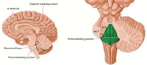
anatomy - Covered by cerebellum and attached via three cerebellar peduncles\n- Remove cerebellum by slicing across the peduncles to expose the 4th ventricle and rhomboid fossa\n
-
Do pontocerebellar fibers pass through the tegmentum to reach the cerebellum?
No, they do NOT cut across the tegmentum; they run within the basis.
-
What is characteristic of the superior medullary velum?
- Very thin, fragile, adherent to the cerebellum; often torn when the cerebellum is removed
velum -
Why is the pontocerebellar tract not considered strictly ascending or descending?
Because it runs transversely within the basis to the cerebellum rather than up or down the brainstem.
-
What is the rhomboid fossa and what are its rostral and caudal limits?
- Floor of the 4th ventricle, rhomboidal in shape\n- Rostral limit: exit of CN IV at ponto-midbrain junction dorsally\n- Caudal limit: stria medullaris of the 4th ventricle
rhomboidfossa -
Which tracts are listed in the basis of the midbrain?
- Corticospinal tract
- Corticobulbar tract
- Corticopontine tract
-
How is the pontomedullary junction identified on the rhomboid fossa?
- The rhomboid fossa is bisected along the plane of the tallest point of the 4th ventricle marking the pontomedullary junction
pontomedullary -
Which tracts are listed in the basis of the pons?
- Corticospinal tract
- Corticobulbar tract
- Corticopontine tract
- Pontocerebellar tract
-
Along which plane does the pontomedullary junction lie dorsally?
- Along the tallest point of the 4th ventricle
- At the widest diameter of the 4th ventricle
pons ventricle -
-
What is the locus coeruleus and what does it contain?
- Deeply pigmented group of cells
- Produces norepinephrine (a catecholamine)
- Contains neuromelanin
locus neurochemistry -
Why is the medullary basis called the 'medullary pyramids'?
Because it contains the corticospinal fibers arranged as prominent pyramidal bundles.
-
What is the substantia ferruginea and where is it located?
- Part of the sulcus limitans that overlies the locus coeruleus
- Produces a bluish-gray discoloration rostral to the median sulcus
substantia ferruginea -
What surface of the pons is referred to as the 'basis'?
The ventral surface of the pons is called the basis.
-
How can the substantia ferruginea be exposed anatomically?
- Follow the sulcus limitans rostrally towards the midbrain
- Remove part of the superior cerebellar peduncle covering it
dissection landmarks -
What is the anatomical relation of corticobulbar fibers in the brainstem?
They descend in the basis and terminate in motor cranial nerve nuclei located in the tegmentum.
-
What are the roles of the stria medullaris of the 4th ventricle?
- Transversely crossing fibers
- Mark the pontomedullary junction dorsally and the posterior border between pons and medulla
- Indicate the caudal limit of the pons dorsally and rostral limit of the open medulla of the 4th ventricle
stria junction -
Which cranial nerve motor nuclei are explicitly mentioned as corticobulbar targets?
Facial motor nucleus (CN VII) and hypoglossal nucleus (CN XII).
-
What does the median sulcus do in the rhomboid fossa?
- Divides the rhomboid fossa into two symmetrical halves
median rhomboid -
What role do pontine nuclei play in cortico-cerebellar communication?
They relay cortical information to the cerebellum via the pontocerebellar tract.
-
Where is the sulcus limitans located and what is its significance?
- Runs lateral and parallel to the median sulcus (paired)
- Its rostral extension becomes discolored as the substantia ferruginea
sulcus brainstem -
Do corticopontine fibers reach the medulla?
No, corticopontine fibers terminate in pontine nuclei and do not go to the medulla.
-
How is the medial sulcus formed?
- By the posterior surface of the pons
- And the cranial half of the medulla oblongata
medial formation -
What is the primary functional division between lateral and ventral corticospinal systems?
Lateral corticospinal system controls distal limb movements; ventral corticospinal influences axial/trunk muscles.
-
Name key landmarks visible on the posterior view of the 4th ventricle.
- Median sulcus
- Sulcus limitans
- Stria medullaris
- Medial eminence
- Facial colliculus
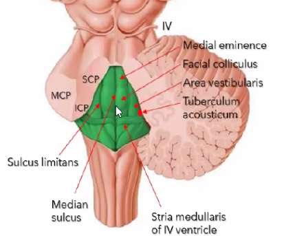 posterior landmarks
 -
Where is the pyramidal decussation located?
In the caudal medulla where most corticospinal fibers cross to the opposite side.
-
Where are the vagal and hypoglossal triangles located?
- On the floor of the 4th ventricle posteriorly, lateral to the medial eminence
vagal hypoglossal -
How do corticospinal fibers travel through the brainstem basis layers?
They descend in the basis of midbrain, basis of pons, and basis of medulla as compact bundles.
-
What causes the dark pigment in catecholamine-producing cells like the locus coeruleus?
- Accumulation of neuromelanin in catecholamine-producing cells
pigment neuromelanin -
What distinguishes basis fibers from tegmental structures in the brainstem?
Basis mainly contains descending/transverse fiber bundles; tegmentum contains nuclei and ascending/descending projections to them.
-
Close-up: which structures are shown around the substantia ferruginea in the posterior 4th ventricle?
- Substantia ferruginea
- Median sulcus
- Sulcus limitans
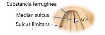 closeup substantia
 -
Why are transverse pontine fibers clinically important?
They relay cortical information to the cerebellum and are essential for coordinated motor planning and timing.
-
anatomy pons
-
What is one functional consequence of lesions in the pontine basis affecting pontocerebellar fibers?
Disrupted communication to the cerebellum leading to ataxia or coordination deficits.
-
Which bulge is present in the upper (rostral) part of the dorsal pons?
- Medial eminence (only enlargement in upper pons).
pons medial -
-
What are the prominences in the lower dorsal pons from medial to lateral?
- Facial colliculus
- Area vestibularis
- Tuberculum acousticum
pons structures -
-
Where are the prominences more prominent on the pons?
They are more prominent caudally (on the lower half of the pons).
pons location -
-
What causes the bulge of the medial eminence internally?
Bulging is formed by the abducens nucleus and the facial genu in the dorsal pons.
nuclei pons -
-
Where does the medial eminence lie relative to the median sulcus and sulcus limitans?
It lies between the median sulcus and the sulcus limitans and is medial to the sulcus limitans.
pons sulci -
-
Where is the area vestibularis located relative to the sulcus limitans and facial colliculus?
The area vestibularis is lateral to the sulcus limitans and the facial colliculus.
vestibular pons -
-
What are the roof, lateral and floor boundaries near these dorsal pons prominences?
- Roof: Superior medullary velum
- Lateral: Cut sections of superior and middle cerebellar peduncles
- Floor: Rhomboid fossa (divided into two planes)
boundaries pons -
-
What are the transverse fibers coursing on the dorsal pons called?
The transverse fibers are the stria medullaris.
fibers pons -
-
Where is the tuberculum acousticum located?
- Lateral to the area vestibularis
- Small bulge at the extreme lateral end of the rhomboid fossa (lower half of pons)
pons tuberculum -
-
How does the prominence of the tuberculum acousticum compare to the facial colliculus?
- Less prominent than the facial colliculus
pons comparison -
Where do the vertebral arteries course prior to forming the basilar artery?
- On the ventral surface of the medulla
-
cranialnerves pons
-
At which anatomical level do the vertebral arteries fuse to form the basilar artery?
- At the pontomedullary junction (near inferior pontine sulcus)
-
Which cranial nerves are contained in the lower half of the pons?
- CN VI (abducens)
- CN VII (facial)
- CN VIII (vestibulocochlear)
cranialnerves pons -
-
pons levels
-
What is the main connection function of the middle cerebellar peduncle (MCP)?
- Connects the pons to the cerebellum laterally
-
pons levels
-
What is the general role of the sulci of the pons?
- They mark junctions that demarcate the pons from rostral and caudal structures
-
How does the fourth ventricle differ between upper and lower pons?
- Upper half: narrower fourth ventricle
- Lower half: wider fourth ventricle
ventricle pons -
Which cranial nerves are commonly labeled at the lateral base of the pons in anatomical views?
- V
- VI
- VII
- VIII
-
In cross-sectional orientation, which surface faces up in anatomical sections?
- Dorsal surface faces up (anatomical orientation)
orientation anatomy -
-
In cross-sectional orientation, which surface faces down in clinical sections?
- Ventral surfaces face down (clinical orientation; opposite to anatomical)
orientation clinical -
What ventral feature indicates the midline position of the basilar artery on the pons?
- The basilar sulcus lodges the basilar artery at midline
-
Name the main cross-sectional regions of the pons.
- Pontine basis
- Pontine tegmentum
- Superior medullary velum (tectum)
- Basilar sulcus
structures pons -
What anatomical view highlights the pons and medulla beneath the forebrain?
- Anterior-inferior view of the brainstem
-
Which cranial nerve uniquely exits from the dorsal surface of the brainstem?
- CN IV (trochlear nerve)
cranialnerves pons -
Why is the inferior pontine sulcus clinically important regarding arterial anatomy?
- It marks where vertebral arteries fuse to form the basilar artery ventrally
-
limits pons
-
Name a visible landmark that helps identify the rostral and caudal borders of the pons on the ventral surface.
- Superior pontine sulcus (rostral)
- Inferior pontine sulcus (caudal)
-
limits pons
-
Which arteries form the basilar artery and where are they located before fusion?
- Two vertebral arteries located inferiorly on the ventral medulla
-
In anatomical orientation when viewing slides, which direction is dorsal?
- Dorsal is above
- Ventral is below
orientation anatomy -
Where does the basilar artery run on the pons?
- On the ventral surface of the pons along the basilar sulcus
-
When viewing MRI or CT scans of a patient in the supine position, which surface faces down and which faces up?
- Dorsal surface faces down
- Ventral surfaces face up
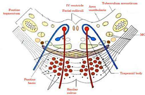 imaging orientation
 -
-
If ventral surfaces face down (anatomical orientation for slides), what structures are found above and below the pons?
- Above: superior medullary velum and IV ventricle
- Below: basilar sulcus
pons orientation -
Where does the basilar artery terminate ventrally?
- At the superior pontine sulcus (ponto-midbrain junction) forming two posterior cerebral arteries
-
If the patient is supine for MRI/CT (dorsal faces down), what structures are placed above and below the pons?
- Above: basilar sulcus
- Below: superior medullary velum and IV ventricle
imaging pons -
-
What are the three prominent bulgings on the lower half of the floor of the 4th ventricle?
- Facial colliculus — medial, motor
- Area vestibularis — lateral, sensory
- Tuberculum acousticum — most lateral, sensory
4thventricle facialcolliculus -
What structure does the basilar sulcus relate to?
- Groove on the ventral pons that houses the basilar artery
-
What are key features of the IV ventricle at the level of the lower pons?
- Open cavity and relatively wider
- Located dorsally; floor is the rhomboid fossa
- Superior medullary velum closely opposed
ventricle anatomy -
-
What defines the pontine tegmentum in the lower pons?
- Most dorsal part of the pons
- Forms the floor of the 4th ventricle
- Contains tegmental nuclei and ascending/descending tracts
tegmentum pons -
Where is the Middle Cerebellar Peduncle located relative to the pons?
- Posterolateral to the ventral surface of the pons
-
Where is the pontine basis located and what is its composition?
- Ventral to the tegmentum at the boundary of tegmentum and basis
- Made of transversely coursing longitudinal fibers (most ventral part)
basis pons -
-
Which major descending motor tracts and related structures are contained in the pontine basis?
- Pyramidal tract
- Corticospinal tract
- Corticobulbar (corticonuclear) tract
- Corticopontine tract
- Pontine nuclei and pontocerebellar tract
tracts motor -
-
What is the orientation and function of the facial colliculus on the 4th ventricle floor?
- Medial bulging on the lower half of the 4th ventricle floor
- Primarily motor in function
facialcolliculus 4thventricle -
Where does the trigeminal nerve (V) emerge on the pons?
- At the junction of the pons proper with the MCP, posterolateral to the pons proper
-
Name common sensory nuclei and roots found lateral to the pontine tegmentum in the lower pons.
- Dorsal & ventral cochlear nuclei
- Vestibular nuclei
- Cochlear root and vestibular root of VIII nerve
sensory nuclei -
What roots does the trigeminal nerve have?
- Large sensory root (portio major)
- Small motor root (portio minor)
-
pons anatomy
-
What is the main function of the trigeminal sensory root?
- Major source of sensory innervation to the face
-
What is the basilar sulcus relation to pontine structures?
- Lies on the ventral surface of the basis
- Continuous posteromedially with the middle cerebellar peduncles
pons basilar -
-
How many middle cerebellar peduncles are there and where are they located?
- One on each side
- Continuous posteromedially to the basilar sulcus
mcp cerebellum -
Which nerves emerge along the inferior pontine sulcus/pontomedullary junction ventrally?
- Abducens nerve (VI)
- Facial nerve (VII)
- Vestibulocochlear nerve (VIII)
-
Where do corticopontine fibers terminate?
- Terminate at the pontine nuclei scattered in the pontine basis
pontine pathways -
-
Where does the corticospinal tract originate?
- Originates from the cerebral motor cortex and descends as the corticospinal tract
corticospinal motor -
What roots compose the facial nerve (VII)?
- Large motor root
- Smaller root called nervus intermedius (intermediate nerve or nerve of Wrisberg)
-
What is the effect of a corticospinal lesion above the decussation?
- Contralateral hemiparesis (weakness of the opposite side of the body)
lesion motor -
What is the main function of the facial nerve large motor root?
- Major source of motor innervation to muscles of facial expression
-
What percent of corticospinal fibers cross at the pyramidal decussation?
- 90% cross at the pyramidal decussation
- 10% remain uncrossed
decussation corticospinal -
-
What are the two corticospinal divisions after the caudal medulla and their general targets?
- Lateral corticospinal tract: after crossing, in the lateral funiculus; synapses to lateral motor neurons → supplies limbs
- Ventral corticospinal tract: uncrossed/bilateral; synapses to medial motor neurons → supplies trunk
tracts motor -
What fiber types are contained in the nervus intermedius?
- Parasympathetic preganglionic efferent (visceromotor)
- Sensory fibers
-
Describe the cortico-ponto-cerebellar pathway from the right cerebral hemisphere.
- Right cerebral hemisphere → Right corticopontine tract → Pontine nuclei → Pontocerebellar fibers decussate → Left middle cerebellar peduncle → Left cerebellar hemisphere
corticopontine cerebellum -
Is it correct to call the facial smaller root purely sensory?
- No. The smaller root is the nervus intermedius and contains parasympathetic and sensory fibers, not purely sensory
-
pons tracts
-
-
What tract connects the pons and the cerebellum and where does it decussate?
- Pontocerebellar tract
- Decussates in the basis of the pons
pons decussation -
-
How do pontine nuclei axons reach the opposite cerebellar hemisphere?
- Axons decussate transversely in the pons
- Enter the opposite MCP and go to the opposite cerebellar hemisphere
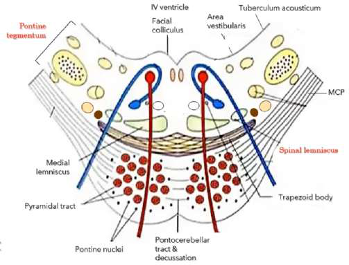 pontine mcp
 -
According to the Law of Lamination, which vestibulocochlear root is more medial and primitive?
- Vestibular root
-
What is the primary role of the cerebellum in movement?
- Coordinates movements
- Receives motor plan from cortex
- Makes movements smooth and precise
cerebellum motor -
-
Which main descending tracts pass through the pons basis?
- Corticospinal tract
- Pyramidal tracts
- Corticobulbar tract
- Cortico-ponto-cerebellar pathway
- Corticopontine tract
tracts basis -
What are the three cerebellar peduncles prominent on the lateral surface of the pons?
- Superior cerebellar peduncle (SCP)
- Middle cerebellar peduncle (MCP)
- Inferior cerebellar peduncle (ICP)
-
What does the pontine tegmentum (lower half) contain?
- A mix of nuclei and fiber tracts
- Includes facial colliculus, vestibular area, pontine nuclei, medial lemniscus, trapezoid body
tegmentum pons -
-
What is the trapezoid body and where is it located?
- Most ventral structure in pontine tegmentum
- Contains transverse auditory fibers
- Lies at the junction of tegmentum and basis
trapezoid auditory -
-
What sensory modalities travel in the medial lemniscus?
- Conscious proprioception
- Vibration sense
- Discriminative (fine) touch
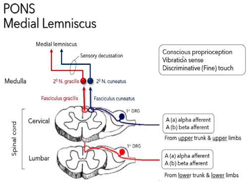 mediallemniscus sensory
 -
-
Where are the second-order neurons for the medial lemniscus located?
- Nucleus cuneatus and nucleus gracilis in the medulla
nuclei medulla -
What are the three pairs of cerebellar peduncles?
- Superior cerebellar peduncle (SCP)
- Middle cerebellar peduncle (MCP)
- Inferior cerebellar peduncle (ICP)
-
Where does the medial lemniscus first form and what is its relation to the trapezoid body?
- First forms in the medulla
- Located dorsal to the trapezoid body close to the midline
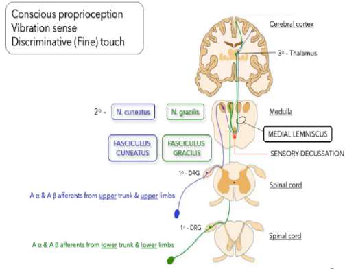 mediallemniscus location
 -
Anatomically, what is the role of the cerebellar peduncles?
- Attach cerebellum to dorsal surface of brainstem
- Only anchors of the cerebellum to the nervous system
-
Through which cerebellar peduncle do pontocerebellar fibers enter the cerebellum?
- Middle cerebellar peduncle (MCP)
mcp cerebellum -
What would happen if you sliced across the cerebellar peduncles?
- The cerebellum would fall off because the peduncles are its only anchors
-
pons lesion sensory
-
Define cerebellar afferents.
- Fibers going toward the cerebellum
- Input from other parts of the nervous system to the cerebellum
-
Why is dorsal column sensation ipsilateral in the spinal cord?
- Because the dorsal column fibers have not yet decussated
dorsalcolumn decussation -
Define cerebellar efferents.
- Fibers leaving the cerebellum
- Output from the cerebellum to other parts of the nervous system
-
After the medullary sensory decussation, the medial lemniscus conveys sensation to which side?
- Contralateral side
medulla mediallemniscus -
What is the primary fiber direction of the SCP?
- Primarily cerebellar efferents
- Contains only a minute amount of afferents
-
How does the medial lemniscus appearance change between medulla and pons?
- Two flattened elliptical discs
- Long anteroposterior diameter in medulla
- Compressed in pons due to surrounding structures
mediallemniscus anatomy -
Which spinocerebellar tract enters the cerebellum via the SCP?
- Ventral spinocerebellar tract (small group of afferents entering via SCP)
-
Which dorsal column fibers enter the spinal cord first?
- Fibers from lower trunk and lower limbs (lumbar and sacral) enter first
dorsalcolumn gracilis cuneatus -
Which spinocerebellar tract enters the cerebellum via the ICP?
- Dorsal spinocerebellar tract (enters via ICP)
-
How is the dorsal column divided at cervical and upper thoracic levels?
- Fasciculus cuneatus: upper trunk and limbs (lateral)
- Fasciculus gracilis: lower trunk and limbs (medial)
cuneatus gracilis dorsalcolumn -
-
Where do dorsal column fibers terminate and where do they decussate?
- Terminate in their sensory nuclei in the medulla
- Axons cross in the sensory decussation and ascend as medial lemniscus
decussation medulla mediallemniscus -
What is the main functional content of the MCP?
- Purely cerebellar afferents
- Mainly the pontocerebellar tract from the pons
-
spinallemniscus spinothalamic
-
-
What modalities does the spinal lemniscus convey?
- Pain and temperature (lateral tract)
- Crude (non-discriminative) touch (ventral tract)
- All from the contralateral side
spinallemniscus sensory pain -
Describe the path of the pontocerebellar tract.
- Runs transversely across the basis of the pons
- Enters the cerebellum via the MCP
-
Why do the spinothalamic tracts convey contralateral sensation?
- Because their axons decussate at the ventral white commissure in the spinal cord before ascending
decussation spinothalamic spinalcord -
-
Where is the medial longitudinal fasciculus (MLF) located and what is its role?
- Most dorsal medial aspect, near floor of 4th ventricle
- Mediates horizontal conjugate eye movements/gaze
mlf ocular brainstem -
-
Where is the inferior cerebellar peduncle (ICP) located in the pons?
- Lateral aspect of pontine tegmentum; contains restiform and juxtarestiform bodies
icp cerebellarpeduncle pons -
What does the ICP primarily contain?
- Primarily cerebellar afferents from spinal cord and medulla
- Also contains a small group of cerebellar efferents
-
Where is the central tegmental tract (CTT) located relative to the medial lemniscus?
- Dorsal to the medial lemniscus
ctt tegmentum -
Which nucleus mediates nonconscious proprioception feeding the dorsal spinocerebellar tract?
- Dorsal nucleus of Clarke
-
What modalities are carried by the descending nucleus and tract of CN V?
- Pain
- Temperature
- Crude touch of the face
trigeminal sensory -
What is the role of the accessory (lateral) cuneate nucleus in the medulla?
- Sends fibers to the cerebellum as the cuneocerebellar tract
- These fibers enter via the ICP
-
trigeminal anatomy
-
Where does the cuneocerebellar tract enter the cerebellum?
- Via the inferior cerebellar peduncle (ICP)
-
Which muscle is innervated by CN VI (abducens nerve) and what is its action?
- Lateral rectus (LR)
- Action: deviates the eyeball laterally
- Innervation type: GSE
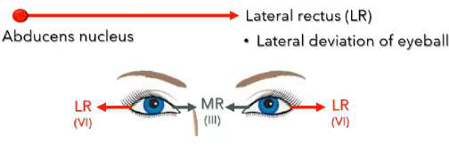 abducens muscles
 -
-
Where does the abducens nucleus lie in relation to the facial colliculus?
- Lies beneath the facial colliculus (dorsal location)
- Nucleus is dorsal but the nerve courses ventrally
abducens anatomy -
-
How does the abducens nerve exit the pons?
- Courses ventrally and medially
- Emerges at ventromedial aspect of pons along the inferior pontine sulcus or pontomedullary junction
abducens anatomy -
Summarize the overall function of the three cerebellar peduncles.
- Contain fiber tracts that connect the cerebellum with other parts of the nervous system
- Provide afferent and efferent routes
-
cranialnerves abducens
-
Which peduncle anatomically and functionally connects the pons with the cerebellum?
- Middle cerebellar peduncle (MCP) via pontocerebellar fibers
-
What is the clinical sign of a left abducens nerve lesion?
- Left eye is medially deviated due to unopposed action of the left medial rectus (antagonist)
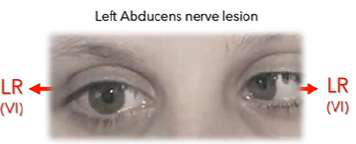 clinical abducens
 -
Name a small group of cerebellar afferents that come from the spinal cord and enter via the SCP.
- Ventral spinocerebellar tract
-
What two structures make the facial colliculus prominent?
- Abducens nucleus
- Genu of the facial motor root
facial anatomy -
Does the ICP contain any cerebellar efferents?
- Yes, a small group of cerebellar efferents are present in the ICP
-
Describe the course of the facial motor root relative to the abducens nucleus.
- Runs dorsally and medially toward the floor of the 4th ventricle
- Ascends medial to the abducens and turns sharply over the abducens nucleus forming the facial 'genu'
facial course -
What is the basis of the pons important component mentioned in the text?
- The pontocerebellar tract arising from pontine nuclei
-
Where is the facial motor nucleus located in the pontine tegmentum relative to the abducens nuclei?
- Located lateral to the abducens nuclei
- Not beneath the facial colliculus
facial nucleus -
What is the function of the Inferior Cerebellar Peduncle (ICP)?
Mostly connects the spinal cord and medulla with the cerebellum (cerebellar afferent); also carries a small amount of cerebellar efferent from vestibular nuclei.
-
What is the facial genu in the pons?
- Bend where the facial motor root turns around the abducens nucleus
- Lies just beneath the facial colliculus
- Emerges ventrolaterally at the inferior pontine sulcus
anatomy pons -
Which cranial nerve emerges at the junction of the ventral pons and the middle cerebellar peduncle?
CN V — the Trigeminal nerve
-
What is the modality and main targets of the facial motor nucleus?
- SVE (special visceral efferent)
- Innervates most facial muscles (except extraocular and mastication)
- Derived from branchiomeric muscles
physiology facial -
Which cranial nerves emerge along the inferior pontine sulcus at the pontomedullary junction?
CN VI, CN VII, CN VIII
-
Which muscles/nerves close and open the eyelids?
- Orbicularis oculi closes the eyelids — innervated by CN VII
- Levator palpebrae opens/raises the eyelids — innervated by CN III
muscles cranialnerves -
-
laterality facial
-
Where do the Facial (CN VII) and Vestibulocochlear (CN VIII) nerves emerge?
Posterolaterally from the cerebello-pontine angle
-
What are the main signs of a peripheral (Bell's) facial nerve lesion?
- Ipsilateral facial sagging
- Flattening of wrinkle lines
- Inability to close the eye (risk of dry cornea)
- Shallow nasolabial fold and drooping angle of mouth
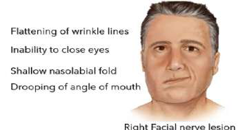 clinical bellspalsy
 -
What is the cerebello-pontine angle?
An angular space between the medulla, pons, and adjoining cerebellum posterior to the pons; site where CN VII and CN VIII emerge
-
How do corticobulbar projections innervate the facial nucleus for upper vs lower face?
- Upper face: bilateral corticobulbar projections
- Lower face: contralateral corticobulbar projections
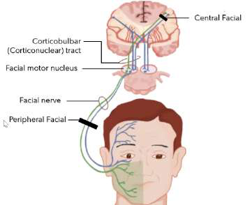 pathways corticobulbar
 -
What covers the dorsal surface of the pons?
The cerebellum, attached via the three pairs of cerebellar peduncles (SCP, MCP, ICP)
-
What facial deficit results from a unilateral central (corticobulbar) lesion?
- Paralysis of the contralateral lower quarter of the face; the upper face is spared due to bilateral input
clinical stroke -
How must the cerebellum be removed to view the dorsal surface of the pons?
Slice across the three cerebellar peduncles to detach and remove the cerebellum
-
Give an example of a facial muscle and its function mentioned in the text.
- Orbicularis oculi — closes the eyelids (innervated by CN VII)
muscles examples -
What structures are exposed after removing the cerebellum?
The fourth ventricle and its floor, the rhomboid fossa
-
Which parasympathetic nuclei are associated with CN VII?
- Superior salivatory nucleus
- Lacrimal nucleus
parasympathetic nuclei -
Describe the superior medullary velum.
Very thin and fragile; dorsal part is adherent to the cerebellum and often tears when the cerebellum is removed
-
What is the 'nervus intermedius' (intermediate nerve)?
- Smaller root of the facial nerve (CN VII)
- Also called the intermediate nerve of Wrisberg
neuroanatomy cn7 -
Why is the superior medullary velum often not seen in dissections?
It usually gets torn or destroyed when the cerebellum is removed, so it is not preserved on many specimens
-
Where is the intermediate nerve located relative to the facial motor nucleus?
- Lies dorsolateral to the facial motor nucleus and lateral branchiomeric nucleus
anatomy pons -
What is the rhomboid fossa?
The rhomboidal floor of the fourth ventricle, revealed after removal of the cerebellum
-
What efferent modality does the intermediate nerve carry?
- GVE (general visceral efferent): parasympathetic preganglionic efferents
physiology parasympathetic -
How is the rhomboid fossa bounded laterally?
By the cut sections of the three pairs of cerebellar peduncles
-
Which nuclei provide the parasympathetic fibers in the intermediate nerve?
- Superior salivary nucleus
- Lacrimal nucleus
nuclei salivation -
What is the rostral limit of the rhomboid fossa?
The exit of CN IV at the ponto-midbrain junction dorsally
-
What are the parasympathetic targets and functions of the intermediate nerve?
- Sphenopalatine ganglion → lacrimal gland (lacrimation)
- Submandibular ganglion → submandibular & sublingual glands (salivation)
glands autonomic -
-
ganglion sensory
-
What feature bisects the rhomboid fossa marking the pontomedullary junction?
The plane at the tallest point of the fourth ventricle bisects the rhomboid fossa
-
Which taste region is carried by the intermediate nerve?
- SVA: taste from the anterior 2/3 of the tongue
taste sva -
-
Where do taste fibers from the intermediate nerve terminate centrally?
- Rostral portion of the Nucleus of Tractus Solitarius (gustatory portion) in the medulla
nts medulla -
What fibers does the Superior Cerebellar Peduncle (SCP) mainly carry?
Mostly cerebellar efferent fibers, with a small cerebellar afferent contribution (ventral spinocerebellar tract)
-
What GVA sensations does the intermediate nerve carry?
- Viscerosensory from nasal cavity, soft palate, and sinus cavity (synapse at caudal Nucleus of Tractus Solitarius)
gva visceroception -
-
What GSA fibers are carried by the intermediate nerve?
- Cutaneous sensation from the external ear and external auditory canal (via geniculate ganglion)
gsa cutaneous -
What fibers does the Inferior Cerebellar Peduncle (ICP) carry?
Mostly cerebellar afferent from spinal cord and medulla; small cerebellar efferent from vestibular nuclei
-
After the geniculate ganglion, which two central pathways do sensory fibers take?
- Nucleus of Tractus Solitarius: rostral = taste, caudal = general visceral sensation
- Descending nucleus of Trigeminal V: somatic sensation from external ear
pathways trigeminal -
-
Which other cranial nerves contribute to taste sensation alongside CN VII?
- CN IX (glossopharyngeal)
- CN X (vagus)
cranialnerves taste -
Which cranial nerves are considered the lateral cranial nerves of the pons?
CN V, CN VI, CN VII, CN VIII
-
Pons cross-section: which structures are near the facial colliculus and facial motor nucleus?
- The diagram shows the facial colliculus, facial motor nucleus, medial lemniscus, pyramidal tract and nearby pontine nuclei
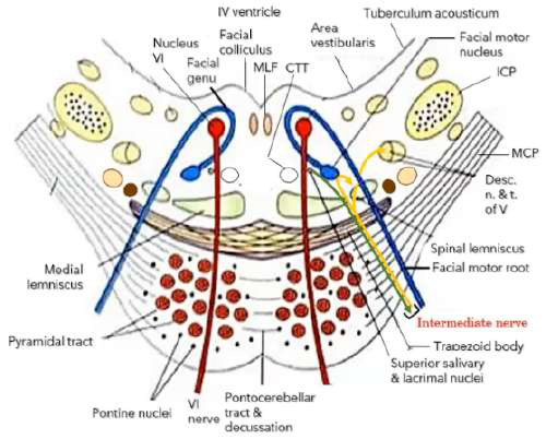
imaging pons - The diagram shows the facial colliculus, facial motor nucleus, medial lemniscus, pyramidal tract and nearby pontine nuclei
-
Why must you slice across all three cerebellar peduncles to remove the cerebellum?
Because the cerebellum is attached to the dorsal pons by the three peduncles (SCP, MCP, ICP), so cutting them frees the cerebellum
-
What is the visceral afferent center in the brainstem?
- Nucleus of Tractus Solitarius
- Convergence site for similar sensory modalities from different cranial nerves
brainstem nucleus -
What does the pontomedullary junction divide the 4th ventricle and rhomboid fossa into?
- Upper half: pons
- Lower half: open medulla
-
Which nerve is CN VIII and what are its two roots?
- Vestibulocochlear nerve (CN VIII)
- Vestibular root
- Cochlear root
cnviii anatomy -
Dorsally, along which plane does the pontomedullary junction lie?
- The tallest point of the 4th ventricle
- The widest diameter of the 4th ventricle
-
What sensory modality does the vestibular root mediate?
- SSA (Special Somatic Afferent)
- Mediates sense of equilibrium and balance
vestibular modality -
Which transversely crossing fibers mark the pontomedullary junction dorsally?
- Stria medullaris of the 4th ventricle
-
What are the peripheral receptors for the vestibular root?
- Semicircular canals
- Otolith organs: utricle & saccule
receptors vestibular -
What posterior border does the stria medullaris of the 4th ventricle mark?
- The posterior border between the pons and the medulla (at the widest part)
-
vestibular nuclei
-
What dorsal limits does the stria medullaris indicate?
- Caudal limit of the pons dorsally
- Rostral limit of the open medulla of the 4th ventricle
-
Where are the vestibular nuclei located relative to the cochlear nuclei?
- Medial to the cochlear nuclei
- Lie beneath the area vestibularis
location vestibular -
-
Which vestibular nuclei are found in the pons versus the medulla?
- In the pons: superior, medial and lateral
- In the medulla (area vestibularis): medial and inferior
vestibular location -
-
What is Scarpa's ganglion and what is unique about its neurons?
- Scarpa's ganglion contains vestibular sensory neurons
- Exceptionally made of bipolar neurons (not pseudounipolar)
ganglion scarpa -
-
What are the roles of the dorsal and ventral cochlear nuclei?
- Process auditory inputs
- Site of synapse for the cochlear root of CN VIII
cochlear auditory -
-
Where do the dorsal cochlear nuclei lie anatomically?
- Lateral to vestibular nuclei
- Dorsal cochlear nuclei lie beneath the tuberculum acousticum
cochlear location -
What is the substantia ferruginea in the rhomboid fossa?
- Part of the sulcus limitans that overlies the locus coeruleus
- Accounts for the bluish-gray discoloration rostral to the median sulcus
-
Which central connection of vestibular nuclei coordinates eye and head movement?
- Cranial nerve nuclei (EOMs)
- Supply extraocular muscles to coordinate eye and head movement
connections eye -
How can the substantia ferruginea be exposed during dissection?
- Follow the sulcus limitans rostrally toward the midbrain and remove part of the superior cerebellar peduncle covering it
-
Which vestibular connection maintains upright posture and balance?
- Spinal cord via vestibulospinal tract
- Maintains posture and balance
connections posture -
What forms the floor of the 4th ventricle (rhomboid fossa)?
- Posterior surface of the pons
- Cranial half of the medulla oblongata
-
How does the cerebellum connect to the vestibular nuclei and what is its role?
- Via the inferior cerebellar peduncle (ICP)
- Regulates movements underlying posture and equilibrium
connections cerebellum -
-
What is the role of the cerebral motor cortex in vestibular function?
- Provides perception of spatial orientation
- Contributes to conscious awareness of balance and position
connections cortex -
What sulcus runs lateral and parallel to the median sulcus?
- Sulcus limitans (paired, one on each side)
-
What type of neurons make up the spiral ganglion in the auditory pathway?
- Bipolar neurons (exception to sensory pseudounipolar)
auditory neuroanatomy -
-
What is the first cell station of the auditory pathway?
- Spiral ganglion (cell bodies of cochlear nerve)
auditory pathway -
What is the medial eminence?
- A raised area on the floor of the 4th ventricle located between the median sulcus and the sulcus limitans
-
Which nerve root carries auditory impulses from the organs of Corti to the brainstem?
- Cochlear root of CN VIII (with cell bodies in spiral ganglion)
auditory cranialnerves -
-
Upon entering the pons, where do cochlear root fibers first synapse?
- Dorsal and ventral cochlear nuclei
pons nuclei -
What are the vagal and hypoglossal triangles?
- Regions on the posterior surface of the medulla representing the surface areas of vagal and hypoglossal nuclei
-
Which nucleus in the pons serves as an early relay nucleus in the auditory pathway?
- Superior olivary nucleus (relay nucleus in pons)
pons relay -
What does SCP, MCP, and ICP stand for?
- SCP: Superior cerebellar peduncle
- MCP: Middle cerebellar peduncle
- ICP: Inferior cerebellar peduncle
-
auditory tracts
-
Name key landmarks visible on the posterior view of the 4th ventricle floor.
- Median sulcus
- Medial eminence
- Facial colliculus
- Sulcus limitans
- Stria medullaris
- Substantia ferruginea
-
Name an acoustic stria that contributes to the formation of the lateral lemniscus.
- Dorsal acoustic stria
auditory tracts -
Why is the substantia ferruginea visible in living and dead tissue?
- Because of neuromelanin in catecholaminergic cells (gives bluish-gray discoloration)
-
auditory tracts
-
What structure lies immediately lateral to the median sulcus?
- The medial eminence (followed laterally by the sulcus limitans)
-
Where is the trapezoid body located within the brainstem?
- Most ventral, at the demarcation of tegmentum and basis
anatomy pons -
Which structure should be specified when referring to the 'stria medullaris' in this region?
- Specify the stria medullaris of the 4th ventricle (other stria medullaris exist elsewhere)
-
What is the principal ascending auditory pathway tract that originates in the pons?
- Lateral lemniscus (ascends to midbrain and beyond)
pathway pons -
Where are the prominent bulges (enlargements) of the pons located?
On the rhomboid fossa of the pons (dorsal surface).
-
Give the main ascending relay sequence from cochlear nuclei to auditory cortex.
- Superior olive
- Lateral lemniscus
- Inferior colliculus
- Medial geniculate body
- Auditory receiving centers (cortex)
pathway sequence -
Which enlargement is present in the upper part of the pons?
The medial eminence is the only enlargement in the upper part of the pons.
-
Where are cochlear nuclei and the main auditory structures in the pons located?
- Pontine tegmentum (lower half of pons)
pons location -
Which three prominences appear in the lower part of the dorsal pons from medial to lateral?
- Facial colliculus
- Area vestibularis
- Tuberculum acousticum
-
anatomy nuclei
-
Are the dorsal prominences of the pons more prominent rostrally or caudally?
They are more prominent caudally, i.e., on the lower half of the pons.
-
What is the main projection of the dorsal cochlear nucleus?
Sends axons that cross via the dorsal acoustic stria and ascend contralaterally as part of the lateral lemniscus.
auditory cochlear dorsal -
What lies between the median sulcus and the sulcus limitans on each side?
The medial eminence lies between the median sulcus and the sulcus limitans.
-
How does the ventral cochlear nucleus send crossed fibers?
Axons cross via the intermediate and ventral acoustic striae and ascend contralaterally as part of the lateral lemniscus.
auditory cochlear ventral -
What is the median sulcus on the dorsal pons?
A midline longitudinal groove of the rhomboid fossa that helps mark medial structures.
-
What uncrossed projection arises from the ventral cochlear nucleus?
Axons synapse with the ipsilateral superior olivary nucleus and can ascend ipsilaterally in the lateral lemniscus.
auditory cochlear ventral -
Where is the facial colliculus located relative to the median sulcus and sulcus limitans?
It lies between the median sulcus and the sulcus limitans, medial to the sulcus limitans.
-
What fibers form the trapezoid body (ventral acoustic stria)?
Transversely coursing crossing auditory fibers from ventral cochlear nucleus and contralateral superior olivary nuclei at the ventral pons.
trapezoid acoustic crossing -
What internal structures form the bulge of the facial colliculus?
The facial colliculus is formed by the abducens nucleus and the genu (loop) of facial nerve fibers.
-
What is the main function of the lateral lemniscus?
Conveys bilateral auditory information (stronger contralateral input); contains both crossed and uncrossed auditory fibers.
lateral_lemniscus auditory -
What is the facial genu?
The genu is the internal loop of the facial nerve fibers that bend around the abducens nucleus.
-
Which nuclei provide the crossed auditory fibers in the lateral lemniscus?
- Contralateral dorsal cochlear nucleus
- Contralateral ventral cochlear nucleus
- Contralateral superior olivary nucleus
auditory crossed -
What is the abducens nucleus function relevant to the facial colliculus?
It contains motoneurons for the lateral rectus muscle, contributing to eye abduction.
-
Which nucleus provides the uncrossed auditory fibers in the lateral lemniscus?
- Ipsilateral superior olivary nucleus
auditory uncrossed -
Where is the area vestibularis located relative to the sulcus limitans?
The area vestibularis is lateral to the sulcus limitans and lateral to the facial colliculus.
-
What hearing deficit occurs with a central lesion affecting the lateral lemniscus?
- Bilateral partial deafness
- More severe on the contralateral side
clinical central_lesion hearing -
What is the tuberculum acousticum?
A lateral prominence on the dorsal pons associated with cochlear and vestibular nuclei.
-
Why does a central lesion cause bilateral partial deafness more on the contralateral side?
- Bilateral: lateral lemniscus carries crossed and uncrossed fibers
- Contralateral predominance: majority of fibers originate from the opposite side
- Partial: intact opposite lemniscus still transmits some bilateral input
clinical pathophysiology -
What forms the roof of the IV ventricle in the region of the dorsal pons?
The superior medullary velum forms the roof in this superior portion of the IV ventricle.
-
What is the effect of a peripheral lesion destroying the cochlear nerve?
- Ipsilateral complete deafness
- Cochlear nerve conveys only ipsilateral auditory information
clinical peripheral_lesion cochlear_nerve -
What structures form the lateral boundaries near the dorsal pons?
Cut sections of the superior cerebellar peduncles (SCP) and middle cerebellar peduncles (MCP) are lateral.
-
Where is the paramedian pontine reticular formation (PPRF) located relative to the abducens nucleus?
Lies ventrolateral to the abducens nucleus; lesions affect the abducens.
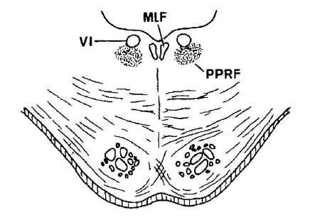 pprf pons vi
 -
What forms the floor in the region of the dorsal pons and IV ventricle?
The floor is the rhomboid fossa, which is divided into two planes at its widest diameter.
-
respiration medulla
-
What are the stria medullaris on the floor of the IV ventricle?
Transversely coursing fibers visible on the rhomboid fossa; they are called the stria medullaris.
-
pons respiration
-
Where is the stria medullaris of the IV ventricle relative to the facial colliculi?
The stria medullaris is caudal/rostral relation: the facial colliculi are rostral to the stria medullaris.
-
pons respiration
-
What is the sulcus limitans and its functional significance?
A longitudinal groove lateral to the medial eminence; it separates motor (medial) and sensory (lateral) nuclei in the brainstem.
-
What is the primary action of the apneustic center?
- Promotes and prolongs inspiration by stimulating the dorsal respiratory group
apneustic respiration -
Which prominence is noted as the only enlargement at the upper portion of the pons?
The medial eminence is the only enlargement at the upper portion of the pons.
-
What happens if the apneustic center is unchecked?
- Very long, deep inspirations (apneustic breathing)
apneustic pathology -
What marks the pontomedullary junction?
The pontomedullary junction marks the transition between the pons and the medulla oblongata.
-
Which structures inhibit the apneustic center?
- Pneumotaxic center (upper pons)
- Pulmonary stretch receptors via the vagus nerve
apneustic inhibition -
What anatomical landmarks are labeled on the dorsal pons dissection described in the lecture?
- Medial eminence
- Facial colliculus
- Stria medullaris
- Superior medullary velum
- SCP
- MCP
- Tuberculum acousticum
-
What is the effect of stretch-receptor impulses at maximal inspiration?
- They inhibit the inspiratory and apneustic centers, allowing exhalation
respiration vagus -
How do facial nerve fibers relate to the facial colliculus externally?
The looping (genu) of facial nerve fibers around the abducens nucleus produces the external bulge called the facial colliculus.
-
respiration physiology
-
Which cerebellar peduncle abbreviations appear on the dorsal pons diagrams?
- SCP: Superior cerebellar peduncle
- MCP: Middle cerebellar peduncle
-
Name the main respiratory control components in the brain stem.
- Pneumotaxic center
- Apneustic center
- Pre-Bötzinger complex
- Dorsal respiratory group
- Ventral respiratory group
- Medullary respiratory center
respiration brainstem -
What is the practical surgical relevance of recognizing dorsal pontine prominences?
They help localize underlying nuclei and fiber tracts during dorsal brainstem surgery and avoid damage to cranial nerve nuclei.
-
What pons function is related to horizontal eye movements?
- Coordinates horizontal conjugate eye movements (coordinating center near abducens nerve)
pons eye -
What is the tuberculum acousticum?
- A small lateral bulge on the floor of the 4th ventricle
- Located at the extreme lateral end of the rhomboid fossa in the lower pons
-
Which cranial nerves are associated with the pons region?
- V (trigeminal)
- VI (abducens)
- VII (facial)
- VIII (vestibulocochlear)
cranialnerves pons -
Where is the tuberculum acousticum located relative to the area vestibularis?
- Lateral to the area vestibularis
-
Name key nuclei related to the facial and lacrimation functions in the pons.
- Facial motor nucleus
- Superior salivatory nucleus
- Lacrimal nucleus
nuclei pons -
How does the tuberculum acousticum compare to the facial colliculus in prominence?
- The tuberculum acousticum is less prominent than the facial colliculus
-
List important ascending and descending tracts found in the upper pons.
- Pyramidal tract
- Medial lemniscus
- Lateral lemniscus
- Trigeminal lemniscus
- Spinal lemniscus
tracts pons -
What does area vestibularis refer to?
- The vestibular area on the lateral floor of the 4th ventricle associated with vestibular nuclei
-
Name cerebellar-related structures visible in the upper pons cross-section.
- Middle cerebellar peduncle
- Superior cerebellar peduncle
- Pontine nuclei
cerebellum pons -
-
Which pontine nuclei relay information to the cerebellum?
- Pontine nuclei (part of pontocerebellar tract)
pontine cerebellum -
-
Which pathways in the pons are involved in auditory and vestibular inputs?
- Dorsal and ventral cochlear nuclei
- Vestibular nuclei and roots
- Lateral lemniscus
auditory vestibular -
Which cranial nerves are present at the lower half level of the pons?
- CN VI, CN VII, CN VIII
- Also contains the facial colliculus
-
How does the IV ventricle in the upper pons compare to the lower half?
- Much narrower compared to the lower half
ivventricle pons anatomy -
What is the median sulcus in the floor of the 4th ventricle?
- A midline groove on the floor of the 4th ventricle separating left and right structures
-
ivventricle superiormedullaryvelum anatomy
-
What is the substantia ferruginea?
- A pigmented area on the floor of the 4th ventricle adjacent to the medial eminence
-
What forms the lateral walls of the IV ventricle at this level?
- Superior cerebellar peduncles (cut portion)
ivventricle superiorcerebellarpeduncle anatomy -
What creates the facial colliculus clinically?
- A dorsal bulge formed by the internal fibers of CN VII looping over the abducens nucleus (CN VI)
-
pontine tegmentum anatomy
-
How does the 4th ventricle width differ between pons halves?
- Upper half: narrower 4th ventricle
- Lower half: wider 4th ventricle
-
rhomboidfossa ivventricle anatomy
-
What do the abbreviations SCP and MCP stand for?
- SCP: Superior cerebellar peduncle
- MCP: Middle cerebellar peduncle
-
What prominent bulge is displayed on the rhomboid fossa?
- Medial eminence
- Facial colliculus located in the lower 1/2 medially
rhomboidfossa medialeminence anatomy -
What is the pontine basis?
- The ventral part of the pons containing corticospinal and corticobulbar fiber tracts
-
medialeminence rhomboidfossa anatomy
-
What is the pontine tegmentum?
- The dorsal pons region containing cranial nerve nuclei, ascending tracts, and reticular formation
-
Where is the trapezoid body located in the pons?
- Most ventral part at the junction of the tegmentum and basis
trapezoid pons anatomy -
What is the superior medullary velum?
- A thin sheet of white matter forming the roof (tectum) of the rostral 4th ventricle
-
Why is the trapezoid body still seen in the upper half of the pons?
- Because auditory fibers have a rostral convexity
trapezoid auditory anatomy -
What structure lies between the pons and medulla at the junction?
- The pontomedullary junction, where pons meets medulla and multiple cranial nerve roots emerge
-
trapezoid pons anatomy
-
Which cranial nerve is the only one to exit the brainstem dorsally?
- CN IV (trochlear nerve) is the only cranial nerve that exits the dorsal surface
-
basilarsulcus basis anatomy
-
What is the stria medullaris of the 4th ventricle?
- A transverse ridge on the floor of the 4th ventricle marking sensory fiber pathways
-
Where is the middle cerebellar peduncle located relative to the basis?
- Posterolaterally to the basis
middlecerebellarpeduncle basis anatomy -
List the main dorsal surface bulges of the pons mentioned in the lecture.
- Facial colliculus
- Tuberculum acousticum
- Area vestibularis
-
Which descending tracts appear as rounded fascicles in the basis?
- Corticospinal tract (pyramidal)
- Corticobulbar tract (corticonuclear)
pyramidal corticospinal anatomy -
Which major cerebellar connection is visible ventrolaterally on the pons?
- The MCP (middle cerebellar peduncle) connects the pons to the cerebellum
-
corticopontine pontinenuclei anatomy
-
Which major cerebellar connection runs from the cerebellum toward the midbrain above the pons?
- The SCP (superior cerebellar peduncle) connects cerebellum to the midbrain
-
How do pontocerebellar fibers reach the cerebellum?
- Course transversely and decussate to enter the opposite middle cerebellar peduncle (MCP)
pontocerebellar mcp anatomy -
What are the functional contents of the pontine basis?
- Descending motor fibers such as corticospinal and corticobulbar tracts
- Fibers that relay cortical input to cerebellum via MCP
-
What is the substantia ferruginea and what does it overlie?
- Bluish-gray discoloration on upper half of sulcus limitans
- Overlies the locus coeruleus (major source of norepinephrine in the CNS)
substantiaferruginea locuscoeruleus anatomy -
What types of structures does the pontine tegmentum contain?
- Cranial nerve nuclei, ascending sensory tracts, and reticular formation
-
Name key structures/components of the basis in the upper half of the pons.
- Corticopontine tract
- Pontine nuclei
- Basilar sulcus
- Pyramidal (corticospinal) tract
- Pontocerebellar tract
- Superior & middle cerebellar peduncles
- Trapezoid body
basis pons anatomy -
What is the clinical orientation used in cross-sections and how does it differ from anatomical orientation?
- Anatomical orientation: dorsal surface facing up
- Clinical orientation: opposite; ventral surfaces face down relative to imaging conventions
-
Which main structures are labeled in the pons diagram?
- Substantia ferruginea
- Superior medullary velum
- IV ventricle
- Medial eminence
- Superior cerebellar peduncle
- Middle cerebellar peduncle
- Pontine nuclei
- Basilar sulcus
- Trapezoid body
- Pyramidal tract
- Pontocerebellar tract
- Lateral lemniscus
- Trigeminal lemniscus
- Spinal lemniscus
- Medial lemniscus
- Locus coeruleus
anatomy pons -
In anatomical orientation for slides, where is 'dorsal' and where is 'ventral'?
- Dorsal is above
- Ventral is below
-
tracts pons
-
How is orientation different for MRI/CT images of supine patients?
- Dorsal surfaces face down
- Ventral surfaces face up
-
anatomy pons
-
In slide anatomical view of the pons, which structures are above and which are below?
- Above: superior medullary velum and IV ventricle
- Below: basilar sulcus
-
What does the medial lemniscus convey?
- Conscious proprioception
- Vibration
- Fine touch
- From the opposite side of the body
sensory lemniscus -
In MRI/CT supine patient view, which pons structures are above and which are below?
- Above: basilar sulcus
- Below: superior medullary velum and IV ventricle
-
anatomy landmarks
-
At what level is the 'lower half of the pons' described in this lesson?
- Level of the facial colliculus
-
What do the MLF and CTT share in the pons?
- They occupy the same positions in both halves of the pons
tracts pons -
Name three prominent bulgings in the lower half of the pontine tegmentum.
- Facial colliculus (medial, motor)
- Area vestibularis (lateral, sensory)
- Tuberculum acousticum (most lateral, sensory)
-
What does the spinal lemniscus convey?
- Pain
- Temperature
- Crude touch
- From the opposite side of the body
sensory spinothalamic -
Where is the IV (fourth) ventricle located relative to the pons?
- It forms the floor of the fourth ventricle at the most dorsal part of the pontine tegmentum
-
What is the trigeminal lemniscus and what does it convey?
- Made of trigeminothalamic fibers (sensory nucleus of V)
- Conveys various sensory modalities from the face
sensory trigeminal -
What is the rhomboid fossa in relation to the pons?
- The floor of the fourth ventricle over the dorsal pons (rhomboid fossa)
-
auditory lemniscus
-
What forms the pontine basis and where is it located?
- Made of transversely coursing fibers (pontocerebellar)
- Located most ventrally in the pons, at boundary of tegmentum and basis
-
Where is the locus coeruleus located?
- Dorsal part of the tegmentum
- Lies at the floor of the 4th ventricle
- Only seen in the upper half of the pons
nuclei locus -
Which major descending motor tracts run in the pontine basis?
- Pyramidal tract
- Corticospinal tract
- Corticobulbar (corticonuclear) tract
- Corticopontine tract
-
neurotransmitter locus
-
Which cerebellar-related tracts and nuclei are found in the pontine basis?
- Pontine nuclei
- Pontocerebellar tract
- Pontocerebellar decussation
-
What key functional roles does the locus coeruleus play?
- Emotion
- Arousal
- Alertness
- Participates in descending pain modulation
function locus -
What are the contents of the pontine tegmentum shown in the lower pons?
- Facial colliculi
- Vestibular nuclei
- Cochlear nuclei
- MLF, medial lemniscus
- Cranial nerve roots (VI, VII, VIII)
- Trapezoid body
-
What pigment and surface change are associated with the locus coeruleus?
- Contains deeply pigmented cells (neuromelanin)
- Overlying sulcus limitans appears bluish-gray (substantia ferruginea)
histology substantia -
What is the facial colliculus anatomically?
- A dorsal bulge on floor of the fourth ventricle formed by the genu of the facial nerve over the abducens nucleus
-
What is the dorsolateral pontine reticular formation and its role?
- Lies ventrolateral to the locus coeruleus
- Plays a role in regulation of REM sleep
- Found at the ponto-midbrain junction (pneumotaxic center)
reticular sleep -
Which nuclei form the tuberculum acousticum and area vestibularis?
- Tuberculum acousticum: cochlear (sensory) structures
- Area vestibularis: vestibular nuclei (sensory)
-
Which ascending tracts lie lateral to the medial lemniscus in the pons?
- Spinal lemniscus
- Trigeminal lemniscus
- Lateral lemniscus
tracts ascending -
What structures are labeled as cochlear and vestibular roots in the lower pons?
- Cochlear root: afferent fibers of VIII nerve to cochlear nuclei
- Vestibular root: afferent fibers to vestibular nuclei
-
respiratory pons anatomy
-
What is the trapezoid body and where is it located?
- A transverse auditory pathway in the ventral pons near the pontine basis and tegmentum junction
-
respiratory function
-
Which cranial nerve nuclei or roots are prominent in the lower pons section?
- VI nucleus/root
- VII nucleus/root (facial motor nucleus and nervus intermedius)
- VIII nerve roots (cochlear and vestibular)
-
How does the pneumotaxic center directly end inspiration?
- By inhibiting the dorsal respiratory group (inspiratory center)
respiratory mechanism -
What sensory ascending tracts are present in the pontine tegmentum?
- Medial lemniscus
- Spinal lemniscus
-
How does the pneumotaxic center limit inspiration via the apneustic center?
- It inhibits the apneustic center to prevent stimulation of the dorsal respiratory group
respiratory apneustic -
What are the superior salivatory nuclei and lacrimal nuclei associated with in the pons?
- Parasympathetic components of the facial nerve located near the facial motor nucleus
-
How does the pneumotaxic center affect respiratory rate when its signals change?
- Stronger signals → faster respiratory rate
- Lesser signals → lower respiratory rate
respiratory physiology -
What is the medial longitudinal fasciculus (MLF) role in the pons?
- A midline tract coordinating eye movements and connecting brainstem ocular motor nuclei
-
What is the respiratory effect of opioids on the pneumotaxic center?
- Opioids depress pneumotaxic signals → apneustic center not inhibited → respiratory rate decreases
respiratory clinical -
Describe the pontine cavity appearance in cross section at the dorsal surface.
- An open cavity (fourth ventricle) that is relatively wider in the pons dorsal surface
-
Which respiratory centers are located in the brainstem?
- Pneumotaxic center (pons)
- Apneustic center (pons)
- Pre-Bötzinger complex
- Dorsal respiratory group (medulla)
- Ventral respiratory group (medulla)
respiratory brainstem -
What neural elements demarcate the boundary between the pontine tegmentum and basis?
- The transition from tegmental structures to ventral transverse fibers of the pontine basis
-
What does the motor root (portio minor) of CN V innervate?
- Muscles of mastication (branchiomeric, SVE)
trigeminal motor -
Which motor nucleus is associated with the facial genu in the lower pons?
- The facial motor nucleus whose fibers loop as the facial genu forming the facial colliculus
-
Where does the motor root of CN V emerge?
- From the ventrolateral aspect of the pons, between ventral pons and middle cerebellar peduncle
trigeminal anatomy -
What is the relationship of the superior medullary velum to the dorsal pons?
- The superior medullary velum is closely opposed to the dorsal surface above the IV ventricle but often destroyed in dissection
-
What are signs of a unilateral lesion of the motor root of CN V?
- Weak mastication muscles
- Reduced bite force
- Jaw deviates toward the injured (ipsilateral) side
trigeminal clinical -
Which structure forms the basilar sulcus and where is it seen?
- The basilar sulcus is seen ventrally on the pontine basis and houses the basilar artery
-
Where is the motor root of V located within the pons?
- Lateral part of the pontine tegmentum in the upper half
trigeminal pons -
Name a compact list of key internal structures found in the lower half of the pons.
- IV ventricle
- Facial colliculi
- Area vestibularis
- Tuberculum acousticum
- Pontine tegmentum
- Pontine basis
-
Where do sensory afferents of CN V synapse and what is the ganglion type?
- Afferents synapse at sensory nuclei in the pons
- Cell bodies in Gasserian (semilunar) ganglion; pseudounipolar neurons
trigeminal sensory -
-
What does the ophthalmic division (V1) of CN V supply?
- Sensation of the upper 1/2 of the face including the forehead
- Pure sensory
trigeminal v1 -
Which tract carries cortical input from the pons to the cerebellum?
- Pontocerebellar tract (transverse fibers from pontine nuclei)
-
What does the maxillary division (V2) of CN V supply?
- Sensation of the middle 1/3 of the face
- Pure sensory
trigeminal v2 -
What fibers make up the most ventral part of the pontine basis?
- Transversely coursing longitudinal fibers (pontocerebellar/pontine transverse fibers)
-
What does the mandibular division (V3) of CN V supply?
- Sensation of the lower 1/3 of the face including the anterior 3 of the tongue
- Both sensory and motor (motor root travels with V3 to muscles of mastication)
trigeminal v3 -
Where is the tegmentum of the pons located?
- Ventral to the 4th ventricle
- Part of the pontine brainstem between the basis and the ventricular floor
-
Which sensory ganglia are exceptions to being pseudounipolar?
- Scarpa's and the spiral ganglion of the vestibular system
neuroanatomy ganglia -
What is the basilar sulcus on the pons?
- A midline groove on the ventral pons
- Harboring the basilar artery
-
Which nucleus of CN V conveys conscious proprioception, fine touch, and vibration from the face?
- Principal (main) nucleus of V
trigeminal sensory neuroanatomy -
What are the middle cerebellar peduncles (MCP)?
- Large lateral fiber bundles connecting pons to cerebellum
- Contain ponto-cerebellar fibers entering the cerebellum
-
Where is the principal nucleus of V located within the pons?
- Lateral to the motor nucleus of V
- Present only in the upper 1½ of the pons
pons anatomy trigeminal -
Where are the pontine nuclei located?
- Scattered in the pontine basis (ventral pons)
- Visible as grey dots within pontine basis
-
What peripheral receptors send input to the principal nucleus of V?
- Muscle spindles
- Golgi tendon organs (GTOs)
- Cutaneous mechanoreceptors
receptors sensory trigeminal -
What is the role of the pontine nuclei?
- Relay cortical motor information to the cerebellum via ponto-cerebellar fibers
- Key node in corticopontocerebellar pathway
-
Where are the sensory cell bodies located for fibers entering the principal nucleus of V?
- Gasserian (trigeminal) ganglion
- Pseudounipolar neurons
gasserian ganglion neurons -
Outline the corticopontine tract pathway
- Cerebral cortex
- Descends as corticopontine tract
- Terminates in pontine nuclei (pontine basis)
-
Which nucleus of V is considered the homologue of the nucleus cuneatus and nucleus gracilis?
- Principal (main) nucleus of V
homologue sensory neuroanatomy -
What happens after cortical fibers terminate in pontine nuclei?
- Pontine neurons send ponto-cerebellar fibers that sweep transversely across the basis and decussate to enter the contralateral MCP
-
What modalities does the descending (spinal) nucleus of V convey?
- Pain
- Temperature
- Crude (non-discriminatory) touch from the face
pain temperature touch -
Describe the corticoponto-cerebellar route from right cortex to cerebellum
- Right cortex → right corticopontine tract → pontine nuclei → decussation → left MCP → left cerebellar hemisphere
-
Where is the descending (spinal) nucleus of V located?
- Lower 1½ of the pons and the medulla
- Not present in the upper 1½ of the pons
pons medulla trigeminal -
Why does each cerebral hemisphere communicate with the opposite cerebellar hemisphere?
- Because ponto-cerebellar fibers decussate, forming the anatomical basis for crossed cortex–cerebellum communication
-
Briefly describe the pathway for facial pain and temperature fibers via CN V.
- Fibers enter via the sensory root whose cell body is in the Gasserian ganglion
- Sensory impulses descend to the descending (spinal) nucleus of V in the lower 1½ pons and medulla
pathway trigeminal pain -
Do corticopontine fibers form fascicles in the basis like corticospinal fibers?
- No
- They separate and terminate in pontine nuclei instead of forming continuous fascicles
-
What is unique about the mesencephalic nucleus of V among CNS neurons?
- It is the only pseudounipolar neuron population located within the CNS
mesencephalic unique neurons -
Where do ponto-cerebellar fibers enter the cerebellum?
- They enter via the middle cerebellar peduncle (MCP) after decussation
-
Which nerve and fiber type carry taste from the anterior 2/3 of the tongue?
- Facial nerve (CN VII)
- SVA (special visceral afferent) — taste
taste facial cn7 -
What structures are part of the pontine dorsal surface (floor of 4th ventricle)?
- Facial colliculi
- Area vestibularis
- Tuberculum acousticum
-
Name the three branches of the trigeminal nerve (CN V). Include the diagram.
- V1 — Ophthalmic
- V2 — Maxillary
- V3 — Mandibular
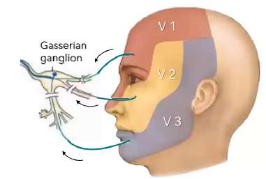 branches v1 v2 v3
 -
What forms the facial colliculus?
- The internal loop of facial nerve fibers around the abducens nucleus on the floor of the 4th ventricle
-
What are the two trigeminothalamic tracts?
- Anterior (ventral) trigeminothalamic tract
- Posterior (dorsal) trigeminothalamic tract
trigeminal pathways neuroanatomy -
What is the trapezoid body in the pons?
- A transverse bundle of fibers in the pontine basis involved in the auditory pathway and decussation of cochlear projections
-
Describe the origin and crossing of the anterior (ventral) trigeminothalamic tract.
- Axons from the principal nucleus of V and descending nucleus of V
- Fibers cross to the opposite side and ascend (majority)
trigeminal ventral crossing -
What is the function of the area vestibularis?
- Region containing vestibular nuclei that process balance and spatial orientation inputs from the inner ear
-
Describe the origin of the posterior (dorsal) trigeminothalamic tract.
- Axons from the principal nucleus of V
- Fibers that do not cross the opposite side and ascend (minority)
trigeminal dorsal principal -
What is the clinical effect of a lesion in the corticospinal tract?
- Contralateral hemiparesis (weakness on the opposite side of the body) when the tract is damaged above decussation
-
What happens when some trigeminal fibers descend?
- They form the descending tract of V
- They synapse in the descending nucleus of V
trigeminal descending nuclei -
At the caudal medulla, what percentage of corticospinal fibers cross at the pyramidal decussation?
- About 90% cross at the pyramidal decussation
- About 10% remain uncrossed
-
Which sensory modalities are conveyed by the ventral trigeminothalamic tract?
- Conscious proprioception
- Discriminative (fine) touch
- Vibration sense
- Modalities from the contralateral face
sensory ventral modalities -
What are the two main divisions of the corticospinal tract after the medulla?
- Lateral corticospinal tract (majority, crossed)
- Ventral (anterior) corticospinal tract (minority, may cross segmentally)
-
Which sensory modalities are conveyed by the dorsal trigeminothalamic tract?
- Pain and thermal sense
- Non-discriminative (crude) touch
- Modalities from the ipsilateral face (uncrossed fibers)
sensory dorsal modalities -
Where does the lateral corticospinal tract descend in the spinal cord?
- In the lateral funiculus of the spinal cord, primarily controlling distal limb muscles
-
How do bilateral and crossed sensory patterns apply to trigeminal modalities?
- Conscious proprioception (fine touch, vibration) is bilateral
- Pain, thermal, and crude touch are stated as always crossed (contralateral)
sensory crossing clinical -
What is the primary role of the ventral (anterior) corticospinal tract?
- Modulates axial and proximal trunk musculature; fibers may cross at segmental levels near their spinal targets
-
Where is the mesencephalic nucleus of V located?
- At the floor of the IV ventricle
- Medial to the superior cerebellar peduncle
- In the upper 1½ of the pons extending into the midbrain
mesencephalic location pons -
Summarize the cortico-ponto-cerebellar system in one line
- Cortex → pontine nuclei (corticopontine) → decussating ponto-cerebellar fibers → contralateral MCP → cerebellar hemisphere
-
What are the function and unique features of the mesencephalic nucleus of V?
- Mediates non-conscious proprioception from muscles of mastication
- Controls the bite reflex
- Contains pseudounipolar neurons, unique in the CNS
- Bypasses the Gasserian ganglion
mesencephalic proprioception reflexes -
What is the pontine basis composed of?
- Ventral pons white matter containing corticospinal and corticopontine fibers intermingled with pontine nuclei
-
Which nuclei are homologues for non-conscious proprioception and their body regions?
- Mesencephalic nucleus — face (mastication muscles)
- Accessory (lateral) cuneate nucleus — upper trunk and limbs
- Dorsal nucleus of Clarke — lower trunk and limbs
proprioception nuclei cerebellum -
-
Which cerebellar tracts arise from the non-conscious proprioception nuclei?
- Mesencephalic n. → trigeminocerebellar tract
- Accessory (lateral) cuneate n. → cuneocerebellar tract
- Dorsal n. of Clarke → dorsal spinocerebellar tract
cerebellar tracts proprioception -
Which tract connects the pons to the cerebellum and decussates in the basis of the pons?
The pontocerebellar tract, which decussates in the basis of the pons
-
Refer to the diagram: which nucleus sends uncrossed fibers ascending as the posterior trigeminothalamic tract?
- The principal sensory nucleus of V sends uncrossed fibers that ascend as the posterior (dorsal) trigeminothalamic tract
image diagram trigeminal -
How do cerebral hemispheres share motor plans via the pons?
Axons of pontine nuclei cross to the opposite side and enter the opposite MCP to reach the opposite cerebellar hemisphere
-
reflex neuro
-
What is the role of the cerebellum in movement?
Coordinates movements by receiving the cortex motor plan to make movements smooth and precise
-
What are the functional components of a basic reflex arc?
- Receptor
- Afferent limb
- Central integrator
- Efferent limb
- Effector organ
reflex anatomy -
Where do axons of pontine nuclei run to reach the cerebellum?
They run transversely, decussate, enter the opposite middle cerebellar peduncle (MCP), and go to the opposite cerebellar hemisphere
-
pons neuroanatomy
-
Name the main descending tracts summarized in the pons section.
- Corticospinal (pyramidal) tract
- Corticobulbar tract
- Corticopontine / cortico-ponto-cerebellar pathway
-
What is the jaw jerk reflex and how is it elicited?
- Masseter reflex: brisk jaw closure in response to tapping the mandible
- Called a 'trigemino-trigeminal' reflex
jaw-jerk reflex -
What does the pontine tegmentum contain?
A mix of nuclei and fiber tracts in the lower half of the pons
-
jaw-jerk physiology
-
List key internal structures found in the pontine tegmentum (lower half).
- IV ventricle
- Facial colliculus
- Tuberculum acousticum
- Area vestibularis
- MCP
- Trapezoid body
- Medial lemniscus
- Pyramidal tract
- Pontine nuclei
- Pontocerebellar tract & decussation
-
jaw-jerk clinical
-
What structure marks the most ventral part of the pontine tegmentum?
The trapezoid body is the most ventral structure in the pontine tegmentum
-
What is the receptor for the jaw jerk reflex?
- Muscle spindles (intrafusal fibers) in the masseter on both sides
jaw-jerk receptor -
What type of fibers does the trapezoid body contain?
Transversely coursing auditory fibers placed ventrally
-
What is the afferent limb and its cell body for the jaw jerk reflex?
- Mandibular division of CN V (V3)
- Cell body in the mesencephalic nucleus of CN V
trigeminal afferent -
What sensory modalities travel in the medial lemniscus?
- Conscious proprioception
- Vibration sense
- Discriminative (fine) touch
-
What special feature does the mesencephalic nucleus of CN V have?
- It is pseudounipolar and has no sensory ganglion
mesencephalic neuroanatomy -
Where do the medial lemniscus fibers first form?
They first form in the medulla from dorsal column nuclei axons
-
pons integration
-
Which nuclei serve as the second-order neurons for dorsal column modalities?
The nucleus cuneatus and nucleus gracilis in the medulla
-
What is the efferent limb and motor cell body for the jaw jerk reflex?
- Mandibular division of CN V (V3)
- Cell body in the motor nucleus of CN V
trigeminal efferent -
Where is the medial lemniscus located relative to the trapezoid body?
Dorsal to the trapezoid body and close to the midline
-
What is the effector and the response in the jaw jerk reflex?
- Extrafusal muscle fibers of the masseter (both sides)
- Response: brisk jaw closure
jaw-jerk effector -
From which primary afferents do the fasciculus cuneatus and gracilis receive input?
- Fasciculus cuneatus: A-alpha & A-beta from upper trunk and upper limbs
- Fasciculus gracilis: A-alpha & A-beta from lower trunk and lower limbs
-
What is the corneal reflex and its pathway type?
- Trigemino-facial reflex producing direct and consensual blinking
- Polysynaptic
corneal reflex -
What is the pathway from DRG to cortex for fine touch and proprioception?
- 1° DRG → Fasciculus cuneatus/gracilis → Nucleus cuneatus/gracilis (2°) → Medial lemniscus → Thalamus (3°) → Cerebral cortex
-
What happens to the corneal reflex with an afferent limb lesion?
- Absent direct and consensual corneal reflexes
corneal lesion -
What is the sensory decussation related to the medial lemniscus?
Axons from nucleus gracilis and cuneatus cross (decussate) to form the medial lemniscus
-
What happens to the corneal reflex with an efferent limb lesion?
- Absent corneal reflex on the side of the lesion
- Present corneal reflex on the side without the lesion
corneal lesion -
What is the role of the pontine nuclei?
They relay cortical motor information to the cerebellum via pontocerebellar fibers
-
What is the afferent limb of the corneal reflex?
- Ophthalmic division (V1) of CN V on the stimulated side
neuro cranialnerves reflex -
Where do pontocerebellar fibers enter the cerebellum?
Via the middle cerebellar peduncle (MCP) on the opposite side after decussation
-
Where is the afferent neuron cell body for the corneal reflex located?
- Gasserian (Semilunar) ganglion
neuro ganglion -
What marks the boundary between the tegmentum and basis of the pons?
A junctional region where the most ventral part lies at the tegmentum-basis junction, marking their boundary
-
Describe the central pathway for the corneal reflex afferent signal.
- Pons (upper 1½) then descends to descending nucleus of CN V in medulla and lower 1½ of pons; sends bilateral projections to both facial motor nuclei
neuro pons pathway -
Which tract is frequently referred to as the pyramidal tract?
The corticospinal tract is commonly called the pyramidal tract
-
neuro cranialnerves
-
What is the cortico-ponto-cerebellar pathway?
A pathway where cortical motor signals travel to pontine nuclei, then via pontocerebellar fibers to the cerebellum
-
neuro nuclei
-
What is contained in the basis of the pons relevant to decussation?
Transverse pontocerebellar fibers that decussate in the basis before entering the MCP
-
What are the effector organs in the corneal reflex?
- Left and right orbicularis oculi (blink muscles)
neuro muscles -
What sensory modalities are lost with medial lemniscus lesions?
- Loss of conscious proprioception
- Loss of vibration sense
- Loss of fine discriminative touch
-
What is the normal response of the corneal reflex to corneal stimulation?
- Direct (stimulated side) and consensual (other side) blinking/closure bilaterally
reflex neuro -
Why is transverse orientation of pontine fibers important?
It allows cortical motor information to be relayed across midline to coordinate bilateral cerebellar processing
-
Through which foramina do the divisions of CN V exit the skull?
- V1: Superior orbital fissure
- V2: Foramen rotundum
- V3: Foramen ovale
anatomy foramina -
What structure is labeled as 'Area vestibularis' in the pontine tegmentum?
A region containing vestibular-related nuclei or fibers involved in balance and spatial orientation
-
anatomy cn7
-
What is the facial colliculus?
A surface landmark in the floor of the IV ventricle formed by the internal genu of the facial nerve over the abducens nucleus
-
anatomy foramina
-
Why does dorsal column sensation appear ipsilateral in the spinal cord but contralateral at the level of the pons?
Because dorsal column fibers ascend ipsilaterally until their second-order neurons decussate in the medulla at the sensory decussation, so above the decussation (pons) sensation is contralateral.
-
vascular pons
-
What is the shape of the medial lemniscus as it appears in the brainstem?
It appears as two flattened elliptical discs; long anteroposteriorly in the medulla and compressed in the pons by surrounding structures.
-
What deficits can result from obstruction of paramedian pontine branches?
- Pyramidal tract interruption = contralateral hemiparesis
- Abducens nerve interruption = ipsilateral impairment
vascular lesion -
How are fibers from lower trunk and limbs organized when entering the dorsal column?
Fibers from lumbar and sacral levels enter first and ascend medially forming fasciculus gracilis, with later-entering upper-limb fibers occupying lateral positions.
-
arteries pons basilar
-
How is the dorsal column divided at the cervical and upper thoracic levels?
It is divided into: - Fasciculus cuneatus for upper trunk and limbs - Fasciculus gracilis for lower trunk and limbs
-
What are the three main branches of the basilar artery supplying the pons?
- Paramedian branches
- Short circumferential branches
- Long circumferential branches
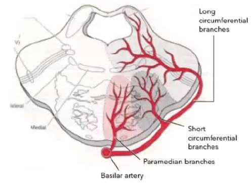 arteries basilar pons
 -
Why is there no cuneatus division in lower thoracic and lumbosacral regions?
Because those segments contain only fibers from the lower trunk and lower limbs, so only fasciculus gracilis is present.
-
Which basilar branch supplies the ventrolateral areas of the pontine basis?
- Short circumferential branches
arteries pons supply -
Where do dorsal column first-order fibers terminate?
They ascend to and terminate in their corresponding sensory nuclei in the medulla: the nucleus gracilis and nucleus cuneatus.
-
Which basilar branch supplies the pontine tegmentum and wedge of the middle cerebellar peduncle?
- Long circumferential branches
arteries pons supply -
What happens to axons of the dorsal column nuclei in the medulla?
They run ventrally and medially and cross to the opposite side via the sensory decussation (internal arcuate fibers) before ascending.
-
Which artery reinforces the long circumferential supply at the caudal pontine level?
- Anterior inferior cerebellar artery (AICA)
arteries aica pons -
After decussation, how do dorsal column second-order axons ascend?
They ascend contralaterally as the medial lemniscus toward the thalamus and then to the sensory cortex for conscious sensation.
-
Which artery reinforces the long circumferential supply at the rostral pontine level?
- Superior cerebellar artery (SCA)
arteries sca pons -
-
Which four cranial nerves exit at the ventral surface / pontine basis?
- Trigeminal nerve (CN V)
- Abducens nerve (CN VI)
- Facial nerve (CN VII)
- Vestibulocochlear nerve (CN VIII)
cranialnerves pons exits -
Which modality does the ventral spinothalamic tract primarily convey?
- Non-discriminative (crude) touch
-
cranialnerves cnvi pons
-
Where do the spinothalamic second-order neurons decussate?
They decussate in the spinal cord via the ventral white commissure soon after synapsing in the dorsal horn.
-
Where do CN VII and CN VIII emerge from the pons?
- Emerge posterolaterally at the cerebellopontine angle
cranialnerves cnvii cnviii -
Why do spinothalamic tracts convey contralateral sensation?
Because their axons have already decussated at the ventral white commissure in the spinal cord before ascending.
-
What are the three cerebellar peduncles visible on the lateral surface of the pons?
- Superior cerebellar peduncle (brachium conjunctivum)
- Middle cerebellar peduncle (brachium pontis)
- Inferior cerebellar peduncle (restiform & juxtarestiform bodies)
anatomy cerebellarpeduncles pons -
What is the spinal lemniscus?
A combined tract in the brainstem made of the lateral and ventral spinothalamic tracts that become closer and function as one pathway.
-
anatomy pons dorsal
-
What sensations does the spinal lemniscus convey?
- Pain
- Temperature
- Crude touch from the contralateral side of the body
-
How is the pons ventrally and dorsally demarcated from midbrain and medulla?
- Ventrally: superior pontine sulcus from midbrain; inferior pontine sulcus from medulla
- Dorsally: line of exit of trochlear (CN IV) from midbrain; stria medullaris of IV ventricle from medulla
anatomy boundaries pons -
Where is the spinal lemniscus located relative to the medial lemniscus?
It is located lateral to the medial lemniscus and is a smaller oval fiber tract in the pontine tegmentum.
-
clinical cnvi signs
-
What is the clinical effect of a lesion in the pons affecting the medial lemniscus?
Hemisensory loss manifested contralaterally because the medial lemniscus carries contralateral discriminative sensation above the decussation.
-
cranialnerves cnvi muscles
-
Summarize the dorsal column-medial lemniscus pathway sequence from periphery to cortex.
- 1st order: DRG fibers ascend in dorsal column
- Terminate: Nucleus gracilis/cuneatus (medulla)
- 2nd order: Cross in sensory decussation
- Ascend: Medial lemniscus to thalamus
- 3rd order: Thalamus to sensory cortex
-
Name major arteries surrounding the brainstem included in the summary
- Anterior communicating artery
- Ophthalmic artery
- Anterior cerebral artery
- Internal carotid artery
- Middle cerebral artery
- Posterior communicating artery
- Posterior cerebral artery
- Superior cerebellar artery
- Basilar artery
- AICA
- PICA
- Vertebral artery
- Dorsal and ventral spinal arteries
arteries brainstem vessels -
Summarize the spinothalamic pathway sequence from periphery to cortex.
- 1st order: DRG to dorsal horn
- 2nd order: Synapse and decussate in ventral white commissure
- Ascend: Lateral and ventral spinothalamic tracts form spinal lemniscus
- 3rd order: Thalamus to cortex
-
What are the three enlargements of the rhomboid fossa of the pons (medial to lateral)?
- Medial eminence (rostral)
- Facial colliculus (caudal)
- Area vestibularis / Tuberculum acousticum
pons anatomy -
Where is the medial longitudinal fasciculus (MLF) located and what is its function?
Location: most dorsal part in the medial aspect near the midline, close to the floor of the fourth ventricle. Function: coordinates horizontal conjugate eye movements and gaze.
-
pons anatomy
-
Where is the central tegmental tract (CTT) located relative to the medial lemniscus?
The CTT is located dorsal to the medial lemniscus in the pontine tegmentum.
-
vestibular pons
-
Where is the inferior cerebellar peduncle (ICP) in the pons and what does it contain?
Location: lateral aspect of the pontine tegmentum. Contains: the restiform and juxtarestiform bodies connecting cerebellum and brainstem.
-
auditory pons
-
Which tract conveys discriminative touch and proprioception to cortex?
The dorsal column-medial lemniscus system conveys discriminative touch and proprioception to the thalamus and sensory cortex.
-
What is the substancia ferruginea associated with in the pons?
- Overlies locus coeruleus
- Locus coeruleus = major source of norepinephrine in the CNS
pons neurochemistry -
Where is the descending nucleus and tract of the trigeminal nerve located in the pons?
- Ventromedial to the ICP (inferior cerebellar peduncle)
-
What are the constant contents of the pontine basis?
- Pyramidal tract
- Corticopontine tract
- Pontine nuclei
- Pontocerebellar tract
pons tracts -
What modalities are carried by the descending nucleus and tract of V?
- Pain, temperature, and crude touch of the face
-
motor pons
-
What muscle does the abducens nerve (CN VI) innervate?
- Lateral rectus (extraocular skeletal muscle, GSE)
-
Where do the corticopontine fibers terminate and what do they give rise to?
- Terminate at pontine nuclei
- Give rise to pontocerebellar tract
pons pathways -
What is the primary action of the lateral rectus muscle?
- Abduction of the eyeball (lateral deviation)
-
What is the role of the cortico-ponto-cerebellar pathway?
- Connects one cerebral hemisphere with the opposite cerebellar hemisphere
cerebellum connectivity -
What happens to the eye in a lesion of the abducens nerve?
- Unopposed medial rectus causes the eye to be medially deviated (cannot abduct)
-
Name three key auditory-related structures in the pontine tegmentum and their roles.
- Superior olivary nucleus: auditory relay nucleus
- Trapezoid body: crossed auditory fibers
- Lateral lemniscus: principal ascending auditory pathway (bilateral, more contralateral)
auditory pons -
Why does a unilateral abducens nerve lesion cause medial deviation rather than lateral?
- The antagonist (medial rectus via CN III) is unopposed and dominates
-
What is the function of the medial longitudinal fasciculus (MLF) and the PPRF in the pons?
- MLF: pathway for conjugate gaze
- PPRF: coordinating center for horizontal conjugate gaze
eye_movements pons -
Where is the abducens nucleus located relative to the facial colliculus?
- Beneath the facial colliculus (dorsal location in the floor of the fourth ventricle)
-
What respiratory centers are located in the pons and their functions?
- Apneustic center: promotes inspiration
- Pneumotaxic center: automatic switching off of inspiration
respiration pons -
Where does the abducens nerve emerge from the brainstem?
- Ventrally at the inferior pontine sulcus / pontomedullary junction after coursing ventrally and medially
-
What sensory pathways in the pons carry somatosensory information from the body?
- Medial lemniscus: conscious proprioception, vibration, fine touch (opposite side)
- Spinal lemniscus: crude touch, pain, temperature (opposite side)
sensory somatosensory -
-
trigeminal sensory
-
Which cranial nerve is the only one that decussates among the ocular motor nerves?
- CN IV (trochlear nerve)
-
Which cranial reflexes are elicited at the level of the pons?
- Jaw jerk reflex (trigemino-trigeminal; monosynaptic)
- Corneal reflex (trigemino-facial; polysynaptic)
reflexes cranial -
-
What are the afferent and efferent limbs of the jaw jerk reflex?
- Afferent: Mandibular division (V3) of trigeminal nerve
- Efferent: Mandibular division (V3) of trigeminal nerve
reflexes trigeminal -
Which muscle is the antagonist to lateral rectus and which nerve supplies it?
- Medial rectus — supplied by CN III (oculomotor)
-
What are the afferent and efferent limbs of the corneal reflex?
- Afferent: Ophthalmic division (V1) of trigeminal nerve
- Efferent: Motor root of facial nerve (VII)
reflexes facial -
What two structures make the facial colliculus prominent?
- Abducens nucleus
- Genu of the facial motor root
-
What is the main blood supply of the pons?
- Pontine branches of the basilar artery
- Reinforcements: AICA and superior cerebellar artery
blood_supply pons -
Where is the facial motor nucleus located in the pons?
- In the pontine tegmentum, lateral to the abducens nucleus (not beneath the facial colliculus)
-
What is the main muscle innervated by the Abducens (VI) nerve?
- Lateral rectus (causes lateral deviation of the eyeball)
abducens cranial_nerve ocular -
Describe the initial course of the facial motor root in the pons.
- Runs dorsally and medially toward the floor of the 4th ventricle
-
abducens fiber_type cranial_nerve
-
Why does the facial motor root form a 'genu' over the abducens nucleus?
- Because it ascends medial to the abducens and turns sharply over the abducens nucleus, forming the facial 'genu'
-
abducens foramina cranial_nerve
-
How does the facial genu relate to the facial colliculus?
- The genu (and the underlying abducens nucleus) bulge into the floor of the 4th ventricle forming the facial colliculus
-
facial motor cranial_nerve
-
What is a clinical sign of a left abducens nerve lesion specifically affecting the nerve (not the nucleus)?
- Left eye cannot abduct; left eye is medially deviated at rest; right eye abduction intact
-
What is the branch of facial nerve that carries autonomic fibers for lacrimation and salivation?
- Intermediate nerve / Nervus intermedius (GVE for lacrimation and salivation)
facial gve autonomic -
What additional function do abducens nucleus neurons have besides innervating lateral rectus?
- Contain interneurons that project via the MLF to the contralateral oculomotor nucleus to coordinate conjugate horizontal gaze
-
facial taste sva
-
What major pontine structures are near the facial motor nucleus and genu (as labeled in cross-sections)?
- IV ventricle, facial colliculus, medial lemniscus, pyramidal tract, pontine nuclei, MCP, trapezoid body
-
What cutaneous sensation does the Facial (VII) nerve contribute to?
- Cutaneous sensation of external ear and external auditory canal (GSA)
facial sensory gsa -
How does a lesion of the abducens nucleus differ clinically from a lesion of the abducens nerve?
- Nucleus lesion: ipsilateral horizontal gaze palsy (both eyes) due to loss of interneurons; nerve lesion: isolated lateral rectus paralysis (eye medially deviated)
-
What visceral sensation is carried by the Facial (VII) nerve?
- Visceral sensation from nasal & sinus cavities and soft palate (GVA, minor)
facial gva sensory -
What does 'GSE' stand for in cranial nerve modalities?
- General Somatic Efferent — motor to skeletal muscles derived from somites (e.g., extraocular muscles)
-
Through which foramina does the Facial (VII) nerve exit?
- Internal auditory meatus and stylomastoid foramen
facial foramina cranial_nerve -
What is the facial genu?
The bend formed when the facial motor root turns sharply around the abducens nucleus; 'genu' means knee or bend.
-
What are the two roots of the Vestibulocochlear nerve and their functions?
- Vestibular root: equilibrium and balance
- Cochlear root: hearing
vestibulocochlear hearing balance -
Where does the facial motor root emerge from the pons?
It continues ventrally and laterally to emerge at the ventrolateral pons along the inferior pontine sulcus as the facial motor root.
-
What is the fiber type for both roots of the Vestibulocochlear nerve?
- SSA (Special Somatic Afferent) for both vestibular and cochlear roots
vestibulocochlear ssa sensory -
Why does the facial nerve lie beneath the facial colliculus?
The sharp bend (genu) of the facial motor root places it just beneath the facial colliculus.
-
vestibulocochlear foramina cranial_nerve
-
-
What is the motor function of the Trigeminal (V) motor root (Portio minor)?
- Muscles of mastication (SVE)
trigeminal motor mastication -
Which muscles does CN VII innervate?
Most facial muscles derived from branchiomeric muscles; excludes extraocular muscles and muscles of mastication.
-
Through which foramen does the mandibular division (V3) that carries trigeminal motor fibers exit?
- Foramen ovale (Mandibular div. V3)
trigeminal v3 foramina -
Give an example of a muscle innervated by CN VII and its function.
Orbicularis oculi — closes the eyelids.
-
What sensory modalities are carried by the Trigeminal (V) sensory root (Portio major)?
- Conscious proprioception, fine touch, vibration sense
- Pain, thermal sense, crude touch (GSA)
trigeminal sensory gsa -
Which nerve closes the eyes and which opens them?
- CN VII closes the eyes via orbicularis oculi
- CN III opens the eyes via levator palpebrae
-
Which trigeminal division carries non-conscious proprioception from muscles of mastication?
- Trigeminal sensory fibers (non-conscious proprioception from muscles of mastication)
trigeminal proprioception mastication -
List common signs of a peripheral facial nerve lesion.
- Flattening of wrinkle lines
- Inability to close the eye
- Shallow nasolabial fold
- Drooping angle of the mouth
- Ipsilateral facial sag
-
trigeminal v1 foramina
-
What is Bell's palsy in simple terms?
An acute peripheral facial nerve (CN VII) palsy causing ipsilateral facial weakness and signs such as inability to close the eye.
-
trigeminal v2 foramina
-
Why does a peripheral CN VII lesion risk a dry cornea?
Inability to close the eyelid exposes the cornea, causing dryness and irritation.
-
trigeminal v3 foramina
-
How does a CN III lesion differ from a peripheral CN VII lesion at the eyelid?
CN VII lesion: inability to close eye; CN III lesion: ptosis due to levator palpebrae dysfunction.
-
What cortical input does the facial motor nucleus receive?
Corticobulbar (corticonuclear) fibers from the cerebral motor cortex.
-
How are corticobulbar projections to the upper face organized?
Bilateral innervation: upper facial motor neurons receive input from both cerebral hemispheres.
-
How are corticobulbar projections to the lower face organized?
Contralateral innervation: lower facial motor neurons are supplied predominantly by the opposite hemisphere.
-
What is the clinical result of a unilateral lesion of corticobulbar fibers?
Weakness of the contralateral lower one quarter of the face; upper face usually spared due to bilateral supply.
-
-
What additional nuclei are associated with CN VII?
Superior salivatory nucleus and lacrimal-related nuclei (parasympathetic functions).
-
What is the embryological origin of most facial muscles innervated by CN VII?
They are branchiomeric muscles derived from the second pharyngeal (branchial) arch.
-
Name two facial signs mentioned for a right facial nerve lesion.
- Flattening of wrinkle lines
- Drooping of the angle of the mouth
-
What facial sign indicates loss of orbicularis oculi function?
Inability to close the eye (orbicularis oculi paralysis).
-
What facial feature becomes shallow with facial weakness?
The nasolabial fold becomes shallow on the affected side.
-
What facial expression becomes asymmetric in lower facial weakness from corticobulbar lesion?
An asymmetric smile due to contralateral lower facial weakness.
-
What simple mnemonic describes CN VII and CN III roles at the eye?
CN VII acts like a 'hook' to close the eye; CN III acts like 'sticks' (levator palpebrae) to keep the eye open.
-
Where do corticobulbar fibers synapse in relation to facial control?
They synapse on the facial motor nucleus cells that control either the upper or lower face.
-
Summarize the two main patterns of facial weakness and their lesion locations.
- Peripheral (CN VII) lesion: ipsilateral weakness of entire half of face
- Central (corticobulbar) lesion: contralateral weakness of lower face only
-
What is the nervus intermedius (intermediate nerve of Wrisberg)?
- The smaller root of the facial nerve (CN VII)
- Also called the 'intermediate nerve of Wrisberg'
-
Where is the nervus intermedius located relative to the facial motor nucleus?
- Lies dorsolateral to the facial motor nucleus
- Lies lateral to the branchiomeric nucleus
-
Which modalities does the nervus intermedius carry?
- GVE (parasympathetic)
- SVA (taste)
- GVA (viscerosensory)
- GSA (somatosensory)
-
What is the GVE component of the intermediate nerve?
- Preganglionic parasympathetic fibers from superior salivatory and lacrimal nuclei
- Exit the pons as part of the nervus intermedius
-
Where do the GVE fibers of CN VII synapse?
- Synapse at the sphenopalatine and submandibular ganglia
- Relay to lacrimal and salivary glands
-
What is the SVA component of the intermediate nerve?
- Taste fibers from the anterior 2/3 of the tongue
- Cell bodies in the geniculate ganglion
-
Where do taste fibers from CN VII terminate centrally?
- Terminate in the rostral portion of the Nucleus of Tractus Solitarius (NTS) in the medulla
-
What is the function of the rostral NTS?
- The gustatory (taste) portion of the Nucleus of Tractus Solitarius
-
What is the GVA component of the intermediate nerve?
- Viscerosensory fibers from nasal cavity, soft palate, and sinus cavities
- Cell bodies in the geniculate ganglion
-
Where do GVA fibers of CN VII project centrally?
- Project to the caudal portion of the Nucleus of Tractus Solitarius (NTS)
-
What is the GSA component of the intermediate nerve?
- Somatosensory fibers for cutaneous sensation from the external ear and external auditory canal
- Cell bodies in the geniculate ganglion
-
Where do GSA fibers from the intermediate nerve project centrally?
- Project to the descending nucleus of trigeminal V for somatosensory processing
-
Which nuclei provide parasympathetic preganglionic fibers for lacrimation and salivation?
- Superior salivatory nucleus
- Lacrimal nucleus (often grouped with superior salivatory)
-
Which glands are targeted by parasympathetic fibers of CN VII?
- Lacrimal gland
- Submandibular gland
- Sublingual gland
-
What is the role of the geniculate ganglion for CN VII?
- Houses the sensory cell bodies for SVA, GVA, and GSA fibers of CN VII
- Sensory fibers synapse or have cell bodies here before central projection
-
Which central nucleus is the visceral afferent center of the brain?
- The Nucleus of Tractus Solitarius (NTS) in the medulla
-
Is the Nucleus of Tractus Solitarius found in the pons?
- No; the NTS is located in the medulla, not the pons
-
What are the two main central pathways after the geniculate ganglion?
- Nucleus of Tractus Solitarius (NTS) for visceral and taste afferents
- Descending nucleus of trigeminal V for somatosensory (external ear) afferents
-
Which cranial nerves contribute to taste sensation?
- CN VII (anterior 2/3 tongue)
- CN IX (posterior 1/3 tongue)
- CN X (taste from epiglottis and region)
-
Which cranial nerves supply somatosensory input from the external ear?
- CN V, CN VII, CN IX, and CN X provide afferents from the external ear and canal
-
What is the distinction between the nervus intermedius and the facial motor root?
- The nervus intermedius carries sensory and parasympathetic fibers
- The facial motor root carries the branchiomotor (motor) fibers of CN VII
-
-
Name the parasympathetic ganglia associated with CN VII targets.
- Sphenopalatine ganglion
- Submandibular ganglion
-
Which nucleus processes general visceral sensation for CN VII GVA fibers?
- The caudal Nucleus of Tractus Solitarius (NTS) processes general visceral afferents
-
What is the role of the Nucleus of Tractus Solitarius?
- Visceral afferent center in the brainstem
- Convergence point for cranial nerves with similar sensory modality
-
-
-
Which vestibular nucleus is the largest and where is it found?
- Medial (Principal)
- Largest; found in both medulla and pons
-
-
-
Relative position: where are vestibular nuclei compared to cochlear nuclei?
- Medial to the cochlear nuclei
-
What is the primary function of the vestibular nuclei?
- Processing information for equilibrium and balance
-
-
Where do the dorsal cochlear nuclei lie relative to the tuberculum acousticum?
- Beneath the tuberculum acousticum (lateral bulge)
-
Why are cochlear nuclei lateral to vestibular nuclei?
- They develop later phylogenetically than vestibular nuclei
-
-
-
-
What receptors provide input to the vestibular root?
- Semicircular canals
- Otolith organs (utricle & saccule)
-
-
What is Scarpa's ganglion notable for?
- Sensory ganglion of vestibular nerve
- Made of bipolar neurons (exception to pseudounipolar)
-
Which central connections link vestibular nuclei to eye movements?
- Cranial nerve nuclei (EOMs) to coordinate eye and head movement
-
How do vestibular nuclei connect to the spinal cord and why?
- Via vestibulospinal tract
- Maintain upright posture and balance
-
How do vestibular nuclei interact with the cerebellum?
- Via the ICP (inferior cerebellar peduncle)
- Regulate movements underlying posture and equilibrium
-
What is the vestibular nuclei connection to the cerebral cortex for?
- Perception of spatial orientation and motion
-
List the central connections of vestibular nuclei.
- Cranial nerve nuclei (EOMs)
- Spinal cord (vestibulospinal tract)
- Cerebellum (via ICP)
- Cerebral motor cortex
-
What root lies medial relative to the cochlear root?
- The vestibular root lies medial to the cochlear root
-
Summarize the functions of CN VIII roots in one line each.
- Vestibular root: mediates equilibrium and balance
- Cochlear root: mediates auditory sensation
-
-
Where are the cell bodies of cochlear nerve first-order neurons located?
- Spiral ganglion
- Bipolar neurons (exception to pseudounipolar sensory neurons)
-
After receptors in the organ of Corti, through which structure do auditory impulses travel first?
- Cochlear root of CN VIII
- Cell bodies in spiral ganglion
-
Upon entering the pons, to which nuclei do cochlear roots synapse?
- Dorsal cochlear nucleus
- Ventral cochlear nucleus
-
-
What is the superior olivary nucleus role in the auditory system?
- Auditory relay nucleus in the pons
- Located lateral to the spinal lemniscus
-
Name the three main acoustic striae from the cochlear nuclei.
- Dorsal acoustic stria
- Intermediate acoustic stria
- Ventral acoustic stria (trapezoid body)
-
What is the trapezoid body?
- Ventral acoustic stria
- Transversely crossing auditory fibers
- Most ventral; at demarcation of tegmentum and basis
-
-
Where does the lateral lemniscus originate and where is it found?
- Originates in the pons
- Also present in midbrain (ascending) but not in medulla
-
List the main ascending stations of the central auditory pathway.
- Cochlear nuclei
- Superior olivary nucleus
- Lateral lemniscus
- Inferior colliculus
- Medial geniculate body
- Auditory receiving centers
-
Which midbrain structure is a major auditory center before the thalamus?
- Inferior colliculus (inferior quadrigeminal body)
-
-
What is indicated by solid lines versus dotted lines in the lateral lemniscus formation diagram?
- Solid lines indicate fibers that decussated
- Dotted lines indicate fibers that did not cross over
-
Which cranial nerve carries the cochlear and vestibular roots?
- Cranial nerve VIII (vestibulocochlear nerve)
-
-
Do cochlear roots have central connections?
- Yes; cochlear roots form central connections in the auditory pathway
-
Which structures in the pons are specifically associated with the auditory pathway?
- Cochlear root
- Dorsal & ventral cochlear nuclei
- Superior olivary nucleus
- Lateral lemniscus origins
-
How is the trapezoid body positioned relative to tegmentum and basis?
- Most ventrally located; lies at the demarcation of tegmentum and basis
-
-
Name one label found on the pons cross-sectional diagram related to facial nerve anatomy.
- Facial motor nucleus
- Facial colliculus
- Facial genu
-
Which tract connects pontine nuclei to the cerebellum?
- Pontocerebellar tract (via middle cerebellar peduncle, MCP)
-
-
-
-
-
Give an atomic definition of the lateral lemniscus formation.
- A tract formed by ascending auditory fibers from cochlear nuclei and superior olive that projects to the inferior colliculus
-
What is the relationship between superior olivary nucleus and lateral lemniscus in the pons?
- Superior olivary nucleus is a relay lateral to the spinal lemniscus and contributes fibers to the lateral lemniscus
-
What is the main projection pattern of the dorsal cochlear nucleus?
- Sends axons that cross via the dorsal acoustic stria
- Ascends contralaterally as part of the lateral lemniscus
-
Name the two main projection options of the ventral cochlear nucleus.
- Cross via intermediate/ventral acoustic stria and ascend contralaterally
- Not cross: synapse ipsilaterally with superior olivary nucleus
-
If ventral cochlear nucleus axons synapse with the superior olivary nucleus ipsilaterally, what are the two possible subsequent routes?
- Become ventral acoustic stria (trapezoid body), cross and ascend contralaterally
- Remain uncrossed and ascend ipsilaterally in the lateral lemniscus
-
What is the trapezoid body (ventral acoustic stria)?
- Crossing bundle of auditory fibers at ventral pons
- Formed by fibers from ventral cochlear nucleus and contralateral superior olives
-
What is the primary function of the lateral lemniscus?
- Conveys bilateral auditory information, with stronger contralateral representation
-
Which sources provide crossed auditory fibers to the lateral lemniscus?
- Contralateral dorsal cochlear nucleus
- Contralateral ventral cochlear nucleus
- Contralateral superior olivary nuclei
-
Which structure provides uncrossed auditory fibers to the lateral lemniscus?
- Ipsilateral superior olivary nucleus
-
What is the auditory deficit pattern in a central lesion of the lateral lemniscus?
- Bilateral partial deafness, more severe on contralateral side
-
Why does a central lesion cause bilateral but predominantly contralateral hearing loss?
- Lateral lemniscus carries both crossed and uncrossed fibers (bilateral)
- Majority of fibers are contralateral, so contralateral deficit is greater
-
What is the auditory deficit pattern in a peripheral lesion that destroys the cochlear nerve?
- Ipsilateral complete deafness of the affected ear
-
Why does cochlear nerve damage produce ipsilateral complete deafness?
- Cochlear nerve transmits only ipsilateral auditory input from the ear
- Destruction abolishes all afferent input from that ear
-
List the main acoustic striae mentioned in the pathway.
- Dorsal acoustic stria
- Intermediate acoustic stria
- Ventral acoustic stria (trapezoid body)
-
What role does the superior olivary nucleus play in the auditory pathway?
- Acts as a relay nucleus receiving cochlear inputs
- Contributes both crossed and uncrossed fibers to brainstem pathways
-
-
What structures form the ascending auditory pathway from the inner ear to the midbrain in this text?
- Organs of Corti → Cochlear nerve → Cochlear nuclei → Superior olivary nucleus → Lateral lemniscus
-
What is the paramedian pontine reticular formation (PPRF)?
- A pontine reticular formation involved in horizontal gaze control
-
Where is the PPRF located relative to the abducens nucleus?
- Lies ventromedial to the abducens nucleus
-
What is the MLF and its general role in brainstem gaze systems?
- Medial longitudinal fasciculus (MLF)
- Connects ocular motor nuclei to coordinate conjugate eye movements
-
How does damage to the PPRF typically affect eye movements?
- Produces impaired horizontal gaze toward the side of lesion (difficulty initiating saccades)
-
What is the coordinating center for horizontal conjugate eye movements in the pons?
Paramedian Pontine Reticular Formation (PPRF), located close to the abducens nucleus
-
Which cranial nerve is the coordinating center for horizontal gaze located near?
Abducens nerve (VI)
-
What are the main inspiratory and expiratory respiratory centers?
Medulla (medullary respiratory centers)
-
-
-
How does the apneustic center affect breathing?
- Promotes inspiration by constant stimulation of the dorsal respiratory group
- Controls depth of breathing
-
What is 'apneustic breathing'?
Very long and very deep inspirations caused by unchecked apneustic center activity
-
Which center inhibits the apneustic center?
Pneumotaxic center (upper pons) and pulmonary stretch receptor input via the vagus nerve
-
How do pulmonary stretch receptors influence the apneustic center?
At maximal inspiration they send inhibitory signals via the vagus nerve to inhibit inspiratory and apneustic centers, allowing exhalation
-
What is the effect of inhibiting the inspiratory center?
Inhibition of the inspiratory center permits exhalation
-
What happens when the apneustic center is inhibited?
It stops stimulating the inspiratory center, which allows exhalation to occur
-
-
Which cranial nerve nuclei are located in the pons?
- Trigeminal (V)
- Abducens (VI)
- Facial (VII)
- Vestibulocochlear (VIII)
-
Which trigeminal nuclei appear in the upper pons?
- Mesencephalic nucleus of V
- Motor nucleus of V
- Principal (main sensory) nucleus of V
- Sensory root of V
-
Which cerebellar peduncles are visible around the pons?
- Middle cerebellar peduncle (prominent in pons)
- Superior cerebellar peduncle (near upper pons)
- Inferior cerebellar peduncle (adjacent)
-
What is the function of the medial longitudinal fasciculus (MLF) mentioned in the pons?
Interconnects ocular motor nuclei and vestibular nuclei to coordinate conjugate eye movements
-
Which nuclei provide parasympathetic lacrimation and salivation in the pons?
Superior salivatory and lacrimal nuclei (associated with facial nerve VII)
-
What is the Nervus Intermedius (intermediate nerve of Wrisberg)?
A sensory and parasympathetic component of the facial nerve carrying taste and autonomic fibers
-
Name components of the reticular formation present in the pons.
- Reticular formation nuclei
- Paramedian Pontine Reticular Formation (PPRF)
-
What is the locus ceruleus and where is it found?
A noradrenergic nucleus in the upper pons involved in arousal and attention
-
-
List important ascending auditory nuclei in the pons.
- Dorsal cochlear nucleus
- Ventral cochlear nucleus
- Lateral lemniscus (pathway)
-
What key motor pathway runs through the pons?
Pyramidal tract (corticospinal fibers) and pontocerebellar tract
-
Which structure carries pontine input to the cerebellum?
Middle cerebellar peduncle carrying pontocerebellar fibers
-
Which sensory pathway for facial sensation is present in the pons?
Trigeminal lemniscus (carries principal sensory information from V)
-
Give three midline or paramedian structures seen in the upper pons.
- Medial eminence
- Basilar sulcus
- Superior medullary velum
-
What role do the superior cerebellar peduncles play at the level of the upper pons?
Form part of the lateral wall of the upper fourth ventricle and connect midbrain to cerebellum
-
What is the middle cerebellar peduncle primarily composed of?
Pontocerebellar fibers carrying cortical information to the cerebellum
-
Where is the basilar sulcus located and what does it contain?
Midline groove on the ventral pons that accommodates the basilar artery
-
What are the pontine nuclei and their main function?
Collections of relay neurons that receive corticopontine input and project to the cerebellum
-
What is the pyramidal tract in the pons?
Descending corticospinal fibers passing ventrally in the basis of the pons
-
What are pontocerebellar fibers and what do they do?
Transverse fibers from pontine nuclei that decussate to enter opposite middle cerebellar peduncle
-
What defines the pontine tegmentum?
Dorsal part of the pons located ventral to the fourth ventricle containing cranial nerve nuclei and ascending tracts
-
What is the medial eminence visible in the rhomboid fossa?
A dorsomedial bulge of the pontine tegmentum seen in the upper half of the rhomboid fossa
-
What is the substancia ferruginea?
Bluish-gray discoloration of the sulcus limitans overlying the locus coeruleus
-
How does the fourth ventricle change in the upper pons?
Becomes much narrower in the upper half and is roofed by the superior medullary velum
-
Which structure forms the lateral wall of the upper fourth ventricle?
Cut portions of the superior cerebellar peduncles
-
What is the relation of the facial colliculus to the medial eminence?
Facial colliculus is a bulge in the lower half of the rhomboid fossa; medial eminence is the main dorsomedial bulge in the upper half
-
What major neurotransmitter is associated with the locus coeruleus?
Norepinephrine (major source in the CNS)
-
What are the key functions of the locus coeruleus?
Plays roles in emotion, arousal, alertness, and descending pain modulation
-
What causes the bluish discoloration called substancia ferruginea?
Accumulation of pigmented catecholamine cells (neuromelanin) in the locus coeruleus beneath sulcus limitans
-
What is the medial lemniscus responsible for?
Conveying conscious proprioception, vibration, and fine touch from the contralateral body
-
Where is the medial lemniscus located relative to the trapezoid body?
Dorsal to the trapezoid body in the pontine tegmentum
-
What is the central tegmental tract (CTT) position relative to the MLF?
CTT and MLF occupy similar relative positions in both halves of the pons
-
What does the spinal lemniscus carry?
Spinothalamic fibers conveying pain, temperature, and crude touch from the opposite side
-
What fibers form the trigeminal lemniscus?
Trigeminothalamic fibers from sensory nuclei of cranial nerve V conveying facial sensations
-
-
Which ascending tracts lie lateral to the medial lemniscus?
Spinal lemniscus and trigeminal lemniscus lie lateral to the medial lemniscus
-
What are the main components of the basis of the upper pons?
Descending pyramidal tract, corticobulbar (corticocuneal), and corticopontine tracts
-
What is the destination of the corticopontine fibers?
Terminate on pontine nuclei to relay cortical information to the cerebellum
-
How do pontocerebellar fibers reach the cerebellum?
They decussate and enter the opposite middle cerebellar peduncle
-
What distinguishes the tegmentum of the upper pons?
Contains locus coeruleus, cranial nerve nuclei, ascending sensory tracts, and reticular formation
-
What visible feature identifies the locus coeruleus histologically?
Deeply pigmented catecholamine cells containing neuromelanin
-
Which surface discoloration overlies the locus coeruleus?
The substancia ferruginea (bluish-gray area on the sulcus limitans)
-
What is the dorsolateral pontine reticular formation role?
Involved in regulation of REM sleep and lies ventrolateral to the locus coeruleus
-
Where is the pneumotaxic center mentioned in relation to the pons?
Found at the ponto-midbrain junction and involved in respiratory pattern regulation
-
What tracts run ventrally in the pons basis as rounded fascicles?
Descending corticospinal and corticopontine fibers appear as rounded fascicles in the basis
-
What is the function of corticobulbar fibers in the pons?
Provide voluntary motor control to cranial nerve motor nuclei
-
Which tract coordinates eye movements and is located in the pontine tegmentum?
Medial longitudinal fasciculus (MLF) coordinates conjugate eye movements and vestibulo-ocular reflexes
-
What is a concise topographic rule for sensory tracts in the pons?
Medial lemniscus is medial; spinal and trigeminal lemnisci lie more lateral to it
-
Where is the locus coeruleus located in the pons?
- Dorsal part of tegmentum at the floor of the 4th ventricle
- Visible only in the upper half of the pons
-
What is the primary neurotransmitter produced by the locus coeruleus?
- Norepinephrine (noradrenaline)
-
List main functions of the locus coeruleus.
- Regulation of emotion, arousal, and alertness
- Participation in descending pain modulation
-
Where is the IV ventricle located relative to the pons?
- Dorsal to the pons, within the rhomboid fossa
- Communicates rostrally with the cerebral aqueduct and caudally with the central canal
-
-
What does the superior cerebellar peduncle connect?
- Connects cerebellum to midbrain and carries major cerebellar outputs
-
What is the main input carried by the middle cerebellar peduncle?
- Pontocerebellar fibers from pontine nuclei (afferent to cerebellum)
-
What are the pontine nuclei responsible for?
- Relaying cortical motor information to the cerebellum via pontocerebellar fibers
-
-
Where is the lateral lemniscus relative to the trigeminal lemniscus?
- Lateral to the trigeminal lemniscus
-
What does the trigeminal lemniscus convey?
- Trigeminothalamic sensory fibers conveying various facial sensations to the thalamus
-
What is the pontocerebellar tract?
- Transverse fibers from pontine nuclei that enter the cerebellum via the middle cerebellar peduncle
-
What modalities does the medial lemniscus convey?
- Conscious proprioception, vibration, and fine touch from the opposite side of the body
-
Where is the medial lemniscus found relative to the trapezoid body?
- Dorsal to the trapezoid body in the pontine tegmentum
-
What is the central tegmental tract (CTT) notable for in the pons?
- Occupies a consistent position with the MLF in both halves of the pons
- Contains fibers between red nucleus, inferior olive, and ascending taste fibers
-
What is the medial longitudinal fasciculus (MLF) function?
- Interconnects brainstem nuclei for coordinated eye movements and vestibulo-ocular reflexes
-
What is the dorsolateral pontine reticular formation role and location?
- Lies ventrolateral to the locus coeruleus
- Plays a role in regulation of REM sleep; located near the ponto-midbrain junction
-
Where is the pneumotaxic center found?
- At the ponto-midbrain junction; involved in modulation of respiratory rhythm
-
Which structures are noted as common to the lower half of the pons in the text?
- Trapezoid body
- Medial eminence
- Medial lemniscus occupies a dorsal position to the trapezoid body
-
How does the locus coeruleus appearance relate to substantia ferruginea?
- Neuromelanin in locus coeruleus neurons produces a bluish-gray discoloration of the overlying sulcus limitans called substantia ferruginea
-
Where is the pneumotaxic center located in the brainstem?
Located in the upper half of the pons; part of the pontine respiratory centers.
-
What is the primary function of the pneumotaxic center?
Automatically switches off inspiration and limits duration of inspiration.
-
How does the pneumotaxic center limit inspiration?
- Direct inhibition of the dorsal respiratory group (inspiratory center)
- Inhibition of the apneustic center to prevent stimulation of the DRG
-
How does the pneumotaxic center control breathing rate?
- Stronger signals → faster respiratory rate
- Weaker signals → slower respiratory rate
-
What occurs when the pneumotaxic center is depressed by opioids?
- Signals weaken → apneustic center not inhibited → prolonged inspiration
- Net effect: respiratory rate decreases; lowered rate is a sign of opioid toxicity
-
What is the role of the apneustic center?
Promotes and prolongs inspiration by stimulating the dorsal respiratory group; normally inhibited by the pneumotaxic center.
-
Name the main respiratory centers in the brainstem.
- Pneumotaxic center
- Apneustic center
- Pre-Bötzinger complex
- Dorsal respiratory group (medulla)
- Ventral respiratory group (medulla)
-
What is the pre-Bötzinger complex?
A medullary area essential for generating the respiratory rhythm; functions as a respiratory pacemaker.
-
What is the function of the dorsal respiratory group (DRG)?
Main inspiratory center in the medulla; receives sensory input and drives inspiration.
-
What is the function of the ventral respiratory group (VRG)?
Contains expiratory and inspiratory neurons that are active during forced breathing and modulate respiratory effort.
-
Where is the motor root of CN V (Portio minor) located?
At the lateral part of the pontine tegmentum in the upper half of the pons; emerges ventrolaterally between ventral pons and MCP.
-
Which muscles does the motor root of V innervate?
Branchiomeric muscles of mastication via the motor nucleus of V (SVE fibers).
-
What are clinical signs of a unilateral lesion of the motor root of V?
- Weak mastication muscles
- Reduced bite force
- Jaw deviates toward the ipsilateral (injured) side
-
Why does the jaw deviate toward the injured side in a motor root lesion?
Because pterygoid muscles on the normal side act unopposed, pulling the jaw toward the injured (ipsilateral) side.
-
Where is the sensory root (Portio major) of V and what is its role?
Larger and lateral to the motor root; afferent fibers synapse in pontine sensory nuclei and mediate general somatic sensation from the face.
-
What are the three divisions of the trigeminal nerve (CN V) and their modalities?
- Ophthalmic (V1): upper 1/3 of face, pure sensory
- Maxillary (V2): middle 1/3 of face, pure sensory
- Mandibular (V3): lower 1/3 of face, anterior 2/3 tongue, sensory + motor
-
What area does V1 (ophthalmic) supply?
Sensation of the upper one-third of the face including the forehead; pure sensory division.
-
What area does V2 (maxillary) supply?
Sensation of the middle one-third of the face; pure sensory division.
-
What area does V3 (mandibular) supply and why is it motor as well?
Sensation of the lower one-third of the face including the anterior two-thirds of the tongue; motor fibers accompany V3 to innervate muscles of mastication.
-
What type of neurons are found in the Gasserian (semilunar) ganglion?
Cell bodies of pseudounipolar sensory neurons.
-
Which sensory ganglia are exceptions to the pseudounipolar type?
- Scarpa's (vestibular) ganglion
- Spiral (cochlear) ganglion These are bipolar sensory ganglia.
-
What modality class do trigeminal sensory fibers belong to?
General Somatic Afferent (GSA) mediating touch, pain, temperature, and proprioception from the face.
-
From where does the motor root of V emerge on the pons surface?
Emerges from the ventrolateral aspect of the pons between the ventral pons and the middle cerebellar peduncle.
-
-
Which trigeminal nucleus is the only pseudounipolar neuron group in the CNS?
- Mesencephalic nucleus of V
-
-
Which nerve conveys general sensation (touch) from the anterior 2/3 of the tongue?
- Trigeminal nerve (GSA)
-
Where is the principal (main sensory) nucleus of V located?
- Lateral to motor nucleus of V
- Seen only in the upper 1½ of the pons
-
What sensory modalities does the principal nucleus of V convey?
- Conscious proprioception
- Fine touch
- Vibration sense
-
What receptors provide input to the principal nucleus of V?
- Muscle spindles
- Golgi tendon organs (GTOs)
- Cutaneous mechanoreceptors
-
Where are the primary cell bodies for fibers entering the principal nucleus of V?
- Gasserian (trigeminal) ganglion; pseudounipolar neurons
-
Which dorsal column nuclei are functional homologues of the principal nucleus of V?
- Nucleus cuneatus
- Nucleus gracilis
-
What is the alternative name for the descending nucleus of V?
- Descending tract of the trigeminal nerve (spinal trigeminal tract)
-
Where is the descending (spinal) nucleus of V located?
- Lower 1½ of the pons and the medulla
- Not present in the upper 1½ of the pons
-
What sensory modalities does the descending (spinal) nucleus of V convey?
- Pain
- Temperature
- Crude (non-discriminative) touch
-
Describe the pathway for pain and temperature fibers from the face to the descending nucleus of V.
- Enter via sensory root with cell bodies in the Gasserian ganglion and then descend to the spinal trigeminal nucleus
-
Relative location of motor versus sensory nucleus of V during development?
- Motor nucleus is more medial (basal plate derivative)
- Sensory nuclei are more lateral (alar plate derivatives)
-
What are the three major divisions of the trigeminal nerve (CN V)?
- V1: Ophthalmic
- V2: Maxillary
- V3: Mandibular
-
-
After entering the brainstem at the upper 1½ of the pons, where do free nerve ending fibers conveying pain and temperature go?
- They descend to the descending (spinal) nucleus of V in the lower 1½ pons and medulla
-
What is the main function of the trigeminal lemniscus?
- Ascending pathway carrying facial somatosensory information to the thalamus (VPM)
-
-
What are the two main trigeminothalamic tracts?
- Anterior (ventral) trigeminothalamic tract
- Posterior (dorsal) trigeminothalamic tract
-
What fibers form the anterior (ventral) trigeminothalamic tract?
- Axons from principal and descending nucleus of 'V' that crossed to the opposite side (majority)
-
What fibers form the posterior (dorsal) trigeminothalamic tract?
- Axons from the principal nucleus of 'V' that did not cross (minority)
-
Which sensory modalities are carried by the ventral trigeminothalamic tract (contralateral face)?
- Conscious proprioception
- Discriminative (fine) touch
- Vibration sense
- Pain and thermal
-
Which sensory modality is associated with the dorsal trigeminothalamic tract (ipsilateral face)?
- Non-discriminative (crude) touch
-
Are pain, thermal, and non-discriminative touch crossed or uncrossed?
- Pain, thermal, and non-discriminative (crude) touch: contralateral (crossed)
-
How is conscious proprioception from the face represented clinically?
- Conscious proprioception (fine touch, vibration) is bilateral, making lesions hard to localize
-
Where is the mesencephalic nucleus of 'V' located?
- At the floor of the IV ventricle, medial to the superior cerebellar peduncle, in the upper 1½ pons extending to the midbrain
-
What is the primary function of the mesencephalic nucleus of 'V'?
- Mediates non-conscious proprioception from muscles of mastication and mediates reflex control of bite
-
What unique neuronal feature does the mesencephalic nucleus have?
- Contains pseudounipolar neurons (the only nucleus in the CNS with pseudounipolar neurons)
-
What is a consequence of the mesencephalic nucleus having pseudounipolar neurons?
- It bypasses the Gasserian (trigeminal) ganglion because two pseudounipolar neurons do not synapse with each other
-
Which cerebellar tract arises from the mesencephalic nucleus?
- Trigeminocerebellar tract (mesencephalic n. → cerebellum)
-
What are the homologues of the mesencephalic nucleus for upper and lower limbs?
- Mesencephalic nucleus: face (mastication muscles)
- Accessory (lateral) cuneate nucleus: upper trunk and limbs
- Dorsal nucleus of Clarke: lower trunk and limbs
-
Which cerebellar tracts correspond to the accessory (lateral) cuneate nucleus and dorsal nucleus of Clarke?
- Accessory (lateral) cuneate n. → cuneocerebellar tract
- Dorsal nucleus of Clarke → dorsal spinocerebellar tract
-
What happens to some trigeminal fibers that descend?
- They form the descending tract of 'V' and synapse in the descending nucleus of 'V'
-
What is the principal sensory nucleus of 'V' responsible for?
- Processing discriminative touch and fine touch information from the face; source of many trigeminothalamic fibers
-
What is the trigeminal (Gasserian) ganglion?
- Sensory ganglion containing primary sensory neuron cell bodies for cranial nerve 'V'
-
Which tract and nucleus relate to the spinal trigeminal system?
- Spinal trigeminal tract and spinal trigeminal nucleus (pars caudalis)
-
To what spinal level does the spinal trigeminal nucleus extend?
- Extends to approximately the C1 spinal level
-
Which trigeminothalamic tract primarily conveys contralateral face information to the thalamus?
- Anterior (ventral) trigeminothalamic tract (majority crossed fibers)
-
Which trigeminothalamic tract primarily conveys ipsilateral face information to the thalamus?
- Posterior (dorsal) trigeminothalamic tract (minority uncrossed fibers)
-
What is the role of the descending nucleus of 'V' in sensory processing?
- Processes pain and temperature information from the face and receives inputs from the descending tract of 'V'
-
Name the major ascending pathway for discriminative facial sensation to the cortex.
- Trigeminothalamic system (primarily via the anterior/ventral trigeminothalamic tract to the contralateral thalamus and then to postcentral gyrus)
-
-
What are the functional components of a basic reflex arc?
- Receptor
- Afferent limb
- Central integrator
- Efferent limb
- Effector organ
-
-
-
What is the receptor for the jaw jerk reflex?
- Muscle spindles (intrafusal fibers) in the masseter on both sides
-
-
Where are the cell bodies of the afferent fibers for the jaw jerk reflex located?
- Mesencephalic nucleus of CN V
-
What is notable about the mesencephalic nucleus of CN V in the jaw jerk pathway?
- It is pseudounipolar and there is no separate sensory ganglion; it directly synapses with the motor nucleus
-
Where is the central integrator for the jaw jerk reflex?
- Mid-pons (motor nucleus of CN V receives the input)
-
Which efferent pathway mediates the jaw jerk response?
- Motor nucleus of CN V via V3 to masseter extrafusal fibers
-
What is the effector organ and observable response in the jaw jerk reflex?
- Extrafusal fibers of masseter
- Brisk bilateral jaw closure
-
How is the jaw jerk reflex classified physiologically?
- Monosynaptic
- A muscle stretch reflex
- Called a trigemino-trigeminal reflex
-
What is the corneal reflex also called and what does it produce?
- A trigemino-facial reflex that causes direct and consensual blinking of both eyes when the cornea is stimulated
-
How is the corneal reflex synaptically classified compared to the jaw jerk?
- Polysynaptic (unlike the monosynaptic jaw jerk)
-
What is the effect of a lesion in the afferent limb of the corneal reflex?
- Absent direct and absent consensual corneal reflexes on both eyes
-
What is the effect of a lesion in the efferent limb of the corneal reflex?
- Absent corneal reflex on side of lesion and present reflex on the opposite side
-
Summarize the jaw jerk pathway from receptor to effector.
- Muscle spindle (masseter)
- Afferent: V3 (cell body in mesencephalic nucleus)
- Direct synapse to motor nucleus of V in mid-pons
- Efferent: V3 to masseter extrafusal fibers
-
Why does the jaw jerk reflex produce a bilateral response?
- Because afferent input from mesencephalic nucleus projects to motor nuclei on both sides, causing bilateral masseter contraction
-
-
-
Where is the afferent cell body for the trigeminal input in the corneal reflex?
- Gasserian (Semilunar) ganglion
-
To which nucleus do afferent trigeminal fibers synapse for the corneal reflex?
- Descending nucleus of CN V
-
-
After the upper pons, where do trigeminal afferents travel for integration?
- Descend to descending nucleus of CN V in medulla and lower 1½ of pons
-
What projection pattern does the descending nucleus of CN V send to facial nuclei?
- Bilateral projections to both facial motor nuclei
-
-
-
What are the effector muscles in the corneal blink reflex?
- Left and right orbicularis oculi (bilateral)
-
What is the observable response of the corneal reflex?
- Direct and consensual blinking/closure of both eyes
-
What is the role of the Gasserian (Semilunar) ganglion?
- Contains trigeminal sensory cell bodies for CN V divisions
-
-
-
-
-
-
-
-
-
What deficit occurs if paramedian pontine branches interrupt the pyramidal tract?
- Contralateral hemiparesis
-
What is a possible effect of paramedian branch interruption on the abducens nerve?
- Ipsilateral abducens nerve dysfunction (lateral rectus palsy)
-
-
Why does corneal stimulation produce bilateral blinking?
- Descending nucleus of CN V sends bilateral projections to both facial nuclei, activating both orbicularis oculi
-
What causes lateral rectus weakness producing medial deviation?
Lesion of abducens nerve (CN VI) causing impaired lateral rectus function
-
What do short circumferential branches of the basilar artery supply?
- Ventrolateral areas of the pontine basis
-
What do long circumferential branches of the basilar artery supply?
- Pontine tegmentum
- Wedge of the middle cerebellar peduncle
-
Which arteries reinforce the long circumferential supply at the caudal pontine level?
- Anterior inferior cerebellar artery (AICA)
-
Which arteries reinforce the long circumferential supply at the rostral pontine level?
- Superior cerebellar artery (SCA)
-
-
Name the three main branch groups of the basilar artery supplying the pons.
- Paramedian branches
- Short circumferential branches
- Long circumferential branches
-
What do paramedian pontine arteries primarily supply?
- Medial portions of the pons (medial basis and tegmentum)
-
Which four cranial nerves exit at the junction of the pontine basis and the middle cerebellar peduncle?
- Trigeminal nerve (CN V)
- Abducens nerve (CN VI)
- Facial nerve (CN VII)
- Vestibulocochlear nerve (CN VIII)
-
-
-
-
-
What demarcates the dorsal border of the pons from the midbrain?
- Line of exit of the trochlear nerve (CN IV)
-
What demarcates the dorsal border of the pons from the medulla?
- Stria medullaris of the fourth ventricle
-
Which structure marks the ventral surface of the pons?
- Basilar groove containing the basilar artery and cranial nerve exits
-
Which three pairs of cerebellar peduncles are visible on the lateral surface of the pons?
- Superior cerebellar peduncle
- Middle cerebellar peduncle
- Inferior cerebellar peduncle
-
What are the alternative names for the superior and middle cerebellar peduncles?
- Superior: Brachium conjunctivum
- Middle: Brachium pontis
-
What are the alternative names for the inferior cerebellar peduncle?
- Restiform body
- Juxtarestiform body
-
-
Which artery often gives rise to the labyrinthine artery supplying the inner ear?
- Anterior inferior cerebellar artery (AICA) is the usual source
-
Name common arteries shown in brainstem vascular diagrams relevant to the pons.
- Anterior communicating artery
- Anterior cerebral artery
- Internal carotid artery
- Middle cerebral artery
- Posterior communicating artery
- Posterior cerebral artery
-
Name posterior circulation arteries relevant to the pons and cerebellum.
- Basilar artery
- Superior cerebellar artery (SCA)
- Anterior inferior cerebellar artery (AICA)
- Posterior inferior cerebellar artery (PICA)
- Vertebral artery
-
Which spinal arteries are listed in the vascular supply relevant to the brainstem?
- Dorsal spinal artery
- Ventral spinal artery
-
Which artery typically supplies the posterior inferior aspect of the cerebellum?
- Posterior inferior cerebellar artery (PICA) arising from the vertebral artery
-
What is the clinical significance of basilar artery branch occlusion in the pons?
- Can cause focal pontine deficits such as motor weakness, cranial nerve palsies, and altered consciousness depending on branch involved
-
What are the three enlargements of the rhomboid fossa of the pons from medial to lateral?
- Medial eminence (rostral)
- Facial colliculus (caudal)
- Area vestibularis / Tuberculum acousticum
-
-
-
-
What is the substantia ferruginea and what does it overlie?
- Rostral extension of the sulcus limitans
- Overlies locus coeruleus
-
-
Are the contents of the pontine basis consistent at different rostrocaudal levels?
- Yes — contents are constant at all levels
-
Name the main contents of the pontine basis.
- Pyramidal tract
- Corticopontine tract
- Pontine nuclei
- Pontocerebellar tract
-
-
-
-
What is the functional role of the cortico-ponto-cerebellar pathway?
- Connects one cerebral hemisphere to the opposite cerebellar hemisphere
-
-
-
What is the lateral lemniscus?
- Principal ascending auditory pathway
- Conveys auditory information from both ears (more contralateral)
-
-
-
-
What sensations does the medial lemniscus convey?
- Conscious proprioception
- Vibration
- Fine touch
- From trunk and limbs, opposite side
-
-
Where are the apneustic and pneumotaxic centers located?
- At the level of the pons
- They are respiration control centers
-
Which cranial nerve reflexes can be elicited at the level of the pons?
- Jaw jerk reflex (trigemino-trigeminal, monosynaptic)
- Corneal reflex (trigemino-facial, polysynaptic)
-
What is the afferent and efferent limb of the jaw jerk reflex?
- Afferent: Mandibular division (V3) of trigeminal nerve
- Efferent: Mandibular division (V3) of trigeminal nerve
-
What is the afferent and efferent limb of the corneal reflex?
- Afferent: Ophthalmic division (V1) of trigeminal nerve
- Efferent: Motor root of facial nerve (VII)
-
What is the main blood supply to the pons?
- Pontine branches of the basilar artery
- Reinforcing contributions from AICA and SCA
-
What is AICA and what is its role regarding the pons?
- AICA = Anterior inferior cerebellar artery
- Provides reinforcing blood supply to the pons
-
What is SCA and how does it relate to pontine blood supply?
- SCA = Superior cerebellar artery
- Contributes reinforcing blood supply to the pons
-
-
What fibers form the pontocerebellar tract?
- Fibers arising from pontine nuclei that project to the contralateral cerebellum
-
What is the primary motor function of the abducens (VI)?
- Lateral rectus muscle (lateral deviation of the eyeball)
-
-
-
-
-
Which foramina are associated with the facial nerve (VII) motor root?
- Internal auditory meatus
- Stylomastoid foramen
-
What autonomic functions does the facial nerve (GVE) supply?
- Lacrimal and salivary glands (lacrimation and salivation)
-
What taste area does the intermediate nerve (nervus intermedius) supply?
- Anterior 2/3 of the tongue (taste)
-
-
What cutaneous sensations does the intermediate nerve convey (GSA)?
- Cutaneous sensation of external ear and external auditory canal (EAC)
-
What visceral sensations does the intermediate nerve convey (GVA)?
- Visceral sensation from nasal & sinus cavities and soft palate (minor)
-
What are the two main functional roots of the vestibulocochlear nerve (VIII)?
- Vestibular root: equilibrium and balance
- Cochlear root: hearing
-
What fiber type does the vestibulocochlear nerve use for hearing and balance?
- SSA (Special Somatic Afferent)
-
Through which foramen does the vestibulocochlear nerve (VIII) enter the skull base?
- Internal auditory meatus
-
What is the primary motor function of the trigeminal motor root (portio minor)?
- Muscles of mastication
-
-
-
Which foramen does the ophthalmic division (V1) of trigeminal use for exit?
- Superior orbital fissure
-
-
What sensory modalities are transmitted by the trigeminal sensory root (portio major)?
- Conscious proprioception, fine touch, vibration, pain, thermal sense, crude touch
-
What is the role of trigeminal non-conscious proprioception?
- Non-conscious proprioception from muscles of mastication
-
Which cranial nerves pass through the internal auditory meatus according to the summary?
- Facial (VII)
- Vestibulocochlear (VIII)
-
-