Sign up to unlock more features
- Save this deck to your account
- Study flashcards with spaced repetition
- Export to Anki (.apkg) or PDF
- Process documents up to 100 pages
- Images extracted from PDFs and documents
- Better text extraction from your PDFs and documents
- Better flashcards with our more advanced AI model
What is the basic efferent chain structure of the autonomic nervous system?
What spinal levels give rise to the sympathetic division?
Where are sympathetic ganglia located?
What are the main neurotransmitters released by sympathetic pre/postganglionic neurons and effectors?
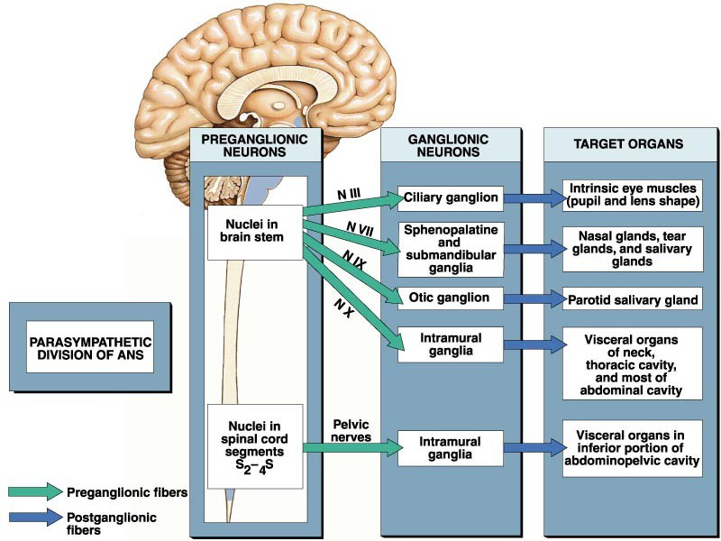
Which cranial nerves and spinal segments comprise the parasympathetic division?
Where are parasympathetic ganglia typically located relative to target organs?
What receptor mediates synaptic transmission at autonomic ganglia?
What receptor mediates parasympathetic effects on target organs?
Name the common pathway options for sympathetic preganglionic fibers after entering the paravertebral chain.
List major organ systems affected by autonomic divisions highlighted in the notes.
Which tissues does autonomic motor (GVE) control?
What are the two divisions of the autonomic nervous system (ANS)?
How many neurons make up each autonomic efferent pathway and what are they called?
Are general visceral afferent (GVA) fibers part of the autonomic nervous system?
What does it mean that target tissues may receive input from both autonomic divisions or only one branch?
What is 'dual control' in autonomic regulation?
What are 'antagonistic effects' in autonomic physiology?
What does 'opposing effects on the same cells' describe?
What does 'opposing effects on different cells' mean?
What are 'cooperative effects' in autonomic control?
What is 'solo control' in the autonomic nervous system?
What does 'antagonistic effects of dual control' mean in the autonomic nervous system?
How do sympathetic and parasympathetic inputs affect the heart when they act on the same effector cells?
Give an example where sympathetic and parasympathetic systems act on different effector cells.
Which iris muscle does the sympathetic system innervate?
In salivation, which gland type is stimulated by the parasympathetic division?
In salivation, which gland type is stimulated by the sympathetic division?
Which physiological variable is listed as 'Control without Dual innervations'?
What does sympathetic innervation alone cause in the context given?
What is the effect of increased sympathetic firing on blood vessels?
What is the effect of decreased sympathetic firing on blood vessels?
What does the label '1* neuron' represent in the diagram of autonomic pathways?
What does the label '2* neuron' indicate in the diagram?
What structure connects the preganglionic and postganglionic neurons?
What term is used for the bulbous ending of the postganglionic axon that interfaces with the target?
What is the space called between the terminal bulb and the receptor on the target?
Which elements are shown between a postganglionic axon and its target in the diagram?
Where are sympathetic preganglionic neuron cell bodies located?
Where are parasympathetic preganglionic neuron cell bodies located?
What neurotransmitter do all preganglionic autonomic neurons release?
What receptor type is found on autonomic ganglionic neurons?
Compare preganglionic axon lengths in sympathetic vs parasympathetic systems.
Compare postganglionic axon lengths in sympathetic vs parasympathetic systems.
What neurotransmitters are released by sympathetic postganglionic neurons?
What neurotransmitter is released by parasympathetic postganglionic neurons?
What receptor type mediates parasympathetic effects on target organs?
Where are sympathetic ganglia typically located?
Where are parasympathetic ganglia typically located?
What are the anatomical names for the two autonomic divisions?
State the basic efferent chain organization of the autonomic nervous system.
Name a sympathetic pathway option that connects to prevertebral ganglia.
Where are the preganglionic sympathetic cell bodies located?
Which cranial nerves contain parasympathetic nuclei?
Which spinal region provides parasympathetic outflow besides cranial nerves?
Name two types of sympathetic ganglia mentioned.
What is the ganglionic (nicotinic) neurotransmitter receptor type listed?
What neurotransmitter and receptor type act at parasympathetic effector organs?
What neurotransmitters and receptor type act at most sympathetic effector organs?
Which chemical transmitter is associated with the sympathetic division?
Which chemical transmitter is associated with the parasympathetic division?
What chemical does the suprarenal gland release according to the diagram?
Which transmitter is shown at sweat glands in the diagram?
In the diagram caption, what does 'N. ep.' abbreviate?
Where do parasympathetic preganglionic neurons originate?
Which cranial nerves carry parasympathetic preganglionic fibers?
Which nerves provide parasympathetic preganglionic fibers to pelvic organs?
Which ganglion supplies parasympathetic input to intrinsic eye muscles (pupil and lens)?
Which ganglia supply parasympathetic innervation to nasal, tear, and salivary glands?
Which parasympathetic ganglion innervates the parotid salivary gland?
What targets receive parasympathetic fibers from intramural ganglia?
What is the primary role of the sympathetic nervous system ('fight or flight')?
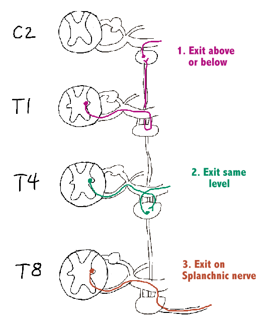
Where is the first (preganglionic) neuron of the sympathetic chain located?
What term describes the spinal outflow of sympathetic preganglionic axons from T1–L2?
Through which root do sympathetic preganglionic axons leave the spinal cord at T1–L2?
After leaving the ventral root, through which structure do preganglionic axons travel to enter the paravertebral ganglia?
What are the three possible continuations of sympathetic fibers after entering a paravertebral ganglion?
What does it mean when sympathetic fibers 'exit above or below' the paravertebral ganglion?
What happens when sympathetic fibers 'exit at the same level' of the paravertebral ganglion?
What does 'exit on a splanchnic nerve' indicate for sympathetic fibers?
What two systems are compared in the figure titled 'Figure 14-1'?
In the somatic arrangement shown, which structure contains sensory (afferent) neurons from the skin?
In the somatic arrangement shown, where do efferent neurons project to?
Name the two spinal roots labeled in the diagram.
Which structure on the autonomic side is labeled as the collection of linked ganglia?
Which two rami are shown connecting the sympathetic trunk and spinal nerves?
Which labeled structure lies between the sympathetic connector neuron and the viscus?
What label identifies the neuron that links spinal cord output to the sympathetic ganglion?
Which splanchnic nerve corresponds to spinal levels T5–T9?
Which splanchnic nerve corresponds to spinal levels T10–T11?
Which splanchnic nerve corresponds to spinal level T12?
Name four major prevertebral ganglia listed for the sympathetic division.
Do some fibers of the greater splanchnic nerve synapse on the adrenal medulla?
What region do the splanchnic nerves supply?
After a sympathetic neuron synapses on a ganglion, where do the postganglionic fibers travel?
What is the approximate ratio of preganglionic to postganglionic sympathetic fibers and what is its functional consequence?
What is the primary function of the parasympathetic nervous system?
Where are the first neurons (parasympathetic nuclei) located?
Describe the typical length and synapse location of preganglionic axons in the parasympathetic system.
What types of ganglia are present in the parasympathetic division?
Which cranial nerves synapse on the head ganglia?
Which nerves supply the thoracic/abdominal organs and which supply the pelvic organs in the parasympathetic system?
What is the approximate preganglionic:postganglionic fiber ratio in the parasympathetic division?
How does the parasympathetic preganglionic:postganglionic ratio affect control compared to the sympathetic system?
Name the four head ganglia listed.
How many inputs does a head ganglion receive?
Name the three types of inputs to a head ganglion.
What is the nature of the sympathetic fibers that enter a head ganglion?
What output does a head ganglion produce?
What is the parasympathetic input to the ciliary ganglion?
Which general sensory nerve contributes input to the ciliary ganglion?
How do sympathetic fibers reach the ciliary region according to the notes?
What is the output pathway from the ciliary ganglion?
What is the target of fibers from the ciliary ganglion?
What is the parasympathetic effect mediated via the ciliary ganglion on the pupil and lens?
What is the sympathetic effect on the pupil mediated in the ciliary region?
List the four main functional categories of input/output described for the ciliary ganglion.
What nerves provide inputs to the ciliary ganglion?
Through which nerves does the ciliary ganglion send outputs to the eye?
Which ocular muscles and structures are listed as targets of the ciliary ganglion outputs?
According to the image transcript, which ganglion is associated with the internal carotid plexus input?
Which gland is explicitly noted as NOT a target of the ciliary ganglion outputs?
What is the parasympathetic input to the sphenopalatine (pterygopalatine) ganglion?
Which nerve provides general sensory input to the sphenopalatine ganglion?
What is the sympathetic input to the sphenopalatine ganglion?
Which nerves carry output fibers from the sphenopalatine ganglion?
Which output nerve from the sphenopalatine ganglion targets the lacrimal gland?
What tissues are targeted by the nasal and palatine output nerves of the sphenopalatine ganglion?
What is the name of the ganglion that receives input from the greater petrosal and deep petrosal nerves?
Which cranial nerve provides parasympathetic input to the pterygopalatine ganglion via the greater petrosal nerve?
Which sympathetic source reaches the pterygopalatine ganglion via the deep petrosal nerve?
Name the main sensory/branchial nerve listed that carries fibers associated with the pterygopalatine ganglion.
What are the primary effector targets of the pterygopalatine ganglion outputs?
What provides the parasympathetic input to the otic ganglion?
Which nerve provides general sensory fibers to the otic ganglion?
Where do the sympathetic fibers to the otic ganglion originate?
Which nerves carry output (postganglionic) fibers from the otic ganglion?
What is the parasympathetic target of the otic ganglion carried by the auriculotemporal nerve?
What target does the mandibular branch of V3 relay from the otic ganglion?
Which cranial nerve provides parasympathetic input to the otic ganglion via the lesser petrosal nerve?
Which branch of the trigeminal nerve provides sensory input to the otic ganglion?
Name two nerve branches that carry outputs from the otic ganglion to the parotid region.
What is a primary target organ of fibers leaving the otic ganglion?
Besides the parotid gland, what tissue is supplied by outputs from the otic ganglion?
Provide the full URL for the diagram image of the otic ganglion used as reference.

What is the parasympathetic input to the submandibular ganglion?
Which nerve provides general sensory input to the submandibular ganglion?
What supplies sympathetic fibers to the submandibular ganglion?
Which nerve carries output fibers from the submandibular ganglion?
What are the target structures innervated via the submandibular ganglion?
Which cranial nerve provides parasympathetic input to the submandibular ganglion via the chorda tympani?
Which trigeminal branch carries the chorda tympani fibers to and from the submandibular ganglion?
What sympathetic structure supplies input to the submandibular ganglion region?
What are the main secretomotor targets of fibers leaving the submandibular ganglion?
Where can a diagram of the submandibular ganglion pathways (facial, lingual, external carotid plexus) be seen?
In the diagram of autonomic efferent pathways, which color represents preganglionic parasympathetic fibers?
In the diagram of autonomic efferent pathways, which color represents postganglionic parasympathetic fibers?
In the diagram of autonomic efferent pathways, which color represents preganglionic sympathetic fibers?
In the diagram of autonomic efferent pathways, which color represents postganglionic sympathetic fibers?
Name three cranial or peripheral ganglia labeled in the efferent ANS diagram.
List five target organs shown in the efferent ANS diagram.
Which cranial nerve-related structures are listed in the diagram transcript besides ganglia and organs?
Provide the full URL of the diagram image for the efferent autonomic pathways (useful for review).

Trace the parasympathetic pathway from the inferior salivatory nucleus to the parotid gland.
Trace the parasympathetic pathway from the lacrimatory nucleus of the facial nerve to the lacrimal gland.
Which cranial nerves are shown supplying parasympathetic pathways to the parotid and lacrimal glands?
Which ganglion mediates parasympathetic innervation to the parotid gland?
Which ganglion mediates parasympathetic innervation to the lacrimal gland?
Which sympathetic structure is shown associated with head autonomic innervation in the diagram?
Which nerve connects the otic ganglion to the parotid gland?

Which ganglion receives parasympathetic fibers from the oculomotor (III) nucleus to innervate the iris?
Which nucleus in the midbrain supplies parasympathetic output for the oculomotor nerve?
Which sympathetic ganglion is listed as supplying structures of the upper eyelid?
Name the muscle of the upper eyelid mentioned in the diagram.
Which brainstem nucleus provides parasympathetic output for the facial nerve to salivary glands?
Through which branch does the facial nerve convey fibers to the submandibular ganglion?
Which ganglion is associated with parasympathetic innervation of the submandibular salivary gland?
Name one arterial plexus mentioned that connects to salivary gland innervation.
Which salivary glands are shown in the diagram (two names)?
View the diagram illustrating autonomic supply to the eyelid, iris, and salivary glands.
What is the basic two-neuron efferent chain of the autonomic nervous system?
What spinal segments give rise to the sympathetic division?
Which neurotransmitters do sympathetic postganglionic effector neurons primarily use?
Which cranial nerves and spinal segments comprise the parasympathetic (craniosacral) division?
What receptor type is found at autonomic ganglia?
Name three pathway options sympathetic preganglionic fibers can take after entering the sympathetic trunk.
Which nerve carries medullary parasympathetic output to the heart and lungs?

List major organ effects included in the core concepts of autonomic divisions.
What is the basic two‑neuron efferent chain of the autonomic nervous system?
Which spinal segments supply sympathetic outflow?
Which cranial nerves and sacral segments carry parasympathetic outflow?
What types of ganglia do sympathetic preganglionic fibers synapse in for abdominal viscera?
Which receptor mediates transmission at autonomic ganglia?
Which neurotransmitters are the primary effectors for sympathetic and parasympathetic divisions?
Name the major prevertebral ganglia that supply sympathetic innervation to the gastrointestinal tract.
List common pathway options taken by sympathetic fibers to reach target ganglia.
Which pathways provide sympathetic innervation to the kidney in the diagram?
Which cranial nerve provides parasympathetic input to the kidney region shown in the diagram?
Name three ganglia or plexuses labeled in the autonomic innervation diagram of the kidney and suprarenal gland.
Which structure is shown alongside the kidney as a target of autonomic innervation in the diagram?
Which spinal levels provide the sympathetic fibers to the sphincters of the anal canal and urinary bladder?
Which spinal segments supply parasympathetic innervation to the sphincter vesicae via the pelvic splanchnic nerves?
Through which plexuses do autonomic fibers travel to reach the urinary bladder and anal canal sphincters?
Name the sympathetic structure shown as a conduit for thoracolumbar autonomic fibers to pelvic organs.
Referencing the diagram, which nerve is explicitly labeled as originating from S2, 3, 4?
What effect does the sympathetic nervous system have on the pupil?
What effect does the parasympathetic nervous system have on the pupil?
How does the sympathetic system affect the ciliary muscle (eye)?
How does the parasympathetic system affect the ciliary muscle (eye)?
What is the sympathetic effect on heart force of contraction?
What is the parasympathetic effect on heart force of contraction?
How does the sympathetic nervous system affect bronchial smooth muscle?
How does the parasympathetic nervous system affect bronchial smooth muscle?
What effect does the sympathetic system have on gastrointestinal (GI) tract peristalsis?
What effect does the parasympathetic system have on gastrointestinal (GI) tract peristalsis?
What sympathetic action occurs in the liver?
How does the sympathetic system affect the gallbladder?
How does the parasympathetic system affect the gallbladder?
What is the sympathetic effect on kidney output and why?
What are the sympathetic actions on the urinary bladder wall and sphincter?
What are the parasympathetic actions on the urinary bladder wall and sphincter?
What parasympathetic action occurs in erectile tissue of the penis and clitoris?
What sympathetic action produces ejaculation?
How do sympathetic and parasympathetic actions differ regarding glandular secretion?
How does parasympathetic activity affect bronchial secretions?
What is the basic efferent chain organization of the autonomic nervous system?
Which spinal segments give rise to the sympathetic division?
Which neurotransmitter do most sympathetic postganglionic neurons release onto effectors?
Which cranial nerves and spinal segments contribute to the parasympathetic division?
What receptor mediates transmission at autonomic ganglia?
Which receptor type mediates parasympathetic effects on target organs?
Name one pathway option for sympathetic preganglionic fibers after entering the sympathetic trunk.
Give three major organ systems affected by autonomic divisions mentioned in the notes.
Flashcards in this deck (232)
-
What is the basic efferent chain structure of the autonomic nervous system?
- Two‑neuron chain: preganglionic → ganglion → postganglionic
ans structure -
sympathetic anatomy
-
sympathetic ganglia
-
What are the main neurotransmitters released by sympathetic pre/postganglionic neurons and effectors?
- Preganglionic: ACh at ganglion (nicotinic)
- Effectors: NE or E at target organs
sympathetic neurotransmitters -
Which cranial nerves and spinal segments comprise the parasympathetic division?
- CN III, VII, IX, X and S2–S4
parasympathetic anatomy -
Where are parasympathetic ganglia typically located relative to target organs?
- Intramural ganglia (within or very near target organs)
parasympathetic ganglia -
ganglia receptors
-
parasympathetic receptors
-
Name the common pathway options for sympathetic preganglionic fibers after entering the paravertebral chain.
- Ascend/descend, synapse at same level, or pass through as splanchnic to prevertebral ganglia
sympathetic pathways -
List major organ systems affected by autonomic divisions highlighted in the notes.
- **Heart
- Pupils
- Lungs
- Gastrointestinal tract
- Bladder
- Adrenal medulla**
ans organs -
ans gve tissues
-
ans divisions
-
How many neurons make up each autonomic efferent pathway and what are they called?
- Two neurons: preganglionic → postganglionic
ans pathway neurons -
Are general visceral afferent (GVA) fibers part of the autonomic nervous system?
GVA fibers can travel with GVE nerves but are not explicitly part of the autonomic nervous system
ans gva afferent -
What does it mean that target tissues may receive input from both autonomic divisions or only one branch?
Some organs get innervation from both sympathetic and parasympathetic divisions (dual control), while others receive input from only one branch (solo control).
autonomic innervation -
What is 'dual control' in autonomic regulation?
Dual control is when a target tissue receives input from both sympathetic and parasympathetic divisions.
autonomic dual -
What are 'antagonistic effects' in autonomic physiology?
Antagonistic effects occur when the two divisions produce opposite actions on the same physiological function.
autonomic antagonistic -
What does 'opposing effects on the same cells' describe?
It describes both autonomic divisions acting directly on the same target cells with opposite effects.
autonomic opposing -
What does 'opposing effects on different cells' mean?
It means sympathetic and parasympathetic divisions produce opposite overall effects by acting on different cell populations within a tissue.
autonomic opposing -
What are 'cooperative effects' in autonomic control?
Cooperative effects occur when sympathetic and parasympathetic actions complement each other to achieve a common outcome.
autonomic cooperative -
What is 'solo control' in the autonomic nervous system?
Solo control refers to a target tissue being innervated and regulated by only one autonomic division.
autonomic solo -
What does 'antagonistic effects of dual control' mean in the autonomic nervous system?
It means the sympathetic and parasympathetic systems produce opposite effects on the same organ or related cells.
autonomic antagonistic -
How do sympathetic and parasympathetic inputs affect the heart when they act on the same effector cells?
- Sympathetic: speeds the heart up
- Parasympathetic: slows the heart down
heart cardiac -
Give an example where sympathetic and parasympathetic systems act on different effector cells.
The iris: sympathetic innervates the pupillary dilator muscle, while parasympathetic innervates the constrictor muscles.
eye pupil -
iris sympathetic
-
In salivation, which gland type is stimulated by the parasympathetic division?
- Serous glands (produce enzymes)
autonomic parasympathetic salivation -
In salivation, which gland type is stimulated by the sympathetic division?
- Mucous cells (produce mucous)
autonomic sympathetic salivation -
autonomics bloodpressure
-
What does sympathetic innervation alone cause in the context given?
Opposite effects (when only sympathetic innervation is present)
sympathetic autonomics -
vasoconstriction sympathetic
-
vasodilation sympathetic
-
What does the label '1* neuron' represent in the diagram of autonomic pathways?
The '1 neuron' is the preganglionic neuron*.
autonomic neuron -
What does the label '2* neuron' indicate in the diagram?
The '2 neuron' is the postganglionic neuron located in a ganglion*.
autonomic ganglion -
What structure connects the preganglionic and postganglionic neurons?
A ganglion connects the preganglionic and postganglionic neurons.
autonomic ganglion -
What term is used for the bulbous ending of the postganglionic axon that interfaces with the target?
The bulbous ending is called the terminal bulb (axon terminal).
synapse axon -
What is the space called between the terminal bulb and the receptor on the target?
That space is the synaptic cleft.
synapse cleft -
Which elements are shown between a postganglionic axon and its target in the diagram?
- Terminal bulb
- Synaptic cleft
- Receptor
- Target
 autonomic synapse
 -
sympathetic ans
-
Where are parasympathetic preganglionic neuron cell bodies located?
Cranial nerve nuclei (III, VII, IX, X) and sacral neurons S2–4
parasympathetic ans -
neurotransmitter ans
-
receptor ans
-
Compare preganglionic axon lengths in sympathetic vs parasympathetic systems.
- Sympathetic: Short
- Parasympathetic: Long
axon ans -
Compare postganglionic axon lengths in sympathetic vs parasympathetic systems.
- Sympathetic: Long
- Parasympathetic: Short
axon ans -
What neurotransmitters are released by sympathetic postganglionic neurons?
Epinephrine and Norepinephrine
sympathetic neurotransmitter -
parasympathetic neurotransmitter
-
parasympathetic receptor
-
sympathetic ganglia
-
Where are parasympathetic ganglia typically located?
- Head ganglia or ganglia on/near target organ (intramural)
parasympathetic ganglia -
What are the anatomical names for the two autonomic divisions?
- Sympathetic: thoracolumbar
- Parasympathetic: craniosacral
ans divisions -
State the basic efferent chain organization of the autonomic nervous system.
- Two‑neuron chain: preganglionic → ganglion → postganglionic
ans structure -
Name a sympathetic pathway option that connects to prevertebral ganglia.
Splanchnic pathway → prevertebral ganglion
sympathetic pathway -
sympathetic anatomy
-
parasympathetic cranial
-
parasympathetic sacral
-
Name two types of sympathetic ganglia mentioned.
- Paravertebral (sympathetic chain) ganglia
- Prevertebral ganglia
sympathetic ganglia -
neurotransmitter receptor
-
What neurotransmitter and receptor type act at parasympathetic effector organs?
- ACh at muscarinic ACh receptors
parasympathetic neurotransmitter -
What neurotransmitters and receptor type act at most sympathetic effector organs?
- NE/E (norepinephrine/epinephrine) at adrenergic receptors
sympathetic neurotransmitter -
autonomic sympathetic neurotransmitter
-
autonomic parasympathetic neurotransmitter
-
autonomic adrenal hormone
-
autonomic sweat neurotransmitter
-
autonomic abbreviation neurotransmitter
-
Where do parasympathetic preganglionic neurons originate?
- Nuclei in the brain stem
- Nuclei in spinal cord segments S2–4S
parasympathetic anatomy -
cranialnerves parasympathetic
-
Which nerves provide parasympathetic preganglionic fibers to pelvic organs?
- Pelvic nerves originating from spinal segments S2–4S
pelvic parasympathetic -
Which ganglion supplies parasympathetic input to intrinsic eye muscles (pupil and lens)?
- Ciliary ganglion
eye ganglion -
Which ganglia supply parasympathetic innervation to nasal, tear, and salivary glands?
- Sphenopalatine and submandibular ganglia
salivation glands -
parotid ganglion
-
What targets receive parasympathetic fibers from intramural ganglia?
- Visceral organs of neck, thoracic cavity, and most of abdominal cavity
- Visceral organs in inferior abdominopelvic cavity
 viscera intramural
 -
What is the primary role of the sympathetic nervous system ('fight or flight')?
To prepare the body for an emergency and intense muscle activity.
sympathetic function -
Where is the first (preganglionic) neuron of the sympathetic chain located?
The intermediolateral nucleus in the lateral horn at spinal levels T1–L2.
sympathetic anatomy -
What term describes the spinal outflow of sympathetic preganglionic axons from T1–L2?
Thoracolumbar outflow.
sympathetic terminology -
Through which root do sympathetic preganglionic axons leave the spinal cord at T1–L2?
They project out the ventral root.
sympathetic pathway -
After leaving the ventral root, through which structure do preganglionic axons travel to enter the paravertebral ganglia?
They travel in the white rami communicantes to enter the paravertebral (sympathetic chain) ganglia.
sympathetic pathway -
What are the three possible continuations of sympathetic fibers after entering a paravertebral ganglion?
- Travel up or down the chain to a different ganglion, then synapse and exit via the gray rami communicantes
- Synapse on the ganglion at that level, then exit via the gray rami communicantes
- Exit the chain on a splanchnic nerve and synapse on a prevertebral ganglion
 sympathetic pathways ans
 -
What does it mean when sympathetic fibers 'exit above or below' the paravertebral ganglion?
- Fibers travel up or down the sympathetic chain to a different ganglion, then synapse there and exit via the gray rami communicantes
sympathetic pathways -
What happens when sympathetic fibers 'exit at the same level' of the paravertebral ganglion?
- Preganglionic fibers synapse on the ganglion at that same level, then postganglionic fibers exit via the gray rami communicantes
sympathetic pathways -
What does 'exit on a splanchnic nerve' indicate for sympathetic fibers?
- Preganglionic fibers leave the sympathetic chain as splanchnic nerves and synapse on prevertebral (collateral) ganglia
sympathetic splanchnic pathways -
What two systems are compared in the figure titled 'Figure 14-1'?
The general arrangement of the somatic part of the nervous system (left) and the autonomic part of the nervous system (right).
overview ans -
In the somatic arrangement shown, which structure contains sensory (afferent) neurons from the skin?
Posterior root contains the afferent neurons from the skin.
somatic afferent -
In the somatic arrangement shown, where do efferent neurons project to?
Efferent neurons project to muscle.
somatic efferent -
spine roots
-
Which structure on the autonomic side is labeled as the collection of linked ganglia?
Sympathetic trunk.
autonomic sympathetic -
sympathetic ramus
-
Which labeled structure lies between the sympathetic connector neuron and the viscus?
Sympathetic ganglion.
sympathetic ganglion -
What label identifies the neuron that links spinal cord output to the sympathetic ganglion?
Sympathetic connector neuron.
sympathetic neuron -
sympathetic splanchnic
-
splanchnic sympathetic
-
splanchnic sympathetic
-
Name four major prevertebral ganglia listed for the sympathetic division.
- Celiac ganglion
- Renal ganglion
- Superior mesenteric ganglion
- Inferior mesenteric ganglion
ganglia sympathetic -
Do some fibers of the greater splanchnic nerve synapse on the adrenal medulla?
Yes, some fibers synapse directly on the adrenal medulla.
adrenal splanchnic -
splanchnic viscera
-
After a sympathetic neuron synapses on a ganglion, where do the postganglionic fibers travel?
To the target cells (effector organs or tissues).
sympathetic pathway -
What is the approximate ratio of preganglionic to postganglionic sympathetic fibers and what is its functional consequence?
About 1:10, producing widespread effects.
sympathetic physiology -
What is the primary function of the parasympathetic nervous system?
To conserve and store energy; described as 'rest and digest'.
parasympathetic function -
Where are the first neurons (parasympathetic nuclei) located?
In cranial nerve nuclei III, VII, IX, and X and in spinal cord levels S2–S4.
anatomy nuclei -
Describe the typical length and synapse location of preganglionic axons in the parasympathetic system.
Preganglionic axons are long and synapse on ganglia in, on, or near the target organ.
pathway axon -
What types of ganglia are present in the parasympathetic division?
There are four head ganglia and other intramural visceral ganglia.
ganglia parasympathetic -
Which cranial nerves synapse on the head ganglia?
Cranial nerves III, VII, and IX synapse on the head ganglia.
cranial ganglia -
Which nerves supply the thoracic/abdominal organs and which supply the pelvic organs in the parasympathetic system?
- Thorax and abdomen: Cranial nerve X (CN X)
- Pelvic organs: Spinal levels S2–S4
organ innervation -
What is the approximate preganglionic:postganglionic fiber ratio in the parasympathetic division?
About 1:3 (or less)
parasympathetic ans -
How does the parasympathetic preganglionic:postganglionic ratio affect control compared to the sympathetic system?
A ~1:3 (or less) ratio allows more fine control compared to the sympathetic nervous system
parasympathetic control -
Name the four head ganglia listed.
- Ciliary ganglion
- Pterygopalatine ganglion
- Otic ganglion
- Submandibular ganglion
neuroanatomy ganglia -
anatomy ganglion autonomic
-
Name the three types of inputs to a head ganglion.
- Parasympathetic fibers that synapse in the ganglion
- Sympathetic postganglionic fibers from the superior cervical ganglion that pass through without synapse
- GSA (general sensory) fibers that pass through without synapse
anatomy inputs ganglion -
What is the nature of the sympathetic fibers that enter a head ganglion?
They are postganglionic fibers from the superior cervical ganglion that pass through the head ganglion without synapsing.
sympathetic ganglion anatomy -
What output does a head ganglion produce?
- One output: the nerve that leaves the ganglion to the target cells
 outputs ganglion anatomy
 -
anatomy parasympathetic
-
Which general sensory nerve contributes input to the ciliary ganglion?
- Nasociliary nerve of V1
- Some from long ciliary nerve
anatomy sensory -
How do sympathetic fibers reach the ciliary region according to the notes?
- Preganglionic neuron synapses at superior cervical ganglion; postganglionic fibers continue along the internal carotid plexus
anatomy sympathetic -
anatomy pathway
-
anatomy eye
-
What is the parasympathetic effect mediated via the ciliary ganglion on the pupil and lens?
- Activates constrictor pupillae and ciliary muscles → pupil constricts and focus shifts to near objects
physiology parasympathetic -
What is the sympathetic effect on the pupil mediated in the ciliary region?
- Activates dilator pupillae → pupil dilates
physiology sympathetic -
List the four main functional categories of input/output described for the ciliary ganglion.
- Input: Parasympathetic (oculomotor)
- Input: General sensory (nasociliary, long ciliary)
- Input: Sympathetic (via superior cervical ganglion/internal carotid plexus)
- Output: Short ciliary nerves to internal eye structures
summary anatomy -
What nerves provide inputs to the ciliary ganglion?
- Oculomotor nerve (III)
- Nasociliary nerve (V1)
- Internal carotid plexus
anatomy ciliaryganglion cranialnerves -
anatomy ciliaryganglion nerves
-
Which ocular muscles and structures are listed as targets of the ciliary ganglion outputs?
- Dilator pupillae
- Sphincter pupillae
- Ciliary muscles
physiology eye ciliaryganglion -
According to the image transcript, which ganglion is associated with the internal carotid plexus input?
- Superior cervical ganglion
anatomy sympathetic cervical -
anatomy eye targets
-
What is the parasympathetic input to the sphenopalatine (pterygopalatine) ganglion?
- Greater petrosal nerve branch of the facial nerve via the nerve of pterygoid canal
neuroanatomy parasympathetic facial -
neuroanatomy sensory v2
-
What is the sympathetic input to the sphenopalatine ganglion?
- Deep petrosal nerve via the nerve of pterygoid canal
neuroanatomy sympathetic -
Which nerves carry output fibers from the sphenopalatine ganglion?
- Zygomatic nerve (V2)
- Nasal and palatine nerves (V2)
neuroanatomy v2 outputs -
Which output nerve from the sphenopalatine ganglion targets the lacrimal gland?
- Zygomatic nerve (V2)
neuroanatomy lacrimal -
What tissues are targeted by the nasal and palatine output nerves of the sphenopalatine ganglion?
- Mucosa of the nose, pharynx, and palate
neuroanatomy mucosa -
What is the name of the ganglion that receives input from the greater petrosal and deep petrosal nerves?
Pterygopalatine (sphenopalatine) ganglion
pterygopalatine ganglion -
Which cranial nerve provides parasympathetic input to the pterygopalatine ganglion via the greater petrosal nerve?
Facial nerve (VII) via the greater petrosal nerve
parasympathetic cranialnerves -
Which sympathetic source reaches the pterygopalatine ganglion via the deep petrosal nerve?
Internal carotid plexus (from superior cervical ganglion) via the deep petrosal nerve
sympathetic internalcarotid -
Name the main sensory/branchial nerve listed that carries fibers associated with the pterygopalatine ganglion.
Maxillary nerve (V2)
v2 trigeminal -
What are the primary effector targets of the pterygopalatine ganglion outputs?
- Lacrimal gland (secretomotor via zygomatic/lacrimal route)
- Mucosa of nose, palate, pharynx (secretomotor via nasal and palatine nerves)
targets secretomotor -
What provides the parasympathetic input to the otic ganglion?
- Glossopharyngeal nerve via the lesser petrosal nerve
otic parasympathetic glossopharyngeal -
otic sensory v3
-
otic sympathetic carotid
-
Which nerves carry output (postganglionic) fibers from the otic ganglion?
- Auriculotemporal (V3) and mandibular (V3)
otic output v3 -
What is the parasympathetic target of the otic ganglion carried by the auriculotemporal nerve?
- Parotid gland
otic parotid auriculotemporal -
otic oral v3
-
Which cranial nerve provides parasympathetic input to the otic ganglion via the lesser petrosal nerve?
- Glossopharyngeal nerve (IX) via the lesser petrosal nerve
otic parasympathetic glossopharyngeal -
Which branch of the trigeminal nerve provides sensory input to the otic ganglion?
- Mandibular nerve (V3)
otic trigeminal v3 -
Name two nerve branches that carry outputs from the otic ganglion to the parotid region.
- Auriculotemporal (V3)
- Mandibular (V3)
otic outputs v3 -
otic parotid gland
-
otic oral mucosa
-
media image otic
-
What is the parasympathetic input to the submandibular ganglion?
The parasympathetic input is the chorda tympani of the facial nerve via the lingual nerve.
anatomy parasympathetic cranialnerve -
Which nerve provides general sensory input to the submandibular ganglion?
General sensory input is provided by the lingual nerve (V3).
anatomy sensory v3 -
What supplies sympathetic fibers to the submandibular ganglion?
Sympathetic fibers are supplied by the external carotid plexus.
anatomy sympathetic plexus -
Which nerve carries output fibers from the submandibular ganglion?
Output fibers travel via the lingual nerve (V3).
anatomy output v3 -
What are the target structures innervated via the submandibular ganglion?
- Submandibular glands
- Sublingual glands
- Oral mucosa
anatomy glands innervation -
Which cranial nerve provides parasympathetic input to the submandibular ganglion via the chorda tympani?
Facial nerve (VII) via the chorda tympani (running with the lingual nerve)
anatomy cranialnerves parasympathetic -
Which trigeminal branch carries the chorda tympani fibers to and from the submandibular ganglion?
Lingual nerve (branch of V3)
lingual anatomy v3 -
What sympathetic structure supplies input to the submandibular ganglion region?
External carotid plexus originating from the superior cervical ganglion
anatomy sympathetic cervical -
What are the main secretomotor targets of fibers leaving the submandibular ganglion?
- Submandibular gland
- Sublingual gland
- Oral mucosa
glands secretomotor anatomy -
Where can a diagram of the submandibular ganglion pathways (facial, lingual, external carotid plexus) be seen?
Diagram:
 Caption: shows facial (VII)/chorda tympani via lingual, external carotid plexus, and outputs to submandibular/sublingual glandsdiagram reference anatomy
 -
In the diagram of autonomic efferent pathways, which color represents preganglionic parasympathetic fibers?
Preganglionic parasympathetic fibers are shown in solid blue.
ans parasympathetic diagram -
In the diagram of autonomic efferent pathways, which color represents postganglionic parasympathetic fibers?
Postganglionic parasympathetic fibers are shown in interrupted blue.
ans parasympathetic diagram -
In the diagram of autonomic efferent pathways, which color represents preganglionic sympathetic fibers?
Preganglionic sympathetic fibers are shown in solid red.
ans sympathetic diagram -
In the diagram of autonomic efferent pathways, which color represents postganglionic sympathetic fibers?
Postganglionic sympathetic fibers are shown in interrupted red.
ans sympathetic diagram -
Name three cranial or peripheral ganglia labeled in the efferent ANS diagram.
- Ciliary ganglion
- Otic ganglion
- Submandibular (and sublingual) ganglia
ans ganglia structures -
ans organs diagram
-
Which cranial nerve-related structures are listed in the diagram transcript besides ganglia and organs?
- Pelvic splanchnic nerve
- Lacrimal gland
- Parotid gland
ans nerves structures -
ans image reference
-
Trace the parasympathetic pathway from the inferior salivatory nucleus to the parotid gland.
- Inferior salivatory nucleus
- Tympanic branch
- Tympanic plexus
- Lesser petrosal nerve
- Otic ganglion
- Auriculotemporal nerve
- Parotid salivary gland
parotid glossopharyngeal parasympathetic -
Trace the parasympathetic pathway from the lacrimatory nucleus of the facial nerve to the lacrimal gland.
- Lacrimatory nucleus (facial nerve)
- Greater petrosal nerve
- Nerve of pterygoid canal
- Pterygopalatine ganglion
- Zygomatic nerve
- Zygomaticotemporal nerve
- Lacrimal gland
lacrimal facial parasympathetic -
Which cranial nerves are shown supplying parasympathetic pathways to the parotid and lacrimal glands?
- Glossopharyngeal nerve (IX)
- Facial nerve (VII)
cranialnerves glossopharyngeal facial -
otic ganglion parotid
-
pterygopalatine ganglion lacrimal
-
Which sympathetic structure is shown associated with head autonomic innervation in the diagram?
- Superior cervical sympathetic ganglion
sympathetic superiorcervical ganglion -
auriculotemporal parotid image
-
Which ganglion receives parasympathetic fibers from the oculomotor (III) nucleus to innervate the iris?
- Ciliary ganglion
ocular parasympathetic -
Which nucleus in the midbrain supplies parasympathetic output for the oculomotor nerve?
- Parasympathetic nucleus of the oculomotor nerve
midbrain ocular -
Which sympathetic ganglion is listed as supplying structures of the upper eyelid?
- Superior cervical sympathetic ganglion
sympathetic eyelid -
anatomy eyelid
-
Which brainstem nucleus provides parasympathetic output for the facial nerve to salivary glands?
- Superior salivatory nucleus of the facial nerve
pons salivary -
Through which branch does the facial nerve convey fibers to the submandibular ganglion?
- Chorda tympani
facial salivary -
Which ganglion is associated with parasympathetic innervation of the submandibular salivary gland?
- Submandibular ganglion
salivary ganglion -
Name one arterial plexus mentioned that connects to salivary gland innervation.
- External carotid arterial plexus
vascular salivary -
Which salivary glands are shown in the diagram (two names)?
- Sublingual salivary gland
- Submandibular salivary gland
salivary glands -
View the diagram illustrating autonomic supply to the eyelid, iris, and salivary glands.
- Diagram:
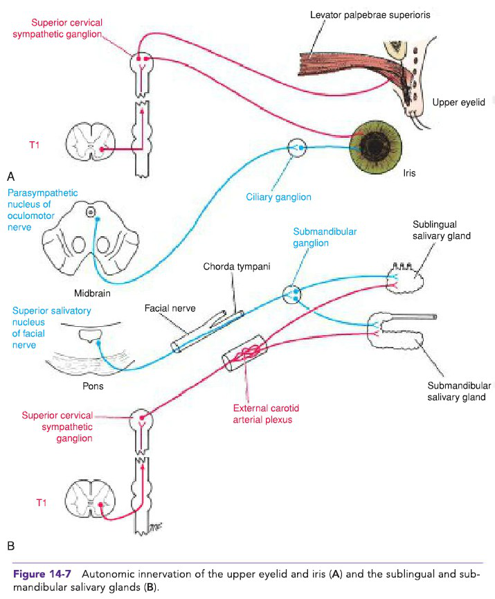
- Labels include: superior cervical sympathetic ganglion, ciliary ganglion, parasympathetic nuclei, chorda tympani, and salivary glands.
diagram illustration - Diagram:
-
What is the basic two-neuron efferent chain of the autonomic nervous system?
- Preganglionic neuron → ganglion → postganglionic neuron
ans pathway -
sympathetic ans
-
Which neurotransmitters do sympathetic postganglionic effector neurons primarily use?
- Norepinephrine (NE) and epinephrine (E)
sympathetic neurotransmitter -
Which cranial nerves and spinal segments comprise the parasympathetic (craniosacral) division?
- CN III, VII, IX, X and S2–S4
parasympathetic ans -
receptors ans
-
Name three pathway options sympathetic preganglionic fibers can take after entering the sympathetic trunk.
- **Ascend or descend to other ganglia
- Synapse at the same level
- Pass through as splanchnic fibers to prevertebral ganglia**
sympathetic pathways -
vagus parasympathetic image
-
List major organ effects included in the core concepts of autonomic divisions.
- Heart, pupil, lungs, gastrointestinal tract, bladder, adrenal medulla
effects organs -
What is the basic two‑neuron efferent chain of the autonomic nervous system?
- Preganglionic neuron → Ganglion → Postganglionic neuron
anatomy autonomic -
sympathetic ans
-
Which cranial nerves and sacral segments carry parasympathetic outflow?
- CN III, VII, IX, X (cranial)
- S2–S4 (sacral)
parasympathetic ans -
What types of ganglia do sympathetic preganglionic fibers synapse in for abdominal viscera?
- Paravertebral (sympathetic trunk) or prevertebral (e.g., celiac, superior/inferior mesenteric) ganglia
sympathetic ganglia -
receptors pharmacology
-
Which neurotransmitters are the primary effectors for sympathetic and parasympathetic divisions?
- Sympathetic effectors: norepinephrine / epinephrine (NE/E)
- Parasympathetic effectors: acetylcholine (ACh) acting at muscarinic receptors
neurotransmitters ans -
Name the major prevertebral ganglia that supply sympathetic innervation to the gastrointestinal tract.
- Celiac ganglion
- Superior mesenteric ganglion
- Inferior mesenteric ganglion
 gi sympathetic
 -
List common pathway options taken by sympathetic fibers to reach target ganglia.
- Ascend/descend in sympathetic trunk
- Synapse at same level
- Pass as splanchnic nerves → synapse in prevertebral ganglia
pathways sympathetic -
Which pathways provide sympathetic innervation to the kidney in the diagram?
Pathways from the thoracic spinal cord via the sympathetic trunk and splanchnic nerves.
sympathetic kidney -
Which cranial nerve provides parasympathetic input to the kidney region shown in the diagram?
The vagus nerve (from the medulla oblongata).
parasympathetic vagus -
Name three ganglia or plexuses labeled in the autonomic innervation diagram of the kidney and suprarenal gland.
- Celiac ganglion
- Renal ganglion in renal plexus
- Renal plexus
ganglia renal -
Which structure is shown alongside the kidney as a target of autonomic innervation in the diagram?
The suprarenal gland (adrenal gland).
suprarenal adrenal -
Which spinal levels provide the sympathetic fibers to the sphincters of the anal canal and urinary bladder?
T11–L2
autonomic sympathetic spinal_levels -
Which spinal segments supply parasympathetic innervation to the sphincter vesicae via the pelvic splanchnic nerves?
S2–S4 via pelvic splanchnic nerves
autonomic parasympathetic pelvic -
Through which plexuses do autonomic fibers travel to reach the urinary bladder and anal canal sphincters?
Hypogastric plexuses
autonomic hypogastric pathway -
Name the sympathetic structure shown as a conduit for thoracolumbar autonomic fibers to pelvic organs.
Sympathetic trunk
autonomic sympathetic anatomy -
Referencing the diagram, which nerve is explicitly labeled as originating from S2, 3, 4?
Pelvic splanchnic nerve
 autonomic diagram pelvic
 -
eye autonomic sympathetic
-
eye autonomic parasympathetic
-
eye ciliary sympathetic
-
How does the parasympathetic system affect the ciliary muscle (eye)?
It contracts the ciliary muscle.
eye ciliary parasympathetic -
What is the sympathetic effect on heart force of contraction?
It increases the force of contraction of cardiac muscle.
heart autonomic sympathetic -
What is the parasympathetic effect on heart force of contraction?
It decreases the force of contraction.
heart autonomic parasympathetic -
How does the sympathetic nervous system affect bronchial smooth muscle?
It relaxes (dilates) the bronchi.
lung bronchi sympathetic -
How does the parasympathetic nervous system affect bronchial smooth muscle?
It contracts (constricts) the bronchi.
lung bronchi parasympathetic -
What effect does the sympathetic system have on gastrointestinal (GI) tract peristalsis?
It decreases peristalsis.
sympathetic gastrointestinal gi -
What effect does the parasympathetic system have on gastrointestinal (GI) tract peristalsis?
It increases peristalsis.
gastrointestinal gi parasympathetic -
liver metabolism sympathetic
-
gallbladder sympathetic
-
gallbladder parasympathetic
-
What is the sympathetic effect on kidney output and why?
It decreases output due to constriction of renal arteries.
kidney renal sympathetic -
What are the sympathetic actions on the urinary bladder wall and sphincter?
Bladder wall (detrusor) relaxes; sphincter vesicae contracts.
bladder urinary sympathetic -
What are the parasympathetic actions on the urinary bladder wall and sphincter?
Bladder wall contracts; sphincter vesicae relaxes.
bladder urinary parasympathetic -
What parasympathetic action occurs in erectile tissue of the penis and clitoris?
It relaxes erectile tissue, causing erection.
erectile sexual parasympathetic -
What sympathetic action produces ejaculation?
It contracts smooth muscle of the vas deferens, seminal vesicles, and prostate.
ejaculation sympathetic reproductive -
How do sympathetic and parasympathetic actions differ regarding glandular secretion?
Sympathetic reduces secretion by vasoconstriction of blood vessels (and increases sweat); parasympathetic increases secretion.
glands secretion autonomic -
lung secretions parasympathetic
-
What is the basic efferent chain organization of the autonomic nervous system?
- Two‑neuron chain: preganglionic → ganglion → postganglionic
ans organization -
sympathetic anatomy
-
Which neurotransmitter do most sympathetic postganglionic neurons release onto effectors?
- Norepinephrine (NE)
sympathetic neurotransmitter -
Which cranial nerves and spinal segments contribute to the parasympathetic division?
- CN III, VII, IX, X and S2–S4
parasympathetic anatomy -
receptors ganglia
-
Which receptor type mediates parasympathetic effects on target organs?
- Muscarinic acetylcholine (ACh) receptor
parasympathetic receptors -
Name one pathway option for sympathetic preganglionic fibers after entering the sympathetic trunk.
- Ascend or descend in the trunk before synapsing
sympathetic pathways -
Give three major organ systems affected by autonomic divisions mentioned in the notes.
- Heart
- Pupil
- Lungs
organs effects
Autonomic Nervous System — Overview
Objectives - Understand function and control of the ANS. - Compare sympathetic and parasympathetic anatomy and pathways. - Trace autonomic innervation to eyes, heart, lungs, GI, kidneys, adrenal medulla, bladder.
What the ANS does - Controls smooth muscle, cardiac muscle, and glands (autonomic motor = GVE). - Uses a two-neuron efferent chain: preganglionic → ganglion → postganglionic → target. - GVA (sensory) fibers may travel with autonomic nerves but are distinct from GVE.
Organization & key features
- Two divisions: Sympathetic (fight/flight) and Parasympathetic (rest/digest).
- Ganglionic relay: all autonomic efferents use a synapse in a ganglion (except some adrenal medulla fibers).
- Ganglionic receptor at autonomic ganglia: nicotinic ACh.
- Effector receptors: parasympathetic uses muscarinic ACh; sympathetic uses adrenergic (α, β) on most targets.
Alt: Two-neuron chain diagram showing pre- and postganglionic pathways.
Modes of autonomic control
- Dual innervation: many organs receive both sympathetic and parasympathetic input.
- Antagonistic effects: opposite actions on same cell (heart rate) or on different cells (iris dilator vs constrictor).
- Cooperative effects: complementary actions (salivation: parasympathetic watery, sympathetic mucous).
- Solo control: some targets get mainly sympathetic input (most blood vessels) and are regulated by changing sympathetic tone.
Sympathetic division (thoracolumbar)
Anatomy & origin - Preganglionic cell bodies: intermediolateral nucleus (lateral horn) of spinal cord T1–L2. - Path out: ventral root → white rami communicantes → paravertebral (sympathetic chain) ganglia.
Three possible pathways after entering chain 1. Ascend/descend the chain, synapse in another ganglion, exit via gray ramus to spinal nerve. 2. Synapse at the same level, exit via gray ramus. 3. Leave chain as a splanchnic nerve → synapse in prevertebral ganglion (e.g., celiac, superior/inferior mesenteric, renal).
 Alt: Diagram comparing origins, ganglia locations, and transmitters.
Splanchnic nerves (examples) - Greater splanchnic: T5–T9; Lesser: T10–T11; Least: T12. - Some fibers (greater splanchnic) synapse directly on adrenal medulla (chromaffin cells) causing release of epinephrine/norepinephrine into blood.
Neurotransmitters & receptors - Preganglionic: ACh → nicotinic receptors on ganglionic neuron. - Postganglionic: mostly norepinephrine (NE) acting on α/β adrenergic receptors on effectors. - Exceptions: sweat glands (sympathetic cholinergic → ACh on muscarinic receptors) and adrenal medulla (releases epinephrine/NE into circulation).
Physiologic features - Divergence ratio ~ 1:10 (preganglionic:postganglionic) → widespread, coordinated effects. - Functions: increase heart rate/contractility, bronchodilation, vasoconstriction in skin/viscera, pupil dilation, glycogenolysis, ↓GI motility, bladder sphincter contraction, ejaculation.
Parasympathetic division (craniosacral)
Anatomy & origin - Preganglionic cell bodies: cranial nerve nuclei III, VII, IX, X and sacral spinal segments S2–S4 (pelvic splanchnics). - Preganglionic fibers are long and synapse in head ganglia or intramural/visceral ganglia near/within target organs. - Divergence ratio ~ 1:3 or less → finer, localized control.
Alt: Parasympathetic pre- and postganglionic fiber distribution.
Ganglia in the head and their main connections
- Ciliary ganglion (CN III): outputs via short ciliary nerves (V1) → sphincter pupillae (pupil constriction) and ciliary muscle (accommodation).
 Alt: Ciliary ganglion and short ciliary nerves to eye.
- Pterygopalatine (sphenopalatine) ganglion (CN VII via greater petrosal): lacrimal gland and nasal/palatal mucosa secretions (via V2 branches).
- Otic ganglion (CN IX via lesser petrosal): parotid gland (auriculotemporal, V3).
- Submandibular ganglion (CN VII via chorda tympani + lingual V3): submandibular and sublingual glands.
Alt: Ciliary ganglion and short ciliary nerves to eye.
- Pterygopalatine (sphenopalatine) ganglion (CN VII via greater petrosal): lacrimal gland and nasal/palatal mucosa secretions (via V2 branches).
- Otic ganglion (CN IX via lesser petrosal): parotid gland (auriculotemporal, V3).
- Submandibular ganglion (CN VII via chorda tympani + lingual V3): submandibular and sublingual glands.
Neurotransmitters & receptors - Preganglionic: ACh → nicotinic receptors on ganglionic neuron. - Postganglionic: ACh → muscarinic receptors on effectors.
Functions - Reduce heart rate, constrict bronchi, stimulate digestion (↑peristalsis and secretion), contract bladder detrusor and relax internal sphincter (promote urination), pupil constriction, accommodation, stimulate glandular secretions, erection (parasympathetic vasodilation of erectile tissue).
Major organ effects — concise comparison
- Eye: Sympathetic = pupil dilation, relax ciliary (far vision); Parasympathetic = pupil constriction, contract ciliary (near focus).
- Heart: Sympathetic = ↑rate & force; Parasympathetic = ↓rate.
- Lungs: Sympathetic = bronchodilation; Parasympathetic = bronchoconstriction and ↑ secretion.
- GI tract: Sympathetic = ↓motility & secretion, contract sphincters; Parasympathetic = ↑motility & secretion, relax sphincters.
- Bladder: Sympathetic = relax detrusor, contract sphincter (storage); Parasympathetic = contract detrusor, relax sphincter (voiding).
- Adrenal medulla: Sympathetic preganglionic fibers stimulate release of epinephrine/NE into blood (systemic adrenergic effects).
(See embedded diagrams for detailed organ maps and pathways.)
Clinical relevance & exam tips
- Lesion in CN X → widespread visceral parasympathetic deficits (heart, lungs, GI up to transverse colon).
- Horner syndrome: lesion of cervical sympathetic chain → ptosis, miosis, anhidrosis.
- Pharmacology: ganglionic blockers affect both divisions (nicotinic antagonists). Muscarinic blockers (eg, atropine) block parasympathetic end-organ responses. Adrenergic agonists/antagonists target sympathetic receptors (α, β).
- Remember exceptions: sympathetic sweat glands use ACh on muscarinic receptors; adrenal medulla releases hormones into circulation.
Quick study checklist
- Know origins: Sympathetic T1–L2; Parasympathetic CN III, VII, IX, X and S2–S4.
- Remember ganglia: sympathetic = paravertebral / prevertebral; parasympathetic = head ganglia / intramural.
- Memorize 4 head ganglia and their cranial nerve inputs (ciliary III, pterygopalatine VII, otic IX, submandibular VII).
- Be able to trace the 3 sympathetic routes after entering chain (ascend/descend, same-level, splanchnic → prevertebral).
- Recall neurotransmitters at ganglia (nicotinic ACh) and at effectors (ACh → muscarinic; NE/E → adrenergic).
Questions
- If uncertain, draw simple pathway sketches: spinal/cranial origin → preganglionic fiber → ganglion → postganglionic fiber → target.