Sign up to unlock more features
- Save this deck to your account
- Study flashcards with spaced repetition
- Export to Anki (.apkg) or PDF
- Process documents up to 100 pages
- Images extracted from PDFs and documents
- Better text extraction from your PDFs and documents
- Better flashcards with our more advanced AI model
What is the Skeletal System?
What are the two main groups of bones in the Skeletal System?
What does the Axial Skeleton include?
What does the Appendicular Skeleton consist of?
What is the primary function of the Skeletal System?
Regulate body temperature
Digest food
Provide structure, support, and protection
Produce hormones
Which bones are part of the Axial Skeleton?
Femur, humerus, radius
Skull, vertebral column, rib cage
Pelvis, scapula, clavicle
Tibia, fibula, patella
What is the Appendicular Skeleton responsible for?
Connecting to the vertebral column
Protecting vital organs
Supporting the skull
Bones of upper and lower limbs
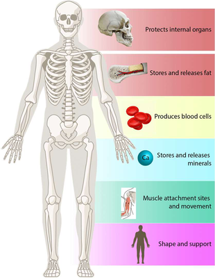
What are the six main functions of bones?
What is the role of bones in blood cell formation?
What minerals are primarily stored in bones?
Where does hematopoiesis occur?
What is one function of bones related to muscle action?
What is stored in bones for energy?
What is the function of bones in support?
What is the function of bones in protection?
Which of the following is NOT a function of bones?
None
Mineral storage
Movement
Blood cell formation
What does the skeletal system primarily protect?
What diagram illustrates the functions of bones?
What are the four main types of bones classified by shape?
Which type of bone is longer than it is wide?
Long bones
Irregular bones
Short bones
Flat bones
What shape are short bones?
Cube-shaped
Complicated shapes
Thin and flat
Longer than wide
Which type of bone is described as thin and flat?
Flat bones
Irregular bones
Long bones
Short bones
Which bone type has complicated shapes?
Irregular bones
Long bones
Flat bones
Short bones
The four main types of bones are: 1) _______ 2) _______ 3) _______ 4) _______.
What is an example of a long bone?
What is an example of a short bone?
What is an example of a flat bone?
What is an example of an irregular bone?
What do sesamoid bones refer to?
What are the characteristics of long bones?
What are the characteristics of short bones?
What are the characteristics of flat bones?
What are the characteristics of irregular bones?
What is illustrated in the diagram of different bone shapes?
What are bone markings?
What is the purpose of projections on bones?
What are depressions in bone structure?
What do openings in bones facilitate?
What is the significance of textural features on bones?
What are the three main categories of bone markings?
What are projections in bone markings?
What do surfaces that form joints refer to in bone markings?
What are depressions and openings in bone markings?
What is a tuberosity in bone markings?
What does a crest refer to in bone markings?
What is a trochanter?
Define a line in bone markings.
What is a tubercle?
What does epicondyle mean in bone markings?
What is the definition of a spine in bone markings?
What is a process in bone markings?
Which of the following is a type of bone projection?
Sinus
Foramen
Tuberosity
Fossa
What is the function of bone projections?
Protection of organs
Blood cell production
Site of muscle and ligament attachment
Storage of fat
The eight main types of projections include: - _______ - _______ - _______ - _______ - _______ - _______ - _______ - _______
What is a tuberosity?

What is a crest?
What is a line?
What is an epicondyle?
What is a spine?
What is a process?
What are the three main types of surfaces in bone markings?
What is a Head in bone markings?
What is a Facet in bone markings?
What is a Condyle in bone markings?
Which of the following describes a Facet?
Bony expansion carried on a narrow neck.
A sharp projection on a bone.
Rounded articular projection; often articulates with a corresponding fossa.
Smooth, nearly flat articular (joint) surface.
Which image illustrates the Head and Facets of a rib?

An illustration of muscle anatomy.
A diagram of a human skull.

What is a groove in bone markings?
What is a fissure in bone markings?
What is a foramen?
What is a meatus?
What is a sinus in bone markings?
What is a fossa?
Which of the following is a type of depression in bone markings?
Head
Groove
Crest
Condyle
What type of opening is a foramen?
Narrow slit
Cavity within a bone
Round or oval opening
Canal-like passageway
What is a fissure?
What is a notch?
What is a sinus?
What is the function of a foramen?
Cavity filled with air
Indentation at the edge of a structure
Canal-like passageway
Round or oval opening through a bone
Which of the following describes a meatus?
Furrow
Indentation at the edge
Shallow depression
Canal-like passageway
What does a fossa represent?
Canal-like passageway
Narrow opening
Cavity filled with air
Shallow, basin-like depression
What are the two major types of bone texture?
What is the characteristic of compact bone?
What is the characteristic of spongy bone?
What fills the gaps in spongy bone?
What are trabeculae in spongy bone?
What does compact bone look like?
What does spongy bone look like?
What is shown in the cross-section of a bone image?
What are the main components of bone structure?
What is the function of osteocytes?
What do osteoblasts do?
What is the role of osteoclasts?
What is the bone matrix composed of?
What are the two major structural components of long bones?
What does the diaphysis of a long bone contain?
What is found in the marrow cavity of adult long bones?
What type of bone is found in the epiphyses of long bones?
What covers the joint surfaces of the epiphyses?
What exists between the diaphysis and epiphysis?
What is the function of the epiphyseal (growth) plate?
What does the epiphyseal line signify?
What does the compact bone collar surround in the diaphysis?
What is the source of the yellow marrow in long bones?
What is the structure of the long bone illustrated by the diagram?
What are the two major membranes of long bones?
What does the term 'periosteum' mean?
What are the two layers of the periosteum?
What is the function of Sharpey's fibers?
What types of cells are found in the inner osteogenic layer of the periosteum?
What is the role of osteoblasts?
What is the role of osteoclasts?
What does the endosteum cover?
What types of cells are contained in the endosteum?
What structures enter the bone via nutrient foramina?
What is depicted in the diagram of a long bone?
What is the structure of short, irregular, and flat bones?
What are the two membranes found in short, irregular, and flat bones?
Where does bone marrow exist in short, irregular, and flat bones?
What is the diploë in flat bones?
What covers the external surfaces of short, irregular, and flat bones?
Spongy bone
Periosteum
Endosteum
Compact bone
What is the delicate membrane on internal spongy bone called?
Endosteum
Periosteum
Diploë
Trabeculae
What is hematopoietic tissue?
Where is adult red marrow primarily found?
Where is newborn red marrow found?
What does the presence of red marrow in newborns indicate?
What are the types of skeletal cartilages?
Where is hyaline cartilage found?
What is the function of elastic cartilage?
Where is fibrocartilage commonly found?
What are skeletal cartilages?
Where are skeletal cartilages found?
Do skeletal cartilages contain blood vessels or nerves?
What does the perichondrium do?
How many major types of skeletal cartilages are there?
Which of the following is NOT a location where skeletal cartilages are found?
In the skin
In the brain
In the heart
None, they are found in joints, rib connections, nose, ear, and other skeletal structures.
Skeletal cartilages provide support, cushioning, and shape to the skeleton and are found in: - _______ - _______ - _______ - _______ - _______.
What are the properties of hyaline cartilage?
What are the features of elastic cartilage?
What are the characteristics of fibrocartilage?
What are the two major ways of cartilage growth?
What does appositional growth involve?
What is the process of interstitial growth?
What is the effect of calcification of cartilage?
What is the basic structural unit of bone?
What type of cells are responsible for bone formation?
What type of cells are involved in bone resorption?
What is the function of osteocytes?
What is the matrix of bone primarily composed of?
What are the small spaces in bone tissue where osteocytes reside called?
What structure connects lacunae to each other?
What is the outer layer of bone called?
What is the central canal in an osteon called?
What type of bone is found at the ends of long bones?
What are the five major cell types in bone?
What do osteogenic cells differentiate into?
What is the primary function of osteoblasts?
Where are osteocytes found?
What is the function of osteoclasts?
What are bone-lining cells called on the external and internal surfaces?
The five major cell types in bone are: - _______ - _______ - _______ - _______ - _______.
What do osteoblasts secrete?
Calcium phosphate
Mineralized bone
Unmineralized osteoid
Collagen fibers
What is the role of osteocytes?
Form new bone
Resorb bone matrix
Maintain bone matrix
Break down bone
What is compact bone also known as?
What are the structural units of compact bone called?
What is the weight-bearing component of osteons?
What runs through the core of an osteon?
What do lacunae contain?
What connects lacunae to each other and the central canal?
What are the canals that connect blood vessels and nerves of the periosteum to the central canal?
What fills gaps between forming osteons?
What extends around the entire surface of the diaphysis?
What helps compact bone resist twisting?
What is the function of osteons?
What structural feature of osteons withstands stress?
The three main components of compact bone are: 1) _______ 2) _______ 3) _______.
What do canaliculi resemble?
Solid rods
Hair-like canals
Flat structures
Large tubes
What is the primary role of the central canal in an osteon?
Supports osteocytes
Holds minerals
Contains blood vessels and nerves
Connects osteons
What type of lamellae fills gaps between osteons?
Interstitial Lamellae
Radial Lamellae
Central Lamellae
Circumferential Lamellae
What are the main structural units of compact bone?
What is the central canal in an osteon responsible for?
What are lamellae?
What are canaliculi?
What are perforating canals?
What is the role of interstitial lamellae?
What is the purpose of the diagram showing compact bone structure?
What type of bone is known as spongy bone?
How is spongy bone organized?
What structures are found in trabeculae of spongy bone?
What supplies nutrients to spongy bone?
What is the function of trabeculae in spongy bone?
Exist along lines of stress
Produce red blood cells
Store fat
Form osteons
What is the appearance of spongy bone?
What is the primary difference between spongy bone and compact bone?
Flashcards in this deck (192)
-
What is the Skeletal System?
The organ system in the human body that provides structure, support, protection, and movement in coordination with the muscular system. It consists of bones, cartilage, ligaments, and joints.
biology skeletal_system -
What are the two main groups of bones in the Skeletal System?
1) Axial Skeleton 2) Appendicular Skeleton
biology skeletal_system -
biology skeletal_system
-
What does the Appendicular Skeleton consist of?
- Bones of upper and lower limbs
- Girdles attaching limbs to axial skeleton
biology skeletal_system -
What is the primary function of the Skeletal System?
Digest food
Regulate body temperature
Produce hormones
Provide structure, support, and protection
biology skeletal_system -
Which bones are part of the Axial Skeleton?
Skull, vertebral column, rib cage
Femur, humerus, radius
Pelvis, scapula, clavicle
Tibia, fibula, patella
biology skeletal_system -
What is the Appendicular Skeleton responsible for?
Protecting vital organs
Bones of upper and lower limbs
Connecting to the vertebral column
Supporting the skull
biology skeletal_system -
What are the six main functions of bones?
1) Support for body/soft organs 2) Protection of brain, spinal cord, vital organs 3) Anchor spots for leverage during muscle action 4) Mineral storage (mainly calcium and phosphorus) 5) Blood cell formation (hematopoiesis) 6) Triglyceride (fat/energy) storage
anatomy skeletal_system -
What is the role of bones in blood cell formation?
Blood cell formation (hematopoiesis) occurs in bone marrow.
anatomy skeletal_system hematopoiesis -
anatomy skeletal_system minerals
-
anatomy skeletal_system hematopoiesis
-
What is one function of bones related to muscle action?
Bones serve as anchor spots for leverage during muscle action.
anatomy skeletal_system muscle_action -
anatomy skeletal_system energy_storage
-
anatomy skeletal_system support
-
What is the function of bones in protection?
Bones protect the brain, spinal cord, and vital organs.
anatomy skeletal_system protection -
Which of the following is NOT a function of bones?
Mineral storage
Movement
None
Blood cell formation
anatomy skeletal_system -
anatomy skeletal_system protection
-
anatomy skeletal_system visual_aid
-
What are the four main types of bones classified by shape?
1) Long bones - Longer than wide (e.g. femur) 2) Short bones - Cube-shaped (e.g. wrist/ankle) 3) Flat bones - Thin and flat (e.g. sternum) 4) Irregular bones - Complicated shapes (e.g. vertebra, hip)
anatomy skeletal_system -
anatomy bones
-
anatomy bones
-
anatomy bones
-
anatomy bones
-
anatomy skeletal_system
-
anatomy bones
-
anatomy bones
-
anatomy bones
-
anatomy bones
-
anatomy bones
-
anatomy bones
-
What are the characteristics of short bones?
They are cube-shaped (e.g. wrist/ankle) and include sesamoid bones.
anatomy bones -
What are the characteristics of flat bones?
They are thin, flat, and slightly curved (e.g. sternum).
anatomy bones -
What are the characteristics of irregular bones?
They have complicated shapes (e.g. vertebra and hip).
anatomy bones -
What is illustrated in the diagram of different bone shapes?
It shows long bone (humerus), irregular bone (vertebra), flat bone (sternum), and short bone (talus).
anatomy skeletal_system -
What are bone markings?
Features on bones that serve as attachment points for muscles, tendons, and ligaments, and help in joint formation.
anatomy bone_markings -
What is the purpose of projections on bones?
To serve as attachment points for muscles and ligaments.
anatomy bone_projections -
What are depressions in bone structure?
Concave areas that allow for the passage of nerves and blood vessels or form joints.
anatomy bone_depressions -
anatomy bone_openings
-
What is the significance of textural features on bones?
They indicate the strength and function of the bone, helping to understand its role in the skeletal system.
anatomy bone_texture -
What are the three main categories of bone markings?
1) Projections (processes) 2) Surfaces that form joints 3) Depressions and openings
anatomy bone_markings -
What are projections in bone markings?
Raised areas that serve as attachment points for muscles and ligaments. Examples include tuberosity, crest, trochanter, tubercle, epicondyle, spine.
anatomy bone_markings -
What do surfaces that form joints refer to in bone markings?
Smooth areas where bones articulate with one another. Examples include head, facet, condyle, ramus.
anatomy bone_markings -
What are depressions and openings in bone markings?
Indentations or holes that allow passage of blood vessels and nerves or accommodate other structures. Examples include foramen, fossa, groove, fissure, notch, meatus, sinus.
anatomy bone_markings -
anatomy bone_markings
-
anatomy bone_markings
-
anatomy bone_markings
-
anatomy bone_markings
-
anatomy bone_markings
-
anatomy bone_markings
-
anatomy bone_markings
-
anatomy bone_markings
-
anatomy bone_markings
-
What is the function of bone projections?
Protection of organs
Site of muscle and ligament attachment
Blood cell production
Storage of fat
anatomy bone_markings -
The eight main types of projections include: - Tuberosity - Crest - Trochanter - Line - Tubercle - Epicondyle - Spine - Process
anatomy bone_markings -
anatomy bone_markings
-
anatomy bone_markings
-
anatomy bone_markings
-
anatomy bone_markings
-
anatomy bone_markings
-
anatomy bone_markings
-
What are the three main types of surfaces in bone markings?
- Head: bony expansion on narrow neck
- Facet: smooth, nearly flat joint surface
- Condyle: rounded articular projection
anatomy bone_markings -
anatomy bone_markings
-
anatomy bone_markings
-
What is a Condyle in bone markings?
Rounded articular projection; often articulates with a corresponding fossa.
anatomy bone_markings -
Which of the following describes a Facet?
A sharp projection on a bone.
Bony expansion carried on a narrow neck.
Smooth, nearly flat articular (joint) surface.
Rounded articular projection; often articulates with a corresponding fossa.
anatomy bone_markings -
Which image illustrates the Head and Facets of a rib?
An illustration of muscle anatomy.
A diagram of a human skull.
anatomy bone_markings -
anatomy bone_markings
-
anatomy bone_markings
-
anatomy bone_markings
-
anatomy bone_markings
-
anatomy bone_markings
-
anatomy bone_markings
-
anatomy bone_markings
-
What type of opening is a foramen?
Round or oval opening
Canal-like passageway
Cavity within a bone
Narrow slit
anatomy bone_markings -
anatomy bone_markings
-
anatomy bone_markings
-
anatomy bone_markings
-
What is the function of a foramen?
Canal-like passageway
Cavity filled with air
Round or oval opening through a bone
Indentation at the edge of a structure
anatomy bone_markings -
Which of the following describes a meatus?
Indentation at the edge
Shallow depression
Canal-like passageway
Furrow
anatomy bone_markings -
What does a fossa represent?
Shallow, basin-like depression
Cavity filled with air
Canal-like passageway
Narrow opening
anatomy bone_markings -
anatomy bones
-
What is the characteristic of compact bone?
- Dense outer layer on every bone
- Appears smooth and solid
anatomy compact_bone -
What is the characteristic of spongy bone?
- Honeycomb/mesh of trabeculae
- Red bone marrow fills gaps between trabeculae
anatomy spongy_bone -
anatomy bone_marrow
-
anatomy trabeculae
-
anatomy compact_bone
-
anatomy spongy_bone
-
What is shown in the cross-section of a bone image?
Compact bone and spongy bone with labeled trabeculae
anatomy bone_structure -
anatomy bones
-
What is the function of osteocytes?
They maintain bone tissue and communicate with other bone cells.
anatomy bones cells -
anatomy bones cells
-
anatomy bones cells
-
What is the bone matrix composed of?
It consists of collagen fibers and inorganic minerals, mainly calcium phosphate.
anatomy bones composition -
What are the two major structural components of long bones?
1) Diaphysis (shaft) 2) Epiphyses (ends of long bones)
anatomy bones -
What does the diaphysis of a long bone contain?
A compact bone collar surrounding the marrow cavity.
anatomy bones -
What is found in the marrow cavity of adult long bones?
Fat (yellow marrow) is contained in the marrow cavity.
anatomy bones -
What type of bone is found in the epiphyses of long bones?
Compact bone externally and spongy bone internally.
anatomy bones -
What covers the joint surfaces of the epiphyses?
Articular (hyaline) cartilage covers the joint surfaces.
anatomy bones -
What exists between the diaphysis and epiphysis?
The epiphyseal line, a remnant of the growth plate.
anatomy bones -
What is the function of the epiphyseal (growth) plate?
It allows for the growth of long bones during childhood.
anatomy bones -
What does the epiphyseal line signify?
It is the remnant of the epiphyseal (growth) plate from childhood.
anatomy bones -
anatomy bones
-
What is the source of the yellow marrow in long bones?
The marrow cavity in adults contains fat (yellow marrow).
anatomy bones -
What is the structure of the long bone illustrated by the diagram?
The diagram shows diaphysis, epiphyses, articular cartilage, spongy bone, compact bone, endosteum, periosteum, epiphyseal line, and medullary cavity.
anatomy bones -
anatomy bones
-
anatomy terminology
-
anatomy bones
-
anatomy bones
-
What types of cells are found in the inner osteogenic layer of the periosteum?
- Osteoblasts
- Osteoclasts
- Osteogenic cells
anatomy cells -
anatomy cells
-
anatomy cells
-
anatomy bones
-
anatomy cells
-
What structures enter the bone via nutrient foramina?
Nerve fibers, nutrient blood vessels, and lymphatic vessels.
anatomy vascular -
What is depicted in the diagram of a long bone?
The diagram shows the proximal and distal epiphysis, diaphysis, articular cartilage, spongy bone, compact bone, endosteum, periosteum, epiphyseal line, and medullary cavity.
anatomy diagrams -
What is the structure of short, irregular, and flat bones?
They consist of thin plates of spongy bone covered by compact bone. In flat bones, spongy bone is called diploë.
anatomy bone_structure -
What are the two membranes found in short, irregular, and flat bones?
1) Periosteum: covers external surfaces 2) Endosteum: delicate membrane on internal spongy bone
anatomy bone_membranes -
Where does bone marrow exist in short, irregular, and flat bones?
Bone marrow exists between the trabeculae; no defined marrow cavity is seen as in long bones.
anatomy bone_marrow -
anatomy flat_bones
-
What covers the external surfaces of short, irregular, and flat bones?
Periosteum
Spongy bone
Endosteum
Compact bone
anatomy bone_membranes -
What is the delicate membrane on internal spongy bone called?
Periosteum
Trabeculae
Endosteum
Diploë
anatomy bone_membranes -
What is hematopoietic tissue?
Blood-forming tissue found mainly in red bone marrow, where stem cells produce red blood cells, white blood cells, and platelets.
biology hematopoiesis -
Where is adult red marrow primarily found?
In trabecular cavities of spongy bone, mainly in heads of femur and humerus, and in the diploë of flat bones.
anatomy bone -
anatomy bone
-
What does the presence of red marrow in newborns indicate?
Highlights the high demand for blood cell formation during early development.
biology development -
anatomy cartilage
-
anatomy hyaline_cartilage
-
anatomy elastic_cartilage
-
anatomy fibrocartilage
-
What are skeletal cartilages?
Strong but flexible connective tissues that provide support, cushioning, and shape to the skeleton.
anatomy cartilage -
anatomy locations
-
Do skeletal cartilages contain blood vessels or nerves?
No, they contain no blood vessels or nerves.
anatomy cartilage -
anatomy perichondrium
-
anatomy cartilage
-
Which of the following is NOT a location where skeletal cartilages are found?
None, they are found in joints, rib connections, nose, ear, and other skeletal structures.
In the heart
In the brain
In the skin
anatomy locations -
Skeletal cartilages provide support, cushioning, and shape to the skeleton and are found in: - joints - rib connections - nose - ear - other skeletal structures.
anatomy locations -
What are the properties of hyaline cartilage?
- Offers support
- Flexibility
- Resilience
- Most common type in the body
- Found in articular, costal, and respiratory cartilages
anatomy cartilage hyaline -
What are the features of elastic cartilage?
- Contains elastic fibers
- Enables repeated bending
- Similar to hyaline cartilage
- Found in external ear and epiglottis
anatomy cartilage elastic -
What are the characteristics of fibrocartilage?
- Contains collagen fibers
- Provides tensile strength
- Supports intervertebral discs, pubic symphysis, and menisci
- Offers shock absorption
anatomy cartilage fibrocartilage -
biology cartilage growth
-
What does appositional growth involve?
Cells secrete matrix against the external face of existing cartilage, increasing its width.
biology cartilage growth -
What is the process of interstitial growth?
Chondrocytes divide and secrete new matrix, expanding cartilage from within.
biology cartilage growth -
What is the effect of calcification of cartilage?
It alters cartilage's flexibility and resilience during normal bone growth and old age.
biology cartilage calcification -
anatomy bone
-
anatomy bone
-
anatomy bone
-
anatomy bone
-
anatomy bone
-
anatomy bone
-
anatomy bone
-
anatomy bone
-
anatomy bone
-
anatomy bone
-
What are the five major cell types in bone?
- Osteogenic cells
- Osteoblasts
- Osteocytes
- Osteoclasts
- Bone-Lining Cells
anatomy bone cells -
anatomy bone cells
-
anatomy bone cells
-
anatomy bone cells
-
anatomy bone cells
-
What are bone-lining cells called on the external and internal surfaces?
- Periosteal cells (external)
- Endosteal cells (internal)
anatomy bone cells -
The five major cell types in bone are: - Osteogenic cells - Osteoblasts - Osteocytes - Osteoclasts - Bone-Lining Cells.
anatomy bone cells -
What do osteoblasts secrete?
Mineralized bone
Calcium phosphate
Collagen fibers
Unmineralized osteoid
anatomy bone cells -
What is the role of osteocytes?
Resorb bone matrix
Break down bone
Form new bone
Maintain bone matrix
anatomy bone cells -
anatomy bone
-
anatomy bone
-
anatomy bone
-
anatomy bone
-
anatomy bone
-
anatomy bone
-
What are the canals that connect blood vessels and nerves of the periosteum to the central canal?
Perforating (Volkmann's) Canal
anatomy bone -
anatomy bone
-
anatomy bone
-
anatomy bone
-
anatomy bone
-
anatomy bone
-
The three main components of compact bone are: 1) Osteon (Haversian System) 2) Central (Haversian) Canal 3) Canals and Canaliculi.
anatomy bone -
anatomy bone
-
What is the primary role of the central canal in an osteon?
Supports osteocytes
Contains blood vessels and nerves
Holds minerals
Connects osteons
anatomy bone -
What type of lamellae fills gaps between osteons?
Radial Lamellae
Central Lamellae
Interstitial Lamellae
Circumferential Lamellae
anatomy bone -
anatomy bone compact_bone
-
anatomy bone compact_bone
-
anatomy bone compact_bone
-
anatomy bone compact_bone
-
anatomy bone compact_bone
-
anatomy bone compact_bone
-
What is the purpose of the diagram showing compact bone structure?
To illustrate the arrangement of osteons and their components
anatomy bone compact_bone -
What type of bone is known as spongy bone?
Spongy (Cancellous) Bone is a lighter, less dense type of bone tissue found mainly at the ends of long bones and inside flat bones.
anatomy bones -
How is spongy bone organized?
Spongy bone is organized into a supportive network called trabeculae, existing along lines of stress.
anatomy bones -
What structures are found in trabeculae of spongy bone?
Trabeculae contain irregularly arranged lamellae, osteocytes, and canaliculi; they do not contain osteons.
anatomy bones -
What supplies nutrients to spongy bone?
Capillaries in the endosteum supply nutrients to spongy bone.
anatomy bones -
What is the function of trabeculae in spongy bone?
Exist along lines of stress
Form osteons
Produce red blood cells
Store fat
anatomy bones -
What is the appearance of spongy bone?
Spongy bone appears poorly organized but is actually organized along lines of stress to help resist any stress.
anatomy bones -
What is the primary difference between spongy bone and compact bone?
Spongy bone is lighter and less dense compared to compact bone, which is denser and forms the outer layer of bones.
anatomy bones