Sign up to unlock more features
- Save this deck to your account
- Study flashcards with spaced repetition
- Export to Anki (.apkg) or PDF
- Process documents up to 100 pages
- Images extracted from PDFs and documents
- Better text extraction from your PDFs and documents
- Better flashcards with our more advanced AI model


What does 'Excitable' mean in muscle tissue?
What does 'Conductive' mean in muscle tissue?
What does 'Contractile' mean in muscle tissue?
What do 'Extensible and Elastic' mean in muscle tissue?
What are the main properties of muscle tissue?
What is the primary role of skeletal muscles in movement?

How do skeletal muscles help maintain posture?
How do skeletal muscles control body entrances and exits?
How do skeletal muscles contribute to temperature regulation?
How do skeletal muscles provide support to the body?
What nutrient-related role do skeletal muscles serve?
How are most skeletal muscle functions accomplished?
What must stimulate skeletal muscle for it to contract?
Is most skeletal muscle control voluntary or involuntary?
What are the key characteristics of skeletal muscle?
What are the key characteristics of cardiac muscle?
What are the key characteristics of smooth muscle?
Which muscle types are striated?
Which muscle type is non-striated (no sarcomeres)?
Which muscle is multinucleated and has large fibers?
Which muscle is autorhythmic and does not require nervous input to beat?
What structures enable electrical coupling between cardiac muscle cells?
Which muscle types are regulated by the autonomic nervous system (ANS)?
What is the nucleus count and cell size of cardiac muscle?
What is the nucleus count and cell size of smooth muscle?
Where is smooth muscle typically located and what is its role?
Where is skeletal muscle attached and what is its main function?
What is the typical shape of smooth muscle cells?
Identify the three muscle tissue types shown in the image: 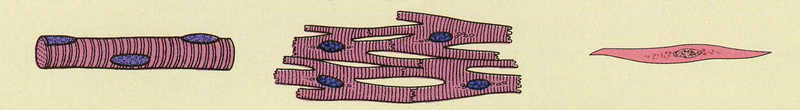
Which feature indicates striated fibers in this microscopic image:  ?
?
What two features supply and control a whole skeletal muscle?
A whole muscle is composed of how many muscle cells?
What are the three connective tissue wrappings of a skeletal muscle?
What does the epimysium wrap?
What does the perimysium wrap?
What does the endomysium wrap?
Identify the hierarchical levels shown in the diagram (largest to smallest). 
Which cells fuse to form skeletal muscle fibers?
From which embryonic germ layer do muscle fibers originate?
How do myoblasts become muscle fibers?
What are two characteristic features of skeletal muscle fibers?
Why are skeletal muscle fibers multinucleated?
Name components shown in a developing muscle fiber diagram.
What is the 'sarcolemma'?
What is the 'sarcoplasm'?
What are transverse (T) tubules and their function?
What membrane event at the sarcolemma initiates contraction?
Name three major internal structures of a skeletal muscle fiber shown in the diagram.
Where are myofibrils located in muscle?
What do myofibrils contain?
What are thin and thick myofilaments made of?
How do myofibrils produce muscle contraction?
Where are myofibrils anchored?
What is a sarcomere?
What does a sarcomere consist of?
Approximately how many sarcomeres are in a myofibril?
Which structures are visible in a detailed muscle fiber cross-section?
What is the primary role of the sarcoplasmic reticulum (SR) in skeletal muscle?
Where is the SR located in relation to myofibrils?
What ion fills the sarcoplasmic reticulum?
How is the SR connected to the T-tubules?
What are terminal cisternae?
What is a 'triad' in skeletal muscle?
 What is shown in this diagram?
What protein makes up the thick filament?
What protein makes up the thin filament?
What protein makes up the elastic filament in the diagram?
What does the I band contain?
What does the A band represent?
What is the H band?
What is the zone of overlap?
What marks the boundary of a sarcomere?
What is the M line in a sarcomere?
What does the Z band mark in a sarcomere?
What is the function of the M line?
What is the H zone in a sarcomere?
What composes the A band?
What is the I band and how does it change during contraction?
In the diagram, which color represents the thick (myosin) filament?
What is the Muscle structural level?
What is a Fascicle?
What is a Muscle fiber (cell)?
What is a Myofibril?
What is a Sarcomere?
What are Myofilaments?
Where are the nuclei located in a muscle fiber?
What is the sarcoplasmic reticulum in muscle?
Which connective tissue surrounds the entire muscle, fascicle, and fiber?
What gives skeletal muscle its striated appearance?
What are the main proteins of the thick and thin filaments?
Name the structural hierarchy shown in the image: 
What are the main filament types in a sarcomere?
What is the sliding filament mechanism?
How do the banding patterns change during contraction?
Compare a relaxed and contracted sarcomere (see diagram): 
What does a TEM image of a sarcomere show? 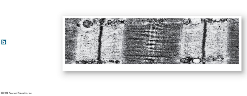
Does the A band change length during contraction?
Which components must be included in the sarcomere drawing?
What is the thick filament to include?
What is the thin filament to include?
Which structure marks the boundary of a sarcomere to include?
Which central line should be included in the sarcomere drawing?
Name the sarcomere band that spans the length of the thick filaments.
Name the sarcomere band that contains only thin filaments.
Which band is the region around the M-line with only thick filaments?
Which elastic filament is listed to include in the drawing?
What is the noise rule for this activity?
How should students be paired for the activity?
How should the drawing process proceed between partners?
How is the winning pair chosen?
Flashcards in this deck (114)
-
-
-
-
-
muscle physiology excitable
-
muscle physiology conductive
-
muscle physiology contractile
-
muscle physiology extensible elastic
-
What are the main properties of muscle tissue?
- Excitable: Can fire an action potential like nervous tissue
- Conductive: Impulse can travel through the muscle cell
- Contractile: Muscle PULLS!
- Extensible and Elastic: Can stretch
muscle physiology properties -
movement muscle
-
posture muscle
-
digestion urinary
-
thermoregulation temperature
-
support anatomy
-
nutrition glycemic
-
contraction physiology
-
nervous_system cns
-
voluntary control
-
What are the key characteristics of skeletal muscle?
- Voluntary control (somatic nervous system)
- Striated (sarcomeres)
- Multinucleated
- Attached to bone
- Large fibers
skeletal muscle anatomy -
What are the key characteristics of cardiac muscle?
- Involuntary
- Regulated by ANS
- Autorhythmic (self-regulated)
- Striated (sarcomeres)
- Mononucleate, small cells
- Branched; intercalated discs; gap junctions
- Found only in heart
cardiac muscle anatomy -
What are the key characteristics of smooth muscle?
- Involuntary (ANS, chemical, or mechanical regulation)
- Non-striated (no sarcomere)
- Mononucleate, small cells
- Visceral; in hollow organs
- Spindle-shaped; gap junctions
smooth muscle anatomy -
striated muscle histology
-
nonstriated smooth muscle
-
nuclei skeletal muscle
-
Which muscle is autorhythmic and does not require nervous input to beat?
- Cardiac muscle
- Autorhythmic (self-regulated)
autorhythmic cardiac heart -
What structures enable electrical coupling between cardiac muscle cells?
- Intercalated discs
- Gap junctions
intercalated gapjunctions cardiac -
Which muscle types are regulated by the autonomic nervous system (ANS)?
- Cardiac muscle
- Smooth muscle
ans regulation muscle -
cardiac nuclei cells
-
smooth nuclei cells
-
Where is smooth muscle typically located and what is its role?
- In hollow organs (visceral muscle)
- Controls organ walls and lumens
location smooth visceral -
Where is skeletal muscle attached and what is its main function?
- Attached to bone
- Produces body movement
attachment skeletal function -
shape smooth cells
-
Identify the three muscle tissue types shown in the image:
- Skeletal muscle
- Cardiac muscle
- Smooth muscle
image identification muscle -
image striations histology
-
muscle anatomy
-
muscle composition
-
What are the three connective tissue wrappings of a skeletal muscle?
- Epimysium
- Perimysium
- Endomysium
connective_tissue muscle -
epimysium connective_tissue
-
perimysium connective_tissue
-
endomysium connective_tissue
-
Identify the hierarchical levels shown in the diagram (largest to smallest).
- Muscle (organ)
- Fascicle
- Muscle fiber
- Myofibril
hierarchy diagram -
myoblasts muscle development
-
mesoderm embryology muscle
-
fusion myoblasts muscle
-
structure muscle histology
-
Why are skeletal muscle fibers multinucleated?
- Allows multiple copies of muscle proteins and enzymes to be produced
multinucleated function muscle -
Name components shown in a developing muscle fiber diagram.
- Nuclei
- Immature muscle fiber
- Myosatellite cell
diagram labels muscle -
What is the 'sarcolemma'?
- Plasma membrane of a muscle fiber
- Change in membrane potential is the first step in triggering contraction
anatomy sarcolemma muscle -
What is the 'sarcoplasm'?
- Cytoplasm of a muscle fiber
- Relatively little cytoplasm because fibers are densely packed with contractile proteins
anatomy sarcoplasm cellular -
What are transverse (T) tubules and their function?
- Tubular extensions of the sarcolemma that penetrate deep into the cell
- Help spread action potentials into the fiber interior
t-tubules physiology muscle -
What membrane event at the sarcolemma initiates contraction?
- A change in sarcolemma membrane potential (action potential) initiates contraction
physiology actionpotential sarcolemma -
Name three major internal structures of a skeletal muscle fiber shown in the diagram.
- Myofibrils
- Sarcoplasmic reticulum
- Mitochondria
 anatomy myofibrils sarcoplasmicreticulum
 -
myofibril muscle
-
myofibril structure
-
myofilament protein
-
contraction physiology
-
myofibril sarcolemma
-
sarcomere muscle
-
What does a sarcomere consist of?
- Repeating thick and thin filaments
- Proteins to stabilize and regulate contraction
sarcomere structure -
sarcomere numbers
-
Which structures are visible in a detailed muscle fiber cross-section?
- Myofibrils
- Thick and thin filaments
- Mitochondria
- Sarcoplasmic reticulum
- T-tubules
- Triads and terminal cisternae
histology image -
What is the primary role of the sarcoplasmic reticulum (SR) in skeletal muscle?
- Regulates muscle contraction by storing and releasing \(Ca^{2+}\)
physiology sarcoplasmicreticulum muscle -
anatomy sarcoplasmicreticulum
-
physiology ions
-
How is the SR connected to the T-tubules?
- Physically connected to T-tubules to coordinate excitation with \(Ca^{2+}\) release
anatomy ttubules sarcoplasmicreticulum -
anatomy sarcoplasmicreticulum reservoirs
-
anatomy triad muscle
-
sarcomere anatomy
-
proteins muscle
-
proteins muscle
-
proteins muscle
-
bands sarcomere
-
bands sarcomere
-
bands sarcomere
-
overlap sarcomere
-
structures sarcomere
-
structures sarcomere
-
sarcomere structure muscle
-
sarcomere myosin structure
-
What is the H zone in a sarcomere?
- Region with only thick (myosin) filaments
- Shrinks or disappears during contraction
sarcomere hzone contraction -
What composes the A band?
- H zone + zone of overlap
- Contains both thick (myosin) and thin (actin) filaments (dark area)
sarcomere aband overlap -
What is the I band and how does it change during contraction?
- Region with only thin (actin) filaments (light area)
- Shrinks during contraction
sarcomere iband contraction -
In the diagram, which color represents the thick (myosin) filament?
- Purple = thick (myosin) filament

sarcomere filaments visual - Purple = thick (myosin) filament
-
What is the Muscle structural level?
- A contractile organ usually attached to bones by tendons
- Composed of bundles (fascicles) of muscle fibers
- Enclosed by epimysium; supplied with nerves and blood vessels
anatomy muscle -
What is a Fascicle?
- A bundle of muscle fibers within a muscle
- Enclosed by perimysium
- Supplied by nerves and blood vessels
anatomy fascicle -
What is a Muscle fiber (cell)?
- Single muscle cell, elongated and threadlike
- Has sarcolemma and multiple nuclei beneath the sarcolemma
- Contains myofibrils and sarcoplasmic reticulum
- Enclosed by endomysium
anatomy cell -
What is a Myofibril?
- Bundle of myofilaments filling most of the cytoplasm
- Surrounded by sarcoplasmic reticulum and mitochondria
- Shows striations from orderly filament overlap
anatomy myofibril -
What is a Sarcomere?
- Segment from one Z disc to the next in a myofibril
- Hundreds end to end make a myofibril
- Functional contractile unit of the muscle fiber
anatomy sarcomere -
What are Myofilaments?
- Fibrous protein strands that carry out contraction
- Two types: thick (mainly myosin) and thin (mainly actin)
- They slide over each other to shorten sarcomeres
anatomy myofilament -
Where are the nuclei located in a muscle fiber?
- Multiple nuclei located immediately beneath the sarcolemma
anatomy nucleus -
What is the sarcoplasmic reticulum in muscle?
- An extensive network of specialized smooth endoplasmic reticulum present in the muscle fiber
anatomy sarcoplasm -
Which connective tissue surrounds the entire muscle, fascicle, and fiber?
- Muscle: epimysium
- Fascicle: perimysium
- Fiber: endomysium
anatomy connective -
What gives skeletal muscle its striated appearance?
- Orderly overlap of protein myofilaments within myofibrils
anatomy striations -
What are the main proteins of the thick and thin filaments?
- Thick filament: mainly myosin
- Thin filament: mainly actin
anatomy proteins -
Name the structural hierarchy shown in the image:
- Muscle
- Fascicle
- Muscle fiber
- Myofibril
- Sarcomere
- Myofilaments
anatomy hierarchy -
anatomy filaments
-
What is the sliding filament mechanism?
- Thin filaments slide past thick filaments via cross-bridge cycling
- Result: sarcomere shortens and muscle contracts
physiology muscle -
How do the banding patterns change during contraction?
- A band: unchanged
- I band: shortens
- H zone: shortens or disappears
- Z discs: move closer
histology muscle -
Compare a relaxed and contracted sarcomere (see diagram):
- Relaxed: wider I bands and H zone; Z discs farther apart
- Contracted: narrower I bands and H zone; Z discs closer
image muscle contraction -
What does a TEM image of a sarcomere show?
- Ultrastructure of filaments and banding pattern
- Visible Z line, A/I bands and M line
microscopy image muscle -
Does the A band change length during contraction?
- No — the A band remains the same length as filaments slide
muscle histology -
Which components must be included in the sarcomere drawing?
- Z-disc
- M-line
- Thick filament (Myosin)
- Thin filament (Actin)
- A-Band
- I-Band
- Titan filament
- H-Band
sarcomere components anatomy -
filaments myosin
-
filaments actin
-
zdisc sarcomere
-
mline sarcomere
-
bands aband
-
bands iband
-
bands hband
-
filaments titan
-
rules activity
-
How should students be paired for the activity?
- Work in pairs with the person seated directly across from you
pairing activity -
How should the drawing process proceed between partners?
- Each person draws one component, labels it, and passes the paper to the person across until the sarcomere is complete
procedure activity -
competition activity