Sign up to unlock more features
- Save this deck to your account
- Study flashcards with spaced repetition
- Export to Anki (.apkg) or PDF
- Process documents up to 100 pages
- Images extracted from PDFs and documents
- Better text extraction from your PDFs and documents
- Better flashcards with our more advanced AI model
What are the three parts of the brainstem?
What structure lies upon the clivus of the occipital bone?
What covers the dorsal aspect of the brainstem?
How is the midbrain connected to the forebrain?
Where is the medulla oblongata continuous with the spinal cord?
What are the inferior and superior colliculi collectively called?
What is the function of the inferior colliculi?
What does the superior colliculi do?
Where is the cerebral aqueduct located?
What is the tectum?
What does the pons consist of?
Where is the pontomesencephalic junction (PMJ) located?
What part of the brainstem does the medulla oblongata represent?
The boundary between the medulla and the spinal cord is at the _______.
The boundary of the medulla and pons is generally marked at the dorsal aspect of the brainstem where the _______ of the floor of the fourth ventricle constitutes the medulla.
The dorsal aspect of the medulla is marked by the dorsal median sulcus and contains the _______ and the _______.
The dorsal columns are ascending tracts that begin in the spinal cord and terminate in the medulla at the nuclei called _______ and _______.
The gracile and cuneate tubercles can be identified in the dorsal aspect of the medulla.
What is the shape of the fourth ventricle?
Where is the fourth ventricle located?
Identify this anatomical diagram.
What forms the floor of the 4th ventricle?
What connects the 4th ventricle to the subarachnoid space?
What is the median aperture of the fourth ventricle also known as?
What forms the lateral walls of the rostral part of the 4th ventricle?
Where do the walls of the 4th ventricle converge?
At what junction does the 4th ventricle become continuous with the cerebral aqueduct?
What is a feature of the ethmoid bone?
What is a feature of the frontal bone?
Name a feature of the sphenoid bone.
What passes through the hypoglossal canal?
What travels through the foramen magnum?
The features of the ethmoid bone include: - _______: where the olfactory nerve exits the skull - _______ - _______ - _______, where the falx cerebri attaches.
The sphenoid bone features: - _______ - _______ - _______: where the pituitary gland lies - _______ - _______
The occipital bone features include: - _______: where the hypoglossal nerve travels through - _______ - _______: articulates with C1 - _______: where musculature attaches.
What is the zygomatic process?
What does the mandibular fossa articulate with?
Which part of the temporal bone separates the middle and posterior cranial fossa?
What is found at the internal auditory meatus?
What is the function of the carotid canal?
What does the styloid process provide attachment for?
What does the stylomastoid foramen transmit?
What do the parietal bones have foramina for?
What is one of the paired bones of the skull?
Name a type of bone that is not specified in the details provided.
Flashcards in this deck (45)
-
anatomy brainstem
-
anatomy skull
-
anatomy cerebellum
-
anatomy brain
-
anatomy spinal_cord
-
anatomy brain
-
What is the function of the inferior colliculi?
Part of the auditory pathway connecting the inner ear to the auditory cortex.
anatomy auditory -
anatomy visual
-
anatomy brain
-
What is the tectum?
The midbrain region posterior to the cerebral aqueduct containing the colliculi.
anatomy brain -
anatomy pons
-
anatomy junction
-
What part of the brainstem does the medulla oblongata represent?
The most caudal part of the brainstem.
anatomy brainstem -
anatomy brainstem
-
The boundary of the medulla and pons is generally marked at the dorsal aspect of the brainstem where the caudal third of the floor of the fourth ventricle constitutes the medulla.
anatomy brainstem -
The dorsal aspect of the medulla is marked by the dorsal median sulcus and contains the fasciculus gracilis and the fasciculus cuneate.
neuroanatomy medulla -
The dorsal columns are ascending tracts that begin in the spinal cord and terminate in the medulla at the nuclei called nucleus gracilis and nucleus cuneatus.
neuroanatomy tracts -
neuroanatomy tubercles
-
neuroanatomy ventricles
-
neuroanatomy ventricles
-
neuroanatomy brainstem
-
neuroanatomy 4th_ventricle
-
What connects the 4th ventricle to the subarachnoid space?
Connecting structure: Cerebellopontine angle
neuroanatomy subarachnoid_space -
neuroanatomy 4th_ventricle foramina
-
What forms the lateral walls of the rostral part of the 4th ventricle?
- Superior cerebellar peduncle
- Inferior cerebellar peduncle
neuroanatomy 4th_ventricle -
neuroanatomy 4th_ventricle
-
At what junction does the 4th ventricle become continuous with the cerebral aqueduct?
Pontomesencephalic junction
neuroanatomy 4th_ventricle -
anatomy ethmoid_bone
-
anatomy frontal_bone
-
anatomy sphenoid_bone
-
anatomy occipital_bone
-
anatomy occipital_bone
-
The features of the ethmoid bone include: - Cribriform plate: where the olfactory nerve exits the skull - Superior and middle concha - Ethmoidal air sinus - Crista galli, where the falx cerebri attaches.
anatomy ethmoid_bone -
The sphenoid bone features: - Greater and lesser wings - Medial and lateral pterygoid plates - Sella turcica: where the pituitary gland lies - Optic canal - Superior orbital fissure
anatomy sphenoid_bone -
The occipital bone features include: - Hypoglossal canal: where the hypoglossal nerve travels through - Foramen magnum - Occipital condyle: articulates with C1 - Superior nuchal line: where musculature attaches.
anatomy occipital_bone -
anatomy bones
-
anatomy bones
-
anatomy bones
-
anatomy ear
-
anatomy blood_vessels
-
anatomy bones
-
anatomy nerves
-
anatomy bones
-
anatomy bones
-
anatomy bones
Introduction to the Brainstem
- The brainstem consists of:
- Midbrain
- Pons
- Medulla oblongata
- Located on the clivus of the occipital bone.
- The dorsal aspect is covered by the cerebellum.
- Caudally continuous with the spinal cord below the foramen magnum.
Midbrain
- Identifying features:
- Inferior and superior colliculi (corpora quadrigemina):
- Inferior colliculi: part of the auditory pathway.
- Superior colliculi: involved in eye movements and visual processing.
- Cerebral aqueduct is located here.
- The tectum contains the inferior and superior colliculi.
Pons
- Noted from dorsal view, consists of the rostral two-thirds of the floor of the 4th ventricle, extending from the pontomesencephalic junction (PMJ).
Medulla Oblongata
- Most caudal part of the brainstem:
- The boundary with pons is not clearly marked.
- The caudal third of the 4th ventricle's floor is the dorsal medulla, while the rostral two-thirds is pons.
- Boundary with spinal cord is at foramen magnum.
Dorsal Columns
- Fasciculus (tract) is a collection of nerve fibers:
- Medial: Fasciculus gracilis.
- Lateral: Fasciculus cuneate.
- Ends in nuclei:
- Nucleus gracilis and nucleus cuneatus, marked by tubercles.
Fourth Ventricle
- Rhomboid-shaped, formed at the
- Caudal: Medulla
- Rostral: Pons
- Connections:
- Median aperture (foramen of Magendie) links to subarachnoid space.
Skull Bones Overview
- Unpaired bones:
- Ethmoid: Cribriform plate, conchae, Crista galli.
- Frontal: Frontal sinus, supra-orbital notch.
- Sphenoid: Sella turcica, optic canal.
- Occipital: Foramen magnum, hypoglossal canal.
Paired Bones of the Skull
- Temporal bones:
- Zygomatic process, mastoid process.
- Parietal bones:
- Foramina for emissary veins.
Brainstem Diagram
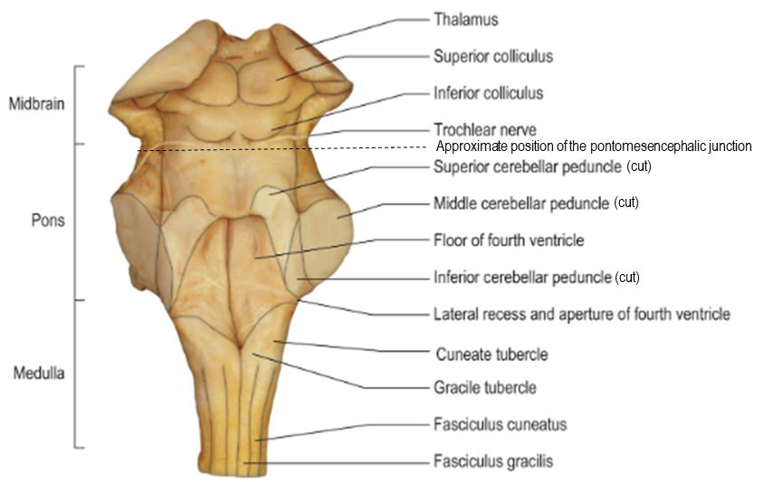 Diagram of the dorsal aspect of the brainstem showing labeled structures.