Sign up to unlock more features
- Save this deck to your account
- Study flashcards with spaced repetition
- Export to Anki (.apkg) or PDF
- Process documents up to 100 pages
- Images extracted from PDFs and documents
- Better text extraction from your PDFs and documents
- Better flashcards with our more advanced AI model
Flashcards in this deck (135)
-
anatomy eye
-
What are the medial and lateral commissures (canthus)?
They are the junctions of the upper and lower eyelids.
anatomy eye -
anatomy eye
-
anatomy eye
-
What is the lacrimal apparatus responsible for?
Secretes tears and contains lysozyme, an antibacterial enzyme.
anatomy eye -
anatomy eye
-
anatomy eye
-
What are the components of the lacrimal apparatus?
- Lacrimal gland
- Lacrimal canaliculus
- Lacrimal sac
- Nasolacrimal duct
anatomy eye -
What are the functions of ciliary glands in the eye?
Ciliary glands are modified sweat glands that lubricate the eye.
anatomy eye glands -
anatomy eye glands
-
List the six extrinsic eye muscles.
- Lateral rectus
- Medial rectus
- Superior rectus
- Inferior rectus
- Inferior oblique
- Superior oblique
anatomy eye muscles -
The functions of ciliary glands in the eye include lubricating the eye and are classified as modified sweat glands.
anatomy eye glands -
The tarsal glands secrete an oily solution to lubricate the eye and are classified as sebaceous glands.
anatomy eye glands -
The six extrinsic eye muscles include: - Lateral rectus - Medial rectus - Superior rectus - Inferior rectus - Inferior oblique - Superior oblique.
anatomy eye muscles -
anatomy eye
-
What is the fibrous layer of the eye?
The outer connective tissue layer that includes the sclera and cornea.
anatomy eye layers -
Identify the two main components of the fibrous layer.
- Sclera: white of the eye
- Cornea: transparent front part that allows light entry.
anatomy eye fibrous_layer -
anatomy eye cornea
-
Describe the vascular layer (Uvea) of the eye.
The middle layer that includes the iris, ciliary body, and choroid.
anatomy eye vascular_layer -
What structures are found in the vascular layer?
- Iris: anterior part
- Ciliary body: controls lens shape and secretes aqueous humor.
- Choroid: nourishes the eye.
anatomy eye vascular_layer -
anatomy eye ciliary_body
-
What is the role of the choroid in the eye?
It acts as a blood-rich layer that nourishes the retina and contains pigments to prevent light scattering.
anatomy eye choroid -
The fibrous layer consists of: - Sclera (the white of the eye) - Cornea (transparent structure for light entry).
anatomy eye fibrous_layer -
anatomy eye vascular_layer
-
The ciliary body consists of: - Ciliary muscle (controls lens shape) - Ciliary processes (secretes aqueous humor).
anatomy eye ciliary_body -
anatomy eye
-
anatomy eye retina
-
anatomy eye vision
-
anatomy eye vision
-
Where are cones concentrated in the eye?
At the macula lutea, specifically the center called fovea centralis
anatomy eye macula -
anatomy eye blind_spot
-
anatomy eye
-
How does ciliary muscle contraction affect vision?
It changes the lens thickness to focus light onto the retina.
anatomy physiology -
anatomy eye
-
What are the two segments of the eye?
- Anterior segment (contains aqueous humor)
- Posterior segment (contains vitreous humor)
anatomy eye -
What is contained in the anterior segment of the eye?
Watery aqueous humor, with two chambers: anterior and posterior.
anatomy eye -
anatomy eye
-
The suspensory ligament (ciliary zonule) holds the lens and enables the ciliary muscle to change lens thickness for focusing light onto the retina. The eye has: 1) Anterior segment containing aqueous humor 2) Posterior segment containing vitreous humor.
anatomy eye -
The eye is divided into two segments: 1) Anterior segment has two chambers: anterior chamber (before iris) and posterior chamber (after iris). 2) Posterior segment contains gel-like vitreous humor.
anatomy eye -
What are the two types of photoreceptors in the retina?
- Cones: color light, bright light, mainly in fovea centralis
- Rods: black/white, dim light, mainly in periphery
anatomy retina photoreceptors -
What is the function of bipolar cells in the retina?
They connect and modulate input from photoreceptors to ganglion cells.
anatomy retina bipolar_cells -
What do ganglion cells do in the retina?
They project axons to the brain via the optic nerve and tract.
anatomy retina ganglion_cells -
anatomy retina optic_disc
-
Photoreceptors in the retina include: - Cones: color light, bright light, mainly in fovea centralis - Rods: black/white, dim light, mainly in periphery.
anatomy retina photoreceptors -
vision anatomy
-
vision anatomy
-
vision anatomy
-
vision anatomy
-
Which structures in the thalamus are involved in the visual pathway?
Superior colliculus & lateral geniculate body.
vision anatomy -
vision anatomy
-
The visual pathway includes the following structures: - optic nerves - optic chiasma - optic tracts - superior colliculus - lateral geniculate body - occipital lobe.
vision pathway -
anatomy vision
-
What happens to images focused onto the retina?
All images are inverted by the lens when focused onto the retina.
anatomy vision -
vision myopia
-
vision hyperopia
-
What is presbyopia?
Presbyopia is far-sightedness caused by age-related decrease in lens elasticity.
vision presbyopia -
What causes astigmatism?
Astigmatism is caused by irregular corneal curvatures that distort the image.
vision astigmatism -
anatomy ear
-
anatomy ear
-
anatomy ear
-
anatomy ear
-
The outer ear consists of the auricle/pinna, which is made of skin covered cartilage, and the external acoustic meatus, which is lined with ceruminous glands.
anatomy ear -
The tympanic membrane is also known as the eardrum, which vibrates with the same frequency as sound waves that enter the canal.
anatomy ear -
anatomy middle_ear auditory
-
What is the function of the ossicles?
They amplify & transmit tympanic membrane vibrations to the oval window.
anatomy middle_ear function -
anatomy middle_ear connections
-
anatomy middle_ear function
-
The middle ear consists of a tympanic cavity containing the auditory ossicles: - Malleus - Incus - Stapes.
anatomy middle_ear -
anatomy middle_ear
-
anatomy middle_ear
-
anatomy middle_ear function
-
anatomy inner_ear fluid
-
What are the three parts of the bony labyrinth?
- Cochlea: involved in hearing
- Vestibule: involved in equilibrium
- Semicircular canals: involved in equilibrium
anatomy inner_ear bony_labyrinth -
anatomy hearing cochlea
-
anatomy equilibrium vestibule
-
What is the key function of the semicircular canals?
The semicircular canals are involved with equilibrium.
anatomy inner_ear semicircular_canals -
What fluid is found inside the membranous labyrinth?
The endolymph is found inside the membranous labyrinth.
anatomy inner_ear membranous_labyrinth -
anatomy inner_ear bony_labyrinth
-
anatomy inner_ear fluid
-
anatomy cochlea
-
anatomy cochlea
-
anatomy cochlea
-
anatomy cochlea
-
anatomy cochlea
-
anatomy cochlea
-
anatomy neuroscience
-
anatomy hearing
-
Which membrane contains a gelatinous structure that stereocilia project into?
The tectorial membrane.
anatomy hearing -
anatomy hearing
-
The membranes of the spiral organ include: - basilar membrane - tectorial membrane - vestibular membrane
anatomy hearing -
What does the stapes do in sound transduction?
It sends vibrations through the perilymph of the scala vestibuli and scala tympani.
anatomy auditory -
anatomy auditory
-
auditory frequency
-
auditory frequency
-
anatomy auditory
-
anatomy auditory
-
auditory frequency
-
auditory frequency
-
hearing tests
-
hearing sensorineural
-
hearing tests
-
What does it indicate if air conduction sounds lower in the Rinne test?
Possible earwax blockage, perforated eardrum, middle ear inflammation, or ossicle damage.
hearing conditions -
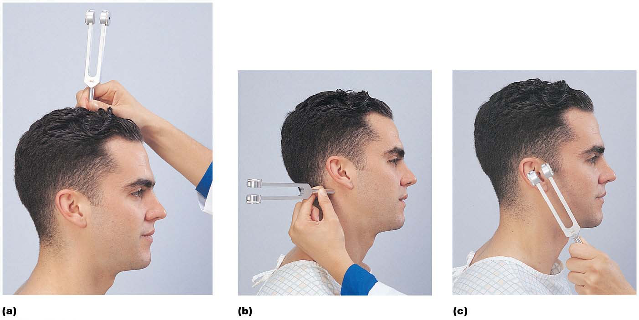
The Weber test checks if sound conduction is equally loud for both ears. A weaker sound can indicate sensorineural deafness.
hearing tests -
The Rinne test compares conduction between bone and air. Lower air conduction may indicate earwax, perforated eardrum, or middle ear inflammation.
hearing conditions -
What does the Weber test assess?
It determines if sound is normal or lateralized, indicating conduction or sensorineural deafness.
auditory testing -
auditory testing
-
Images (b and c) in the auditory tests show the Rinne test comparing:
- Bone conduction
- Air conduction
auditory testing -
The Weber test assesses if sound is normal or lateralized to one side, indicating conduction or sensorineural deafness.
auditory testing -
testing auditory
-
The auditory tests include the Weber test (a) and the Rinne test which compares: - bone conduction - air conduction.
auditory testing -
anatomy vestibular
-
anatomy vestibular
-
anatomy vestibular
-
anatomy vestibular
-
anatomy vestibular
-
anatomy vestibular
-
What do the utricle and saccule contain that helps detect head movement?
They contain hair cells that project stereocilia and kinocilium into the otolithic membrane.
vestibular anatomy -
What is the composition of the otolithic membrane?
It is a gelatinous material containing calcium carbonate crystals called otoliths.
vestibular anatomy -
How does head movement affect the otolithic membrane?
It causes the otolithic membrane to move, stimulating or inhibiting hair cells.
vestibular function -
What happens when hair cells in the utricle and saccule are stimulated?
They alter electrical signals sent along the vestibular nerve to the brain.
vestibular function -
The utricle and saccule contain hair cells that project stereocilia and kinocilium into the otolithic membrane.
vestibular anatomy -
Movement of the head causes the otolithic membrane to move and stimulate or inhibit the hair cells to alter electrical signals sent along the vestibular nerve.
vestibular function -
anatomy vestibular
-
What is the function of the ampullary cupula?
It acts as a gelatinous cap that moves in response to movement.
anatomy vestibular -
How do hair cells in the crista ampullaris send signals?
They send electrical impulses through the vestibular nerve.
nervous_system vestibular -
physiology vestibular
-
anatomy vestibular
-
anatomy vestibular
-
Hair cells send movement information to the brain as electrical impulses through the vestibular nerve.
nervous_system vestibular -
physiology vestibular
-
What should you complete for the lesson assignments?
Complete Canvas Assignment 1 and Mastering A&P Assignment 1.
assignments coursework -
assignments module
-
assignments policies
-
support ta
-
For the lesson, you need to understand the following concepts: External and internal eye anatomy, visual pathways to the brain, auditory test methods, vestibular system functioning, and common vision problems.
anatomy vision hearing
Chapter 23: Special Senses - Vision
External Anatomy of the Eye
- Eyelid (palpebrae): Protects the eye.
- Commissures (canthi): Junctions of upper and lower eyelids.
- Lacrimal caruncle: Produces oily secretion.
- Conjunctiva: Mucus-secreting inner surface of eyelids.
- Lacrimal apparatus: Secretes tears; includes:
- Lacrimal gland: Superior and lateral to the eye.
- Lacrimal canaliculus: Tears enter through the lacrimal punctum.
- Lacrimal sac.
- Nasolacrimal duct.
External Anatomy Continued
- Ciliary glands: Lubricates the eye, modified sweat glands.
- Tarsal glands: Sebaceous glands for oily secretion.
- Extrinsic eye muscles: Six muscles (lateral rectus, medial rectus, superior rectus, inferior rectus, inferior oblique, superior oblique).
Internal Anatomy of the Eye
Three Layers
- Fibrous layer: Outer connective tissue, includes:
- Sclera: White part of the eye.
- Cornea: Transparent, allows light entry.
- Vascular layer (Uvea): Middle layer with:
- Iris: Most anterior part.
- Ciliary body: Contains ciliary muscle for lens shape control and produces aqueous humor.
- Choroid: Blood-rich, absorbs light.
- Sensory layer: Innermost layer, includes the retina:
- Pigmented epithelial layer: Covers ciliary body and iris.
- Neural layer: Contains photoreceptors (rods and cones).
- Rods: Dim light, peripheral vision.
- Cones: Color vision, bright light, concentrated at fovea centralis.
- Blind spot: Where optic nerve exits.
Lens and Cavities
- Suspensory ligament (ciliary zonule): Holds lens, adjusts thickness.
- Lens: Focuses light onto the retina; divides the eye into:
- Anterior segment: Contains aqueous humor; has anterior and posterior chambers.
- Posterior segment: Contains vitreous humor.
Microscopic Anatomy of the Retina
- Photoreceptors: Cones (color, bright light) and rods (black/white, dim light).
- Bipolar cells: Connect photoreceptors to ganglion cells.
- Ganglion cells: Project axons as optic nerve.
Visual Pathway to Brain
- Impulses travel: Optic nerves ⇒ optic chiasma ⇒ optic tracts ⇒ thalamus ⇒ occipital lobe.
Chapter 24: Visual Tests
- Emmetropic eye: Normal focusing.
- Visual acuity issues:
- Myopia: Near-sightedness; image before retina.
- Hyperopia: Far-sightedness; image behind retina.
- Astigmatism: Irregular corneal curvature.
Chapter 25: Hearing & Equilibrium
Anatomy of the Outer Ear
- Auricle: Skin-covered cartilage.
- External auditory canal: Lined with ceruminous glands.
- Tympanic membrane: Vibrates with sound.
Anatomy of the Middle Ear
- Contains ossicles: Malleus, Incus, Stapes. Amplifies sound.
- Auditory tube: Equalizes pressure with outside.
Anatomy of the Inner Ear
- Structure: Bony labyrinth filled with perilymph; includes cochlea, vestibule, semicircular canals.
- Membranous labyrinth: Contains endolymph; housed within perilymph.
Cochlea
- Cochlear duct: Contains endolymph, separated into two chambers.
- Spiral organ: Contains hair cells as sensory receptors.
Sound Transduction
- Stapes vibration stimulates hair cells; frequency detection varies by location in cochlea.
Auditory Tests
- Weber test: Detects lateralization of sound.
- Rinne test: Compares conduction between air and bone.
Vestibular System
- Vestibular apparatus: Monitors equilibrium; includes utricle and saccule.
- Semicircular canals: Detects angular motion.
Utricle and Saccule Function
- Hair cells detect head movement via otolithic membrane.
Semicircular Canals Function
- Hair cells in ampulla send signals based on cupula movement.
Lesson Activities
- Complete Canvas and lab assignments on time. Late submissions not accepted.
Vision and Auditory Tests Overview
Summary
- Eye anatomy and function review, including visual pathways.
- Understanding hearing mechanics and auditory tests for diagnosis.