Sign up to unlock more features
- Save this deck to your account
- Study flashcards with spaced repetition
- Export to Anki (.apkg) or PDF
- Process documents up to 100 pages
- Images extracted from PDFs and documents
- Better text extraction from your PDFs and documents
- Better flashcards with our more advanced AI model
What is the main aim of population-based breast cancer screening?
What is the estimated sensitivity range of mammography?
What is the estimated specificity range of mammography?
What is the positive predictive value of mammography for women under age 50?
What is the positive predictive value of mammography for women age 50-69?
What reduction in breast cancer mortality was shown in RCTs for women aged 50-69?
What is the most demanding X-ray imaging modality?
What parameter is predominantly imaged in mammography?
What are the methods for discovering breast lumps?
What is the relationship between X-ray energy and tissue contrast?
What is the recommended Mean Glandular Dose (MGD) in mammography?
Why is low kVp used in mammography?
What is a key indicator of breast cancer in mammography?
What techniques are used to improve image quality in mammography?
What is the optimal energy band for mammography X-ray tubes?
What does Figure 8.1 illustrate regarding X-ray energy?
What does the graph in Figure 8.1 show?
What materials are typically used in mammographic imaging?
What is the focal spot size for screening in mammography?
What is the focal spot size for magnification mammography?
What is the typical operating potential for mammographic imaging with Mo target?
What effect does the angle of the tube in mammography have?
What is the characteristic radiation contribution in mammography?
What does Figure 8.2 show in mammography?
What does Figure 8.3 illustrate?
What is the typical exposure required for mammographic screen-film detectors?
What is the spatial resolution range for mammographic screen-film detectors?
What are digital detectors replacing in mammography?
What is the purpose of breast compression in mammography?
What force is typically used for breast compression?
How does compression affect scatter in mammography?
What is the Bucky factor of mammographic grids?
What is the effect of using grids in mammography?
What do moving grids do during exposure in mammography?
What happens to radiation dose in magnification radiography?
What is used to achieve breast compression in mammography?
What is the benefit of spot compression in mammography?
What is the purpose of magnification techniques in mammography?
What is an air grid in mammography?
What type of displays are used for mammographic images?
What is the magnification factor when using magnification techniques?
What focal spot size is used in standard screening mammography?
What is the effect of reducing the focal spot size to 0.1 mm?
What is the importance of image display in mammography?
What are the ambient lighting conditions for photographic mammographic images?
What does a mammographic image with microcalcifications indicate?
What is shown in Figure 8.5c?
What is the effect of mammography on radiation dose?
List the factors affecting image quality and radiation dose in mammography.
What is required for good mammographic imaging?
What does DQE stand for in medical imaging?
What does mAs refer to in imaging?
What does kVp stand for in imaging?
What is shown in the diagram related to mammography?
Flashcards in this deck (54)
-
What is the main aim of population-based breast cancer screening?
To reduce the number of deaths from breast cancer.
health cancer screening -
health mammography diagnostics
-
health mammography diagnostics
-
health mammography diagnostics
-
health mammography diagnostics
-
What reduction in breast cancer mortality was shown in RCTs for women aged 50-69?
30% reduction with screening.
health mammography rcts -
health mammography imaging
-
health mammography imaging
-
What are the methods for discovering breast lumps?
- Breast self-examination
- Clinical examination
- Mammogram
health cancer screening -
What is the relationship between X-ray energy and tissue contrast?
Contrast is mainly proportional to the cube of the atomic number of the attenuator at low energies; at higher energies, it is proportional to density.
physics medical_imaging -
What is the recommended Mean Glandular Dose (MGD) in mammography?
MGD should be below 3 mGy per view, typically less than 2 mGy for best practice.
radiation mammography -
Why is low kVp used in mammography?
Low kVp is necessary to obtain good discrimination between normal and abnormal pathology in soft tissues.
radiation mammography -
What is a key indicator of breast cancer in mammography?
Detection of small clusters of microcalcifications, about 100 microns in size, is a key indicator.
cancer mammography -
What techniques are used to improve image quality in mammography?
- Optimal X-ray tube target material and filtration
- Breast compression
- Fast screen/film systems
- Reciprocating grids
- Optimized display and viewing conditions
mammography techniques -
What is the optimal energy band for mammography X-ray tubes?
The optimal energy band is centered between 18 and 20 keV.
radiation mammography -
What does Figure 8.1 illustrate regarding X-ray energy?
It illustrates the contrast between various tissues at different X-ray energies and the percent contrast difference for ductal carcinoma.
figures medical_imaging -
What does the graph in Figure 8.1 show?
The graph shows mammographic contrast vs energy for ductal carcinoma, indicating that contrast increases with energy.
figures medical_imaging -
What materials are typically used in mammographic imaging?
- Molybdenum (Mo) target
- Molybdenum (Mo) filter
- Rhodium (Rh) target-filter combinations
- Aluminium (Al) target-filter combinations
medical imaging mammography -
medical imaging mammography
-
medical imaging mammography
-
medical imaging mammography
-
What effect does the angle of the tube in mammography have?
Utilizes the heel effect to produce maximum intensity at the chest wall and minimum at the nipple.
medical imaging mammography -
What is the characteristic radiation contribution in mammography?
Considerably more than at higher kVps with a tungsten target.
medical imaging mammography -
medical imaging mammography
-
medical imaging mammography
-
medical imaging
-
medical imaging
-
medical imaging
-
What is the purpose of breast compression in mammography?
- Reduces overlapping anatomy
- Decreases tissue thickness
- Reduces scatter
- Reduces geometric blurring
- Lowers radiation dose
medical imaging -
medical imaging
-
medical imaging
-
medical imaging
-
medical imaging
-
What do moving grids do during exposure in mammography?
Oscillate over a distance of about 20 grid lines
medical imaging -
medical imaging
-
medical imaging
-
medical imaging
-
What is the purpose of magnification techniques in mammography?
To obtain more detailed diagnostic images and increase spatial resolution.
medical_imaging mammography -
What is an air grid in mammography?
A technique that reduces scatter in images by allowing some scatter to escape from the field of view.
medical_imaging mammography -
What type of displays are used for mammographic images?
High contrast 5 megapixel displays are mandatory for digital images.
medical_imaging technology -
What is the magnification factor when using magnification techniques?
The image magnification factor is about 2.
medical_imaging mammography -
What focal spot size is used in standard screening mammography?
A focal spot size of 0.3 mm is used.
medical_imaging mammography -
What is the effect of reducing the focal spot size to 0.1 mm?
It compensates for degradation of resolution during image magnification.
medical_imaging mammography -
What is the importance of image display in mammography?
Great attention is paid to ensure visibility of small and subtle abnormalities.
medical_imaging mammography -
What are the ambient lighting conditions for photographic mammographic images?
They are carefully controlled to enhance image visibility.
medical_imaging mammography -
What does a mammographic image with microcalcifications indicate?
It may indicate the presence of breast cancer.
medical_imaging cancer -
What is shown in Figure 8.5c?
Magnification mammography of the breast showing microcalcifications along the lactiferous ducts.
medical_imaging mammography -
What is the effect of mammography on radiation dose?
Mammography results in a higher radiation dose to the patient, justified by high suspicion of cancer or abnormality.
medical_imaging radiation -
List the factors affecting image quality and radiation dose in mammography.
- kVp
- mAs
- Target and filter material
- Image receptor spatial resolution, sensitivity, and DQE
- Compression
- Use of a grid
- Magnification
- Display of the image and viewing conditions
medical_imaging quality_control -
What is required for good mammographic imaging?
Stringent quality control procedures and testing are mandatory to meet regulatory requirements.
medical_imaging quality_control -
medical_imaging abbreviations
-
What does mAs refer to in imaging?
Milliampere-seconds, a measure of the total exposure time and current in imaging.
medical_imaging abbreviations -
What does kVp stand for in imaging?
Kilovolt peak, indicating the peak voltage applied across the x-ray tube.
medical_imaging abbreviations -
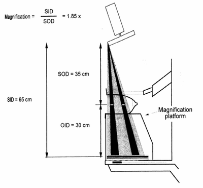
What is shown in the diagram related to mammography?
The diagram shows magnification in mammography with SID = 65 cm, SOD = 35 cm, OID = 30 cm, and a magnification of 1.85x.
 medical_imaging diagrams