Sign up to unlock more features
- Save this deck to your account
- Study flashcards with spaced repetition
- Export to Anki (.apkg) or PDF
- Process documents up to 100 pages
- Images extracted from PDFs and documents
- Better text extraction from your PDFs and documents
- Better flashcards with our more advanced AI model
What hormone does the epithalamus secrete?
How does melatonin affect the brain?
What triggers the epithalamus?
What fluid is linked to the epithalamus?
List the three parts of the brain stem.
What type of matter composes the brain stem?
What automatic behaviors does the brain stem provide?
Which functions does the brain stem control?
How does the epithalamus affect mood?
What image depicts parts of the brain and spine?
What connects the higher and lower brain centers?
What is the role of the midbrain in pain?
What connects the midbrain to the cerebellum?
What functions are associated with the midbrain?
What is the role of the Pons?
What does the medulla oblongata control?
What is the major function of the medulla oblongata?
What is the function of the Corpora Quadrigemina?
How does the nervous system affect heart rate?
What sensory information does the midbrain process?
Which brain structure regulates vomiting, hiccups, swallowing, coughing, and sneezing?
What are the main functions of the cerebellum?
Describe the composition of the cerebellum.
What is the first step in cerebellar processing?
What types of input does the cerebellum receive?
What does the cerebellar cortex do during processing?
How does the cerebellum communicate its findings?
What is represented in the diagram of the brain provided?
What sends signals to muscles for contraction?
What are functional brain systems?
Can functional brain systems be localized?
What is another name for the limbic system?
Which areas contribute input to the limbic system?
What strong link is associated with the limbic system?
Where does output from the limbic system relay?
How can emotions affect physical responses?
What role does the limbic system play in mental conflict?
What is the role of the reticular formation?
What does the Reticular Activation System (RAS) filter?
What does the RAS do when we are awake?
What does an EEG measure?
How does the brain perceive signals from an EEG?
What is the frequency range for Beta waves?
What mental state is associated with Alpha waves?
What occurs right before sleep in relation to Theta waves?
What does Delta wave activity indicate?
What can indicate minor brain damage when awake?
What can indicate significant brain damage in a conscious state?
What is depicted in the provided diagram?
What is the summary of the study notes core concepts?
Define consciousness.
List the levels of consciousness from most to least active.
What happens in stupor?
Describe the state of sleep.
What are the two types of sleep?
What occurs during Non-REM sleep?
What type of brain waves are present during Non-REM sleep?
How long does a Non-REM cycle last?
What characterizes REM sleep?
How often does REM sleep switch during the night?
What is a primary function of REM sleep?
What is memory?
Describe the pathway of information in memory.
What is the gateway to long-term memory?
What factors affect entrance into long-term memory?
How does mood affect memory?
What distinguishes short-term memory from long-term memory?
What protects the brain?
Define working memory.
What is automatic memory?
What are the protective membranes covering the CNS called?
What fluid is contained within the meninges?
What are the three layers of the meninges?
What is the function of the dura mater?
How does the CSF protect the brain?
What are the two layers of the dura mater in the brain?
Which layer of the meninges is closest to the brain?
What is the role of the blood-brain barrier?
What is the relationship between the meninges, CSF, and the blood-brain barrier?
What is the purpose of hair and skin in relation to the brain?
How thick and fibrous are the meninges?
Show a diagram of the cranial meninges.
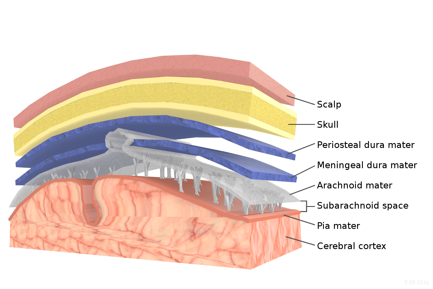
What is the strongest and most superficial covering of the CNS?
What layer is the middle meninx of the CNS?
What space does the Arachnoid Mater create?
What is the deepest layer of the meninges?
What does cerebrospinal fluid provide for the brain and spinal cord?
What is the total volume of cerebrospinal fluid?
How often is cerebrospinal fluid replaced?
By what percentage does cerebrospinal fluid reduce the weight of the brain?
What mechanism protects the brain from physical trauma?
What forms the cerebrospinal fluid?
What is the function of the Blood-Brain Barrier?
What kind of junctions are present in the Blood-Brain Barrier?
What does the capillary endothelium in the blood-brain barrier consist of?
What is the function of cerebrospinal fluid besides cushioning?
What role do astrocytes play in the brain?
Name three substances that can pass through the blood-brain barrier relatively easily.
Where is the blood-brain barrier absent to monitor blood composition?
What triggers the vomiting center in the brain?
What are the functions of the hypothalamus?
Flashcards in this deck (101)
-
brain epithalamus
-
brain melatonin
-
What triggers the epithalamus?
It responds to UV radiation; active at night, inhibited during the day.
brain light -
brain cerebrospinal_fluid
-
brain brain_stem
-
brain matter
-
brain functions
-
brain control
-
brain mood
-
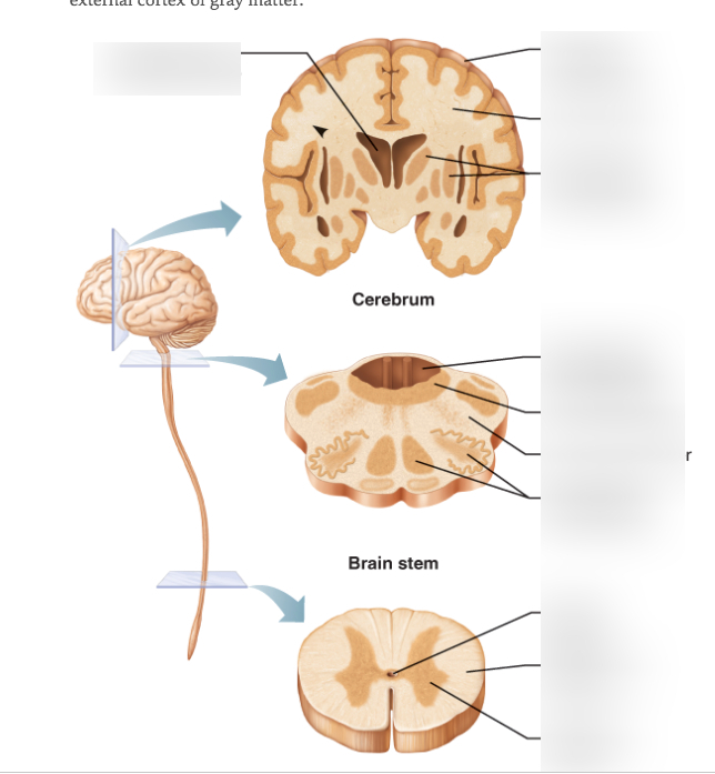
What image depicts parts of the brain and spine?
A diagram showing cross-sections of the brain, brainstem, and spinal cord.
 brain anatomy
 -
What connects the higher and lower brain centers?
The midbrain connects higher and lower brain centers.
anatomy brain -
anatomy pain
-
What connects the midbrain to the cerebellum?
Motor tracts in the midbrain connect to the cerebellum.
anatomy motor -
What functions are associated with the midbrain?
- Pain suppression
- Visual reflex center
- Auditory relay
anatomy functions -
What is the role of the Pons?
It relays information between higher brain centers and the spinal cord.
anatomy pons -
anatomy medulla
-
anatomy functions
-
What is the function of the Corpora Quadrigemina?
It acts as a visual and spatial relay center within the midbrain.
anatomy corpora_quadrigemina -
How does the nervous system affect heart rate?
It usually slows down heart rate but increases it during excitement or fear.
anatomy heart_rate -
What sensory information does the midbrain process?
It processes sound and coordinates eye movement with head movement.
anatomy senses -
Which brain structure regulates vomiting, hiccups, swallowing, coughing, and sneezing?
The medulla oblongata regulates these functions.
brain medulla -
What are the main functions of the cerebellum?
- Refines skeletal muscle contractions
- Involved in cognition, language, and problem-solving
- Coordinates input from other brain regions
brain cerebellum functions -
Describe the composition of the cerebellum.
- Contains white/grey cortex
- Internal white/grey matter masses
- Grey matter arrangement is called arbor vitae.
brain cerebellum anatomy -
What is the first step in cerebellar processing?
The motor association area of the cerebral cortex sends signals to the cerebellum.
brain cerebellum processing -
What types of input does the cerebellum receive?
- Information from visual receptors
- Equilibrium receptors
- Proprioceptors
brain cerebellum input -
What does the cerebellar cortex do during processing?
It calculates the best way to coordinate the force of contraction.
brain cerebellum processing -
brain cerebellum communication
-
What is represented in the diagram of the brain provided?
A diagram showing the major parts of the brain.
brain diagram -
What sends signals to muscles for contraction?
The cerebral cortex sends signals for muscle contraction.
brain muscles -
What are functional brain systems?
They are a network of neurons working together across large distances.
brain systems -
brain localization
-
brain limbic
-
brain input
-
brain memory
-
brain output
-
brain emotions
-
What role does the limbic system play in mental conflict?
It helps resolve mental conflict and interpret gestures.
brain conflict -
brain arousal
-
What does the Reticular Activation System (RAS) filter?
It filters sensory input, determining wakefulness.
brain ras -
What does the RAS do when we are awake?
The RAS sends continual signals to the cerebrum to keep the cerebral cortex active.
brain ras -
eeg neuroscience
-
brain eeg
-
brain_waves beta
-
brain_waves alpha
-
What occurs right before sleep in relation to Theta waves?
They are common in children and uncommon in adults.
brain_waves theta -
brain_waves delta
-
brain neuroscience
-
brain neuroscience
-
What is depicted in the provided diagram?
Diagrams showing Alpha, Beta, Theta, and Delta brain waves over time.
brain_waves visual -
What is the summary of the study notes core concepts?
- Epithalamus and melatonin secretion
- Brain stem components and functions
- Cerebellum’s role in coordination
- Limbic system’s emotional connections
- EEGs and levels of consciousness
- Sleep phases and memory processing
- Brain protection mechanisms
summary study_notes -
psychology consciousness
-
psychology consciousness
-
psychology stupor
-
Describe the state of sleep.
A state of partial or complete unconsciousness; unaware of surroundings.
psychology sleep -
psychology sleep
-
psychology non-rem
-
psychology sleep
-
psychology sleep
-
psychology rem
-
psychology sleep
-
psychology sleep
-
memory neuroscience
-
Describe the pathway of information in memory.
- Stimulus occurs
- Stimulus enters buffer
- If used → Short-term memory
- If not used → Forgotten
memory process -
What is the gateway to long-term memory?
Short-term memory acts as the gateway into long-term memory.
memory long-term -
memory learning
-
memory mood
-
What distinguishes short-term memory from long-term memory?
Short-term memory is temporary, while long-term memory can be more permanent.
memory comparison -
What protects the brain?
- The skull surrounds the brain
- Acts as a helmet
- Flat compact bone with a spongy center.
brain protection -
memory working
-
What is automatic memory?
Circumstances leading to information immediately moving into long-term memory.
memory automatic -
anatomy cns
-
What fluid is contained within the meninges?
Cerebrospinal fluid (CSF) is contained within the meninges.
anatomy csf -
anatomy meninges
-
What is the function of the dura mater?
It covers and protects the CNS and is composed of fibrous connective tissue.
anatomy meninges -
How does the CSF protect the brain?
Cushions brain structures and protects blood vessels supplying nervous tissue.
anatomy csf -
What are the two layers of the dura mater in the brain?
- Periosteal layer (superficial)
- Meningeal layer (deeper)
anatomy dura_mater -
Which layer of the meninges is closest to the brain?
The pia mater is the closest layer to the brain.
anatomy meninges -
What is the role of the blood-brain barrier?
It helps to protect the brain from potentially harmful substances in the blood.
anatomy blood-brain_barrier -
What is the relationship between the meninges, CSF, and the blood-brain barrier?
They work together to protect the brain and maintain its environment.
anatomy protection -
What is the purpose of hair and skin in relation to the brain?
They provide protection and cushion for brain structures.
anatomy protection -
anatomy meninges
-
anatomy diagrams
-
anatomy cns
-
anatomy cns
-
What space does the Arachnoid Mater create?
The subarachnoid space, filled with cerebrospinal fluid.
anatomy cns -
anatomy cns
-
anatomy cns
-
anatomy cns
-
anatomy cns
-
anatomy cns
-
anatomy cns
-
anatomy cns
-
What is the function of the Blood-Brain Barrier?
To protect nervous tissue from bloodborne substances.
anatomy cns -
What kind of junctions are present in the Blood-Brain Barrier?
Tight, impermeable junctions between endothelial cells.
anatomy cns -
What does the capillary endothelium in the blood-brain barrier consist of?
A thick and relatively non-porous layer.
anatomy cns -
What is the function of cerebrospinal fluid besides cushioning?
It helps nourish and carry chemical signals to the brain.
anatomy cns -
What role do astrocytes play in the brain?
They attach neurons to blood vessels and act as a selective filter between neurons and capillaries.
biology neuroscience -
Name three substances that can pass through the blood-brain barrier relatively easily.
- Glucose
- Amino acids
- Electrolytes
biology neuroscience blood-brain_barrier -
biology neuroscience brain_structures
-
What triggers the vomiting center in the brain?
Toxins and poisons, such as alcohol, circulating in the blood.
biology neuroscience vomiting_center -
What are the functions of the hypothalamus?
- Monitors body temperature
- Detects water balance issues
- Feedback on hormone levels
biology neuroscience hypothalamus
Epithalamus
• Secretes melatonin via the pineal gland, regulating the sleep-wake cycle. • Inhibits gland activity during daylight and activates it at night. • Influences mood; sleep deprivation can lead to unhappiness. • Linked to cerebrospinal fluid production by ependymal cells.
Brain Stem
• Comprised of the pons, midbrain, and medulla oblongata. • Contains deep grey matter surrounded by white matter. • Essential for programmed automatic behaviors (breathing, heart rate, blood pressure). • Connects higher and lower brain centers, providing crucial pathways for information.
Midbrain
• Connects motor tracts to the spinal cord and cerebellum. • Functions include pain suppression and sensory coordination. • Houses the corpora quadrigemina, important for visual and spatial relay.
Pons
• Primarily consists of conduction tracts. • Relays information between higher brain centers and the spinal cord.
Medulla Oblongata
• Major autonomic reflex center managing heart rate and blood vessel diameter. • Controls the rate and depth of breathing, as well as reflex actions like vomiting and coughing. • Overlaps significantly with the hypothalamus.
Cerebellum
• Functions: Refines skeletal muscle contractions, contributes to cognition and problem-solving, operates both consciously and subconsciously. • Similar composition to the cerebrum: grey cortex and white matter with a distinctive arbor vitae pattern.
Cerebellar Processing: 1. Motor signals from the cerebral cortex initiate contractions. 2. Receives inputs from visual, equilibrium, and proprioceptive receptors. 3. Coordinates muscle contraction force and dispatches messages back to the cortex.
Functional Brain Systems
• Networks of neurons spanning large distances. • Limbic System: Emotional brain links memory to strong odors, affecting hormonal releases. • Reticular Formation: Governs brain arousal; the Reticular Activation System (RAS) filters sensory inputs.
EEGs and Consciousness
• EEGs measure electrical activity patterns in the brain (action potentials), classified as: - Beta: > 13 waves/sec (focus). - Alpha: 8-13 waves/sec (alert, relaxed). - Theta: 4-8 waves/sec (pre-sleep). - Delta: < 4 waves/sec (deep sleep).
• Consciousness is the awareness of sensations, showcasing various levels from alert to coma.
Sleep and Memory
• Sleep includes Non-REM (restorative) and REM (dreaming) stages, crucial for recovery and analysis of daily events. • Memory: Involves storage and retrieval through linked neural pathways; pathways include stimuli leading to short-term or potentially long-term memory.
Brain Protection
• Physical Protection: - Skull: Encases the brain. - Meninges: Three protective layers (dura mater, arachnoid mater, pia mater) that cushion the brain and spinal cord.
• Cerebrospinal Fluid (CSF): Protects and nourishes the brain, reducing its weight by 97% and absorbing shocks.
Blood-Brain Barrier
• Blood-brain barrier prevents harmful substances in the blood from entering the brain while allowing selective passage of essential nutrients (e.g., glucose, amino acids). • Maintains stable conditions for neural function.