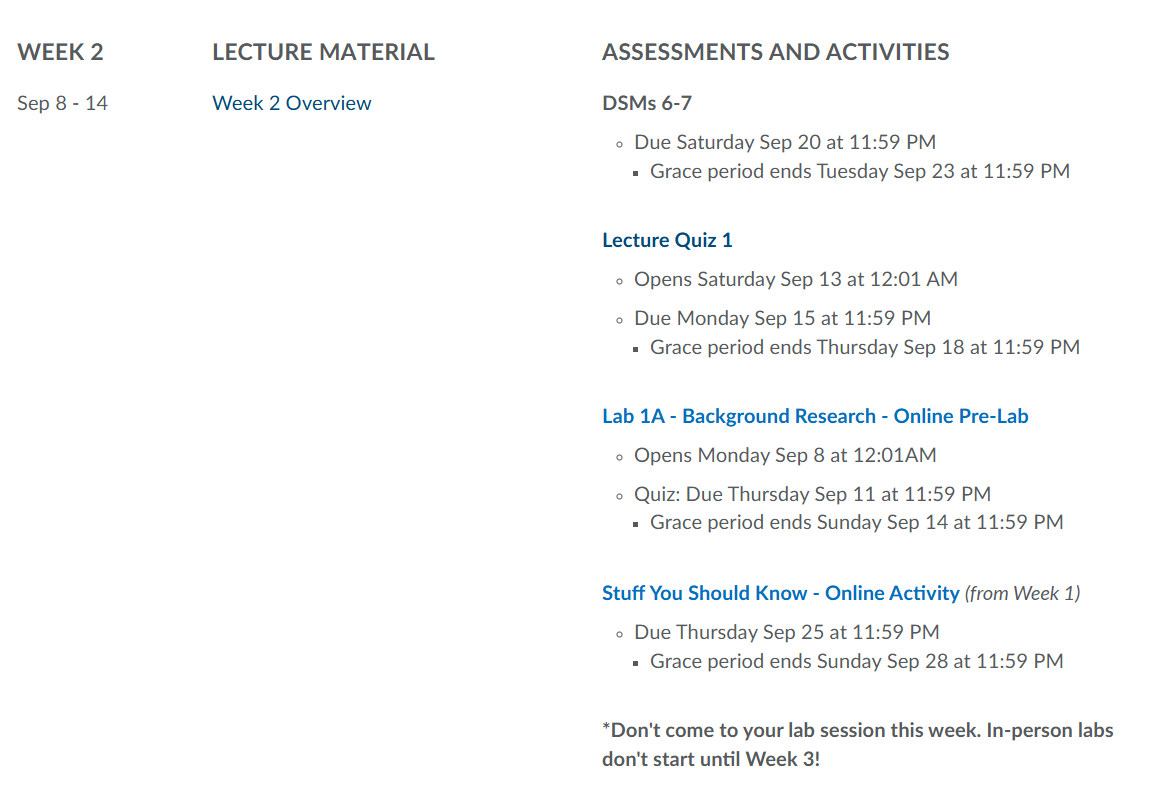
Sauvegarde ton deck avant qu'il ne disparaisse
Ces flashcards ne sont pas encore sauvegardées — elles seront perdues si tu quittes. Crée un compte gratuit pour les garder et débloquer tout ce qui suit.
- Save this deck to your account
- Study with spaced repetition
- Export to Anki (.apkg) or PDF
- Process documents up to 100 pages
- Images extracted from your PDFs
- Sharper text extraction & a more advanced AI model
What is the focus of Molecular and Cell Biology?
What university offers BIOL 102?
Which week is referred to in the lecture title?
What is the logo of Queen's University used for?
What is the due date for DSMs 6-7?
When does Lecture Quiz 1 open?
What is the due date for Lecture Quiz 1?
When does Lab 1A - Background Research quiz open?
What is the due date for Lab 1A quiz?
When is the due date for Stuff You Should Know activity?
When do in-person labs start?
What does the schedule include?
What is light microscopy based on?
What does electron microscopy use?
Define resolution in microscopy.
What is the formula for magnification?
What does contrast refer to?
What does this diagram illustrate?
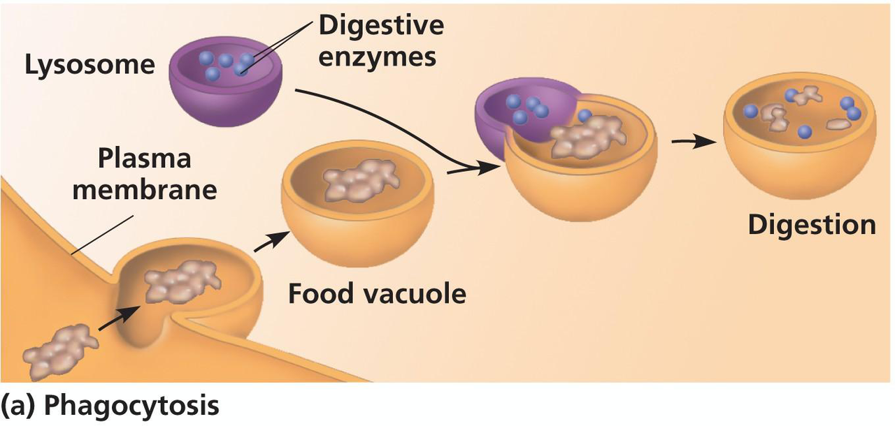
What is the process of phagocytosis? 
What structures are shown in the microscopic view? 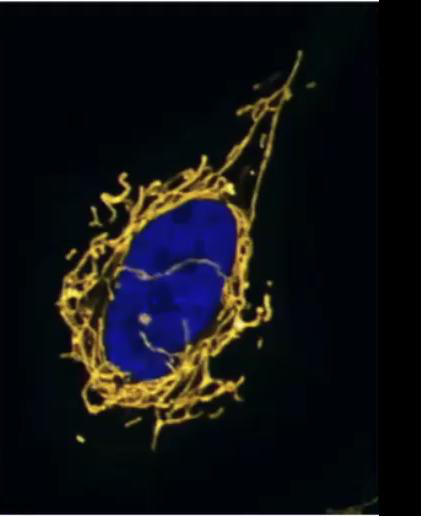
Where in the cell would a culture of rapidly growing animal cells have the greatest concentration of radioactivity when adding non-toxic radioactive dTTP?
Where is the DNA found in eukaryotic cells?
Eukaryotic cells are subdivided into which two types?
What is the interior substance of a cell called?
What jelly-like substance is found in the cytoplasm?
What is present in plant cells but absent in animal cells?
What is unique about the vacuole in plant cells?
Which structure is common to all animal cells?
What organelle is responsible for energy production in both cell types?
Which organelle performs photosynthesis in plant cells?
In which type of cell are lysosomes mostly found?
Which structure is absent in most plant cells?
What structure supports the plant cell and is made of cellulose?
What diagram shows the organelles in a typical animal cell?
What diagram shows the organelles in a typical plant cell?
What do most plant cells look like?
What is a distinguishing feature of plant cells?
What can be seen clearly in most plant cells?
What is shown in the microscopic image of onion cells?
What is the shape of epidermal pavement cells?
What organelles are labeled in the epidermal pavement cell diagram?
How do epidermal pavement cells contribute to plants?
Describe the appearance of green fluorescent plant epidermal cells.
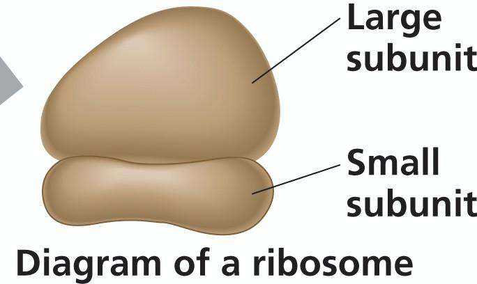
What are ribosomes composed of?
What is the primary role of ribosomes?
Where are free ribosomes located?
What are bound ribosomes associated with?
What does a ribosome consist of?
What is the structure of a ribosome?
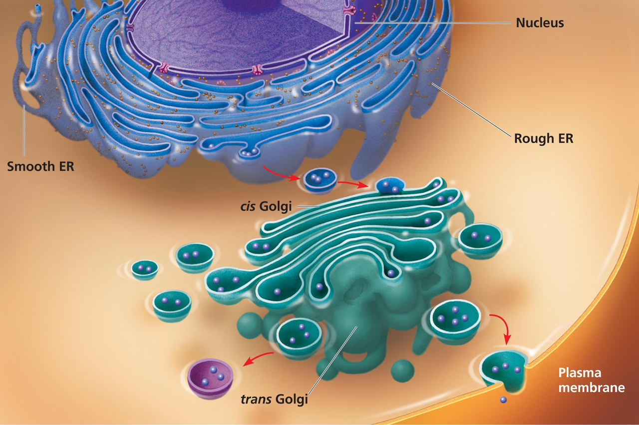
What are the components of the endomembrane system?
How are the components of the endomembrane system connected?
What does the diagram of the endomembrane system include?
What does the nucleus contain?
What is the function of the nucleus?
What is the nuclear envelope?
What provides the stability to the nuclear envelope?
What occurs in the nucleolus?
What accompanies the structure of the nucleus in diagrams?
What does the label in the nuclear envelope diagram indicate?
What is the main function of the Smooth ER?
What does the Rough ER synthesize proteins for?
What processes is the Rough ER involved in?
What does the diagram illustrate?
What is the function of the Golgi apparatus?
What does the Golgi apparatus alter?
What does the Golgi apparatus synthesize?
What are the two faces of the Golgi apparatus?
What are lysosomes?
What is the function of hydrolytic enzymes in lysosomes?
What process involves the fusion of a food vacuole with a lysosome?
What is autophagy?
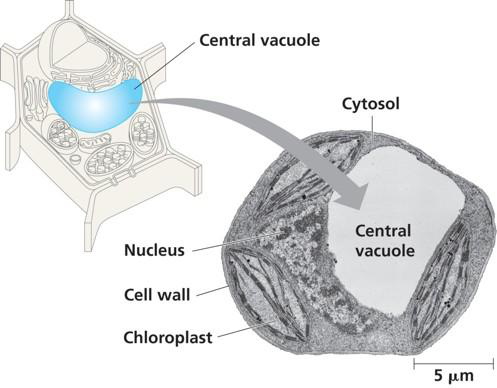
What are vacuoles?
What is the function of the central vacuole?
What does the central vacuole contain?
Where is the central vacuole found?
What is shown in the electron micrograph related to vacuoles?
What are the primary sites of cellular respiration?
What are the main functions of chloroplasts?
What genetic material do mitochondria and chloroplasts contain?
How do the ribosomes of mitochondria and chloroplasts compare?
What does the Endosymbiont Theory describe?
What are the components involved in the Endosymbiont Theory?
What is illustrated in the diagram of the Endosymbiont Theory?
Do alternative theories to the Endosymbiont Theory exist?
A mutation affecting polysaccharide modifications to proteins would likely disrupt which cellular structures?
What are the possible structures affected by the mutation?
What is the cytoskeleton?
What are the three main components of the cytoskeleton?
What does the cytoskeleton provide for the cell?
What role does the cytoskeleton play in cell motility?
What colors represent the components of the cytoskeleton in the micrograph?
What are the main components of the cytoskeleton?
What is the structure of Microtubules?
What are the protein subunits of Microtubules?
What is the main function of Microtubules?
What is the structure of Microfilaments?
What are the main functions of Microfilaments?
What is the structure of Intermediate Filaments?
What is the main function of Intermediate Filaments?
What does the microtubule diagram represent?
What does the microfilament diagram illustrate?
What does the intermediate filament diagram show?
What structure is involved in the cytoskeleton?
What are microtubules made of?
What are microfilaments mainly composed of?
What do you call the fibers that help maintain cell shape?
What are plasmodesmata?
What do gap junctions do?
What is the function of tight junctions?
What do desmosomes do?
What structure is labeled in this diagram? 
What is the purpose of the tight junctions indicated in this diagram? 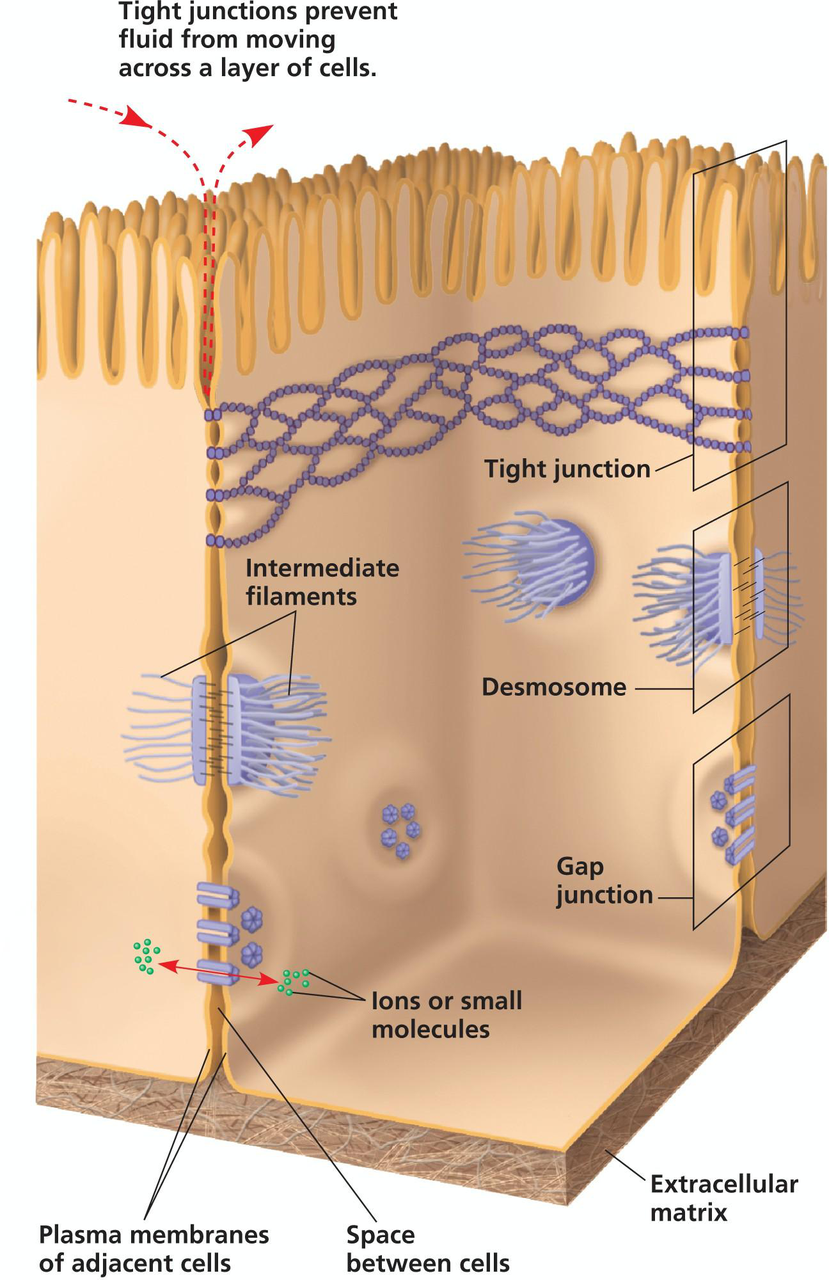
Which organs would dysfunction due to abnormal microtubules?
Which choice includes areas of contraction affected by microtubule dysfunction?
What tissues have projections that might be affected by abnormal microtubules?
Which ducts may experience dysfunction due to abnormal microtubules?
Which cells showing movement may be affected by abnormal microtubules?
What type of cells exhibit amoeboid movement that may be impacted?
What is the primary function of hepatocytes?
What sub-cellular structures do hepatocytes have?
How many mitochondria do hepatocytes contain?
What happens to hepatocytes under chronic exposure to toxins?
What does the smooth ER in hepatocytes assist with?
What does the rough ER in hepatocytes mainly function for?
Show an image of hepatocyte ER structures.
Do erythrocytes (red blood cells) have a nucleus?
What type of energy production do erythrocytes rely on?
What cellular structures are absent in erythrocytes?
Why can't erythrocytes be targeted by viruses?
What is important for erythrocyte deformability?
How does the blood type get determined?
What are the types of blood glycoproteins?
What is shown in this electron micrograph?
What is illustrated in the diagram of blood cells?
What are keratinocytes?
What happens to keratinocytes as they mature?
What provides rigidity to keratinocytes?
What role do desmosomes play in keratinocytes?
Do keratinocytes continue to divide?
What is shown in the skin layers diagram?
What is the structure composition of skeletal muscle cells?
What is the basic contractile unit of muscle?
What connects the sarcomere to other organelles?
What is the role of sarcoplasmic reticulum in muscle cells?
What condition is related to calcium leak in muscle cells?
What do skeletal muscle cells require to produce energy?
What is the role of actin and myosin?
What do you observe in light micrograph of muscle cells?
Flashcards in this deck (148)
-
What is the focus of Molecular and Cell Biology?
Study of cells, their structures, functions, and interactions.
biology molecular_biology -
What university offers BIOL 102?
Queen's University
biology education -
Which week is referred to in the lecture title?
Week 2
lecture biology -
What is the logo of Queen's University used for?
Visual representation of the university.
branding university -
What is the due date for DSMs 6-7?
Due Saturday Sep 20 at 11:59 PM. Grace period ends Tuesday Sep 23 at 11:59 PM.
deadlines assessments -
When does Lecture Quiz 1 open?
Opens Saturday Sep 13 at 12:01 AM.
deadlines quizzes -
What is the due date for Lecture Quiz 1?
Due Monday Sep 15 at 11:59 PM. Grace period ends Thursday Sep 18 at 11:59 PM.
deadlines quizzes -
When does Lab 1A - Background Research quiz open?
Opens Monday Sep 8 at 12:01 AM.
deadlines labs -
What is the due date for Lab 1A quiz?
Due Thursday Sep 11 at 11:59 PM. Grace period ends Sunday Sep 14 at 11:59 PM.
deadlines labs -
When is the due date for Stuff You Should Know activity?
Due Thursday Sep 25 at 11:59 PM. Grace period ends Sunday Sep 28 at 11:59 PM.
deadlines activities -
When do in-person labs start?
In-person labs don’t start until Week 3.
labs schedule -
What does the schedule include?
It includes lecture material, quizzes, and assignment deadlines.
 schedule lectures
 -
What is light microscopy based on?
Passing a beam of light through a specimen
microscopy light -
What does electron microscopy use?
A beam of electrons over or through a specimen
microscopy electron -
Define resolution in microscopy.
Minimum distance two points can be separated and still distinguishable
microscopy resolution -
What is the formula for magnification?
Ratio of an object's image size to its real size
microscopy magnification -
What does contrast refer to?
Difference in brightness between light and dark areas
microscopy contrast -
What does this diagram illustrate?
Scales of biological entities and microscopy ranges
 microscopy diagram
 -
What is the process of phagocytosis?
Macrophage digesting bacteria.
biology cellular_processes -
What structures are shown in the microscopic view?
- Nucleus (blue)
- Mitochondria (yellow)
biology cell_structure -
Where in the cell would a culture of rapidly growing animal cells have the greatest concentration of radioactivity when adding non-toxic radioactive dTTP?
- A. Nucleus
- B. Nucleoid
- C. Endoplasmic reticulum
- D. Cytoplasm
- E. Ribosomes
cell_biology radioactivity nucleus -
Where is the DNA found in eukaryotic cells?
In the nucleus
biology cell_structure -
Eukaryotic cells are subdivided into which two types?
- Animal cells
- Plant cells
biology cell_types -
What is the interior substance of a cell called?
The cytoplasm
biology cell_structure -
What jelly-like substance is found in the cytoplasm?
The cytosol
biology cell_structure -
What is present in plant cells but absent in animal cells?
- Cell wall
- Chloroplasts
- Plastids
biology cell_comparison -
What is unique about the vacuole in plant cells?
One large central vacuole
biology cell_comparison -
Which structure is common to all animal cells?
Presence of centrioles
biology cell_structure -
What organelle is responsible for energy production in both cell types?
Mitochondrion
biology cell_structure -
Which organelle performs photosynthesis in plant cells?
Chloroplast
biology cell_structure -
In which type of cell are lysosomes mostly found?
Animal cells
biology cell_structure -
Which structure is absent in most plant cells?
Cilia
biology cell_structure -
What structure supports the plant cell and is made of cellulose?
Cell wall
biology cell_structure -
What diagram shows the organelles in a typical animal cell?
 biology cell_diagram
 -
What diagram shows the organelles in a typical plant cell?
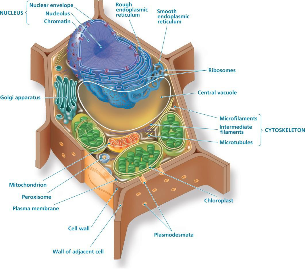 biology cell_diagram
 -
What do most plant cells look like?
They resemble the standard textbook image of cells.
biology plant_cells -
What is a distinguishing feature of plant cells?
They have visible cell walls.
biology plant_cells -
What can be seen clearly in most plant cells?
Visible nuclei within the cells.
biology plant_cells -
What is shown in the microscopic image of onion cells?
Rows of onion cells with distinct cell walls and nuclei.
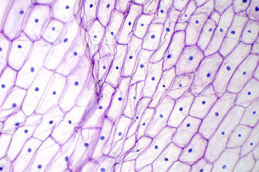 biology microscopy onion_cells
 -
What is the shape of epidermal pavement cells?
These cells have an interlocking, irregular shape.
biology plant_cells -
What organelles are labeled in the epidermal pavement cell diagram?
- Nucleus
- Chloroplast
- Vacuole
- Plasma membrane
- Peroxisome
biology organelles -
How do epidermal pavement cells contribute to plants?
They provide protection and add strength & stability.
biology plant_cells -
Describe the appearance of green fluorescent plant epidermal cells.
They have a characteristic jigsaw puzzle shape.
biology plant_cells -
What are ribosomes composed of?
- Protein
- rRNA
biology ribosomes -
What is the primary role of ribosomes?
Protein synthesis
biology cell -
Where are free ribosomes located?
Cytosol
biology cell -
What are bound ribosomes associated with?
- Endoplasmic reticulum
- Nuclear envelope
biology cell -
What does a ribosome consist of?
- Large subunit
- Small subunit
biology ribosomes -
What is the structure of a ribosome?
3D model with complex structure.
biology ribosomes -
What are the components of the endomembrane system?
- Nucleus
- Endoplasmic reticulum
- Golgi apparatus
- Lysosomes
- Vacuoles
- Plasma membrane
biology cell_structure -
How are the components of the endomembrane system connected?
Through direct physical continuity or the transfer of vesicles.
biology cell_structure -
What does the diagram of the endomembrane system include?
- Nucleus
- Rough ER
- Smooth ER
- Golgi apparatus
biology diagrams -
What does the nucleus contain?
- Chromosomes
- Chromatin (DNA + proteins)
cell_biology nucleus -
What is the function of the nucleus?
- mRNA synthesis
- Export
cell_biology nucleus -
What is the nuclear envelope?
- A double membrane
- Each membrane is a lipid bilayer
cell_biology nucleus -
What provides the stability to the nuclear envelope?
- Nuclear lamina (protein filaments)
cell_biology nucleus -
What occurs in the nucleolus?
- Transcription of ribosomal RNA (rRNA)
- Ribosome assembly
cell_biology nucleus -
What accompanies the structure of the nucleus in diagrams?
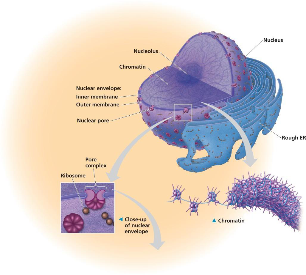 cell_biology nucleus
 -
What does the label in the nuclear envelope diagram indicate?
 cell_biology nucleus
 -
What is the main function of the Smooth ER?
- Lipid synthesis
- Drug detoxification
- Calcium storage
biology cell -
What does the Rough ER synthesize proteins for?
- Exporting proteins
biology cell -
What processes is the Rough ER involved in?
- Protein glycosylation
- Membrane synthesis
biology cell -
What does the diagram illustrate?
The structure of the rough and smooth endoplasmic reticulum
biology cell -
What is the function of the Golgi apparatus?
- Sorts molecules
- Releases vesicles for transport
cell_biology organelles -
What does the Golgi apparatus alter?
- Structure of macromolecules
cell_biology macromolecules -
What does the Golgi apparatus synthesize?
- Secreted polysaccharides
cell_biology synthesis -
What are the two faces of the Golgi apparatus?
- Cis face (receiving)
- Trans face (shipping)
cell_biology golgi_apparatus -
What are lysosomes?
Membranous sacs with an acidic interior containing hydrolytic enzymes for digestion.
cell_biology lysosomes -
What is the function of hydrolytic enzymes in lysosomes?
They are used for the hydrolysis of macromolecules.
cell_biology enzymes -
What process involves the fusion of a food vacuole with a lysosome?
This process is called phagocytosis.
cell_biology phagocytosis -
What is autophagy?
A process in which a vesicle containing cellular debris fuses with a lysosome for digestion.
cell_biology autophagy -
What are vacuoles?
Large vesicles with an internal solution differing in composition from the cytoplasm.
biology cell_structure -
What is the function of the central vacuole?
Important for maintaining cell structure.
biology cell_structure -
What does the central vacuole contain?
- Ions
- Toxins
biology cell_structure -
Where is the central vacuole found?
In plant cells.
biology cell_structure -
What is shown in the electron micrograph related to vacuoles?
The structure of the central vacuole within a plant cell.
biology cell_structure -
What are the primary sites of cellular respiration?
Mitochondria produce ATP from fuels and oxygen.
biology cellular_respiration -
What are the main functions of chloroplasts?
Sites of photosynthesis, producing sugars from carbon dioxide, water, and light.
biology photosynthesis -
What genetic material do mitochondria and chloroplasts contain?
Both contain their own DNA and ribosomes.
biology genetics -
How do the ribosomes of mitochondria and chloroplasts compare?
They are more related to bacteria than to eukaryotic versions.
biology ribosomes -
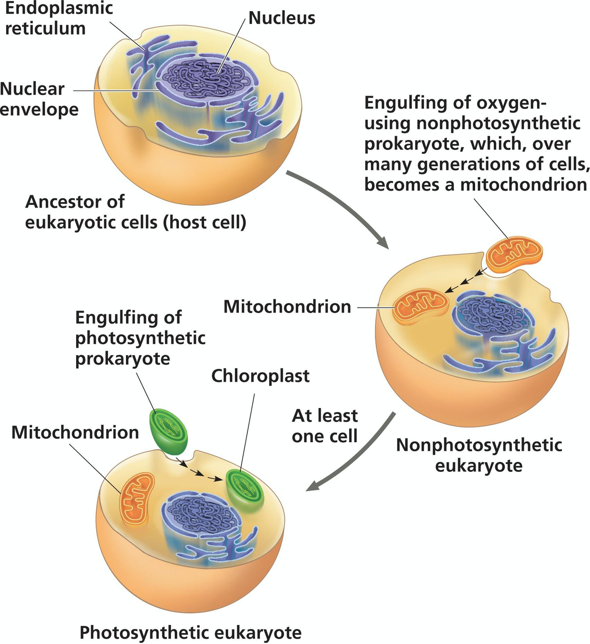
What does the Endosymbiont Theory describe?
The evolutionary origins of mitochondria and chloroplasts.
biology theory -
What are the components involved in the Endosymbiont Theory?
- Asgard archaeon
- alphaproteobacterium
- cyanobacterium
biology evolution -
What is illustrated in the diagram of the Endosymbiont Theory?
An ancestral eukaryotic cell engulfing a nonphotosynthetic prokaryote to become a mitochondrion, and a photosynthetic prokaryote to become a chloroplast.
 biology visual
 -
Do alternative theories to the Endosymbiont Theory exist?
Yes, alternate theories exist.
biology theory -
A mutation affecting polysaccharide modifications to proteins would likely disrupt which cellular structures?
- Golgi apparatus
- Extracellular matrix
cell_biology mutations -
What are the possible structures affected by the mutation?
- Nuclear lamina
- Nuclear matrix
- Mitochondria
- Nuclear pores
- Secretory vesicles
cell_biology structures -
What is the cytoskeleton?
A dynamic network of fibers extending throughout the cytoplasm.
biology cell_structure -
What are the three main components of the cytoskeleton?
- Microtubules
- Microfilaments (actin filaments)
- Intermediate filaments
biology cell_structure -
What does the cytoskeleton provide for the cell?
- Mechanical support
- Structure
- Anchorage for organelles
biology cell_structure -
What role does the cytoskeleton play in cell motility?
Involved in the movement of the cell location and the movement of cell parts.
biology cell_motility -
What colors represent the components of the cytoskeleton in the micrograph?
- Microtubules: Green
- Microfilaments: Red
- Nucleus: Blue
biology cell_structure microscopy -
What are the main components of the cytoskeleton?
- Microtubules
- Microfilaments
- Intermediate Filaments
biology cytoskeleton -
What is the structure of Microtubules?
- Hollow tubes
- Diameter: 25 nm
biology structure -
What are the protein subunits of Microtubules?
- Tubulin (α-tubulin and β-tubulin)
biology proteins -
What is the main function of Microtubules?
- Maintenance of cell shape
- Cell motility
- Chromosome movements in cell division
biology functions -
What is the structure of Microfilaments?
- Two intertwined strands of actin
- Diameter: 7 nm
biology structure -
What are the main functions of Microfilaments?
- Maintenance of cell shape
- Muscle contraction
- Cytoplasmic streaming
biology functions -
What is the structure of Intermediate Filaments?
- Fibrous proteins coiled into cables
- Diameter: 8-12 nm
biology structure -
What is the main function of Intermediate Filaments?
- Anchorage of nucleus
- Form nuclear lamina
biology functions -
What does the microtubule diagram represent?
Diagram of a column of tubulin dimers, structural unit of microtubules.
 biology diagrams
 -
What does the microfilament diagram illustrate?
Diagram of an actin filament, composed of two intertwined strands of actin subunits.
biology diagrams -
What does the intermediate filament diagram show?
Diagram of keratin proteins coiled together to form a fibrous subunit of intermediate filaments.
biology diagrams -
What structure is involved in the cytoskeleton?
- Microtubules
- Microfilaments
- Intermediate Filaments
biology cytoskeleton -
What are microtubules made of?
Tubulin Polymers
biology cell_structure -
What are microfilaments mainly composed of?
Actin Filaments
biology cell_structure -
What do you call the fibers that help maintain cell shape?
Intermediate Filaments
biology cell_structure -
What are plasmodesmata?
Channels connecting plant cells, allowing transfer of compounds.
cell_biology plant -
What do gap junctions do?
Allow passage of ions/molecules between animal cells.
cell_biology animal -
What is the function of tight junctions?
Create seals between cells to prevent extracellular fluid movement.
cell_biology animal -
What do desmosomes do?
Fasten cells together using intermediate filaments.
cell_biology adhesion -
What structure is labeled in this diagram?
Plasmodesmata, allowing cell-to-cell communication.
cell_biology plant -
What is the purpose of the tight junctions indicated in this diagram?
Prevent intracellular fluid movement.
cell_biology animal -
Which organs would dysfunction due to abnormal microtubules?
- Limbs and heart: areas with contraction
- Microvilli, alveoli, glomeruli: increase surface area
- All ducts: transport fluids
- Sperm, larynx, trachea: contain flagella or cilia
- Phagocytic cells: show amoeboid movement
biology microtubules dysfunction -
Which choice includes areas of contraction affected by microtubule dysfunction?
- Limbs
- Heart
biology microtubules contraction -
What tissues have projections that might be affected by abnormal microtubules?
- Microvilli
- Alveoli
- Glomeruli
biology microtubules surface_area -
Which ducts may experience dysfunction due to abnormal microtubules?
- Salivary glands
- Sebaceous glands
biology microtubules ducts -
Which cells showing movement may be affected by abnormal microtubules?
- Sperm
- Larynx
- Trachea
biology microtubules movement -
What type of cells exhibit amoeboid movement that may be impacted?
- Phagocytic cells
- White blood cells
biology microtubules amoeboid -
What is the primary function of hepatocytes?
Drug detoxification, lipid synthesis/export, protein secretion.
biology hepatocytes -
What sub-cellular structures do hepatocytes have?
- Extensive smooth ER
- Rough ER
- Golgi apparatus
biology cell_structure -
How many mitochondria do hepatocytes contain?
1000-2000 mitochondria per cell.
biology mitochondria -
What happens to hepatocytes under chronic exposure to toxins?
Proliferation of smooth ER and increased drug tolerance.
biology toxins -
What does the smooth ER in hepatocytes assist with?
- Drug detoxification
- Lipid synthesis/export
biology smooth_er -
What does the rough ER in hepatocytes mainly function for?
Protein secretion.
biology rough_er -
Show an image of hepatocyte ER structures.
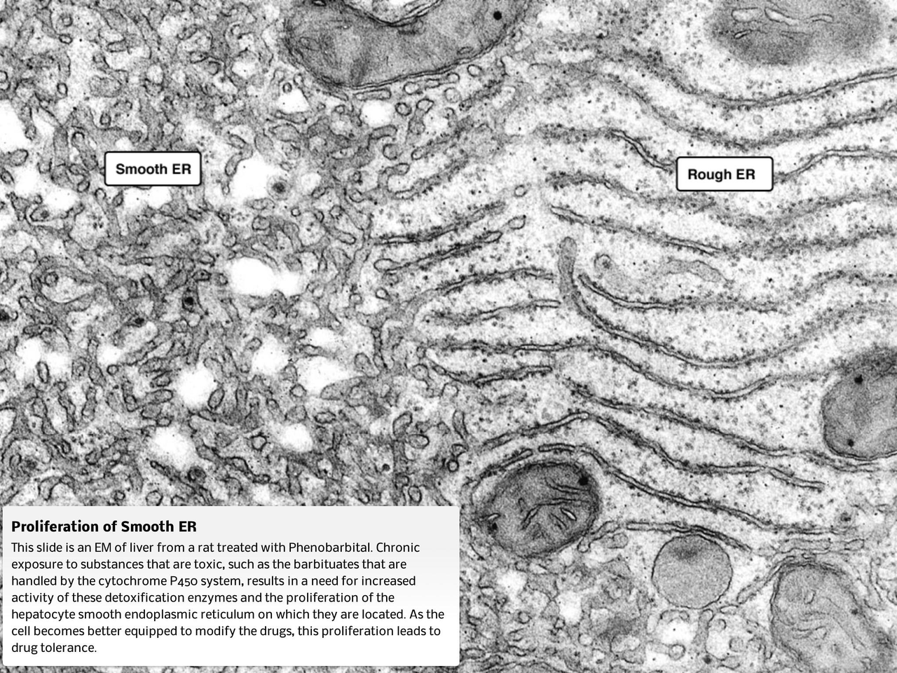 biology microscopy
 -
Do erythrocytes (red blood cells) have a nucleus?
No, they do not have a nucleus in mammals.
biology erythrocytes -
What type of energy production do erythrocytes rely on?
ATP is produced from fermentation, as they lack mitochondria.
biology metabolism -
What cellular structures are absent in erythrocytes?
- DNA
- RNA
- Golgi apparatus
- ER
biology cell_structure -
Why can't erythrocytes be targeted by viruses?
They lack DNA and RNA, preventing protein synthesis.
biology viruses -
What is important for erythrocyte deformability?
The actin cytoskeleton and fluid cell membrane are crucial.
biology cell_structure -
How does the blood type get determined?
Blood type is determined by the presence of glycoproteins.
biology blood_type -
What are the types of blood glycoproteins?
- O
- A
- B
- AB
biology blood_type -
What is shown in this electron micrograph?
An erythrocyte (red blood cell).
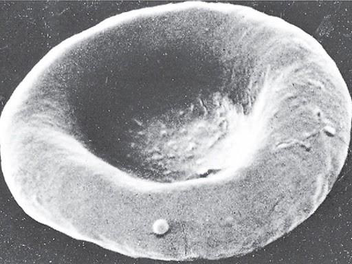 biology microscopy
 -
What is illustrated in the diagram of blood cells?
Different types of blood cells: O, A, B, AB.
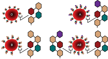 biology blood_type
 -
What are keratinocytes?
Cells that form the epidermis of the skin.
biology skin cells -
What happens to keratinocytes as they mature?
They lose their nucleus and other organelles.
biology skin cells -
What provides rigidity to keratinocytes?
An extensive network of intermediate filaments (keratins).
biology skin -
What role do desmosomes play in keratinocytes?
They provide adhesion and help in wound healing.
biology skin -
Do keratinocytes continue to divide?
They permanently withdraw from the cell cycle.
biology cells -
What is shown in the skin layers diagram?
The surface, upper keratinocytes, melanocytes, and basal keratinocytes.
biology anatomy -
What is the structure composition of skeletal muscle cells?
- Multinucleated
- Lots of mitochondria
- Actin and myosin for contraction
biology muscle_cells -
What is the basic contractile unit of muscle?
The sarcomere
biology muscle_cells -
What connects the sarcomere to other organelles?
Intermediate filaments
biology muscle_cells -
What is the role of sarcoplasmic reticulum in muscle cells?
Stores calcium, important for contraction
biology muscle_cells -
What condition is related to calcium leak in muscle cells?
Rigor mortis
biology muscle_cells -
What do skeletal muscle cells require to produce energy?
Lots of ATP from mitochondria
biology muscle_cells -
What is the role of actin and myosin?
Required for contraction of muscle cells
biology muscle_cells -
What do you observe in light micrograph of muscle cells?
Striated appearance
biology muscle_cells
Upcoming Deadlines
Schedule
| WEEK 2 | LECTURE MATERIAL |
|---|---|
| Sep 8 - 14 | Week 2 Overview |
Assessments and Activities
DSMs 6-7 * Due: Saturday Sep 20 at 11:59 PM * Grace period: Ends Tuesday Sep 23 at 11:59 PM
Lecture Quiz 1 * Opens: Saturday Sep 13 at 12:01 AM * Due: Monday Sep 15 at 11:59 PM * Grace period: Ends Thursday Sep 18 at 11:59 PM
Lab 1A - Online Pre-Lab * Opens: Monday Sep 8 at 12:01 AM * Quiz Due: Thursday Sep 11 at 11:59 PM * Grace period: Ends Sunday Sep 14 at 11:59 PM
Stuff You Should Know * Due: Thursday Sep 25 at 11:59 PM * Grace period: Ends Sunday Sep 28 at 11:59 PM
Note: No in-person lab this week; labs start in Week 3.
Principles of Microscopy
Light microscopy: Uses light beam through specimen.
Electron microscopy: Uses beam of electrons.
Key Concepts
- Resolution: Minimum distance to distinguish two points.
- Magnification: Image size to real size ratio.
- Contrast: Brightness difference between areas.
Microscopy Examples
Notable Examples
-
Phagocytosis: Macrophage digesting bacteria.
-
Cell Nucleus and Mitochondria: Microscopic view.
Eukaryotic Cells
Definition: Cells with DNA in a nucleus. - Cytoplasm: Contains organelles in cytosol.
Cell Comparison
| Property | Animal Cell | Plant Cell |
|---|---|---|
| Cell Wall | Absent | Present (cellulose) |
| Vacuole | Small vacuoles | Large central vacuole |
| Centrioles | Present | Lower plant forms only |
| Chloroplast | Absent | Present |
| Lysosomes | Present | Usually absent |
Nucleus
Components: - Contains chromosomes made of chromatin (DNA + proteins). - Site of mRNA synthesis and export.
Nuclear Envelope
- Double membrane with nuclear lamina for structure.
Nucleolus: - Site of ribosomal RNA transcription.
Ribosomes
Definition: Complexes of protein and rRNA for protein synthesis. - Free Ribosomes: In cytosol. - Bound Ribosomes: On endoplasmic reticulum.
Endomembrane System
Components: - Nucleus, ER, Golgi apparatus, lysosomes. - Physically connected or via vesicles.
Cytoskeleton
Description: Dynamic fiber network providing support and motility. - Components: Microtubules, microfilaments, intermediate filaments.
Functions:
- Cell shape and movement.
Cell Junctions
Types: - Plasmodesmata: Plant cell channels. - Gap Junctions: Enable communication in animal cells. - Tight Junctions: Seals preventing fluid passage. - Desmosomes: Anchor cells together.