Save your deck before it's gone
These flashcards aren't stored yet — they'll disappear when you leave. Create a free account to keep them, and unlock everything below.
- Save this deck to your account
- Study with spaced repetition
- Export to Anki (.apkg) or PDF
- Process documents up to 100 pages
- Images extracted from your PDFs
- Sharper text extraction & a more advanced AI model
What is the primary function of the cheeks and lips during mastication?
What are the two primary functions of saliva secreted by the salivary glands?
_______ is an enzyme that splits starch into smaller fragments, specifically _______, _______, and _______.
What is the role of extrinsic tongue muscles?
What is the function of intrinsic tongue muscles?
How is the secretion of saliva regulated by taste buds?
_______ secrete the enzyme _______, which breaks down triglycerides into _______ and _______.
Define a bolus.
Why must carbohydrates be broken down into monosaccharides?
What happens to starch digestion after food is swallowed?
When does lingual lipase become active?
Describe the composition of a diglyceride.
Describe the location and structure of the pharynx.
What are the three parts of the pharynx?
How do the functions of the nasopharynx differ from the oropharynx and laryngopharynx?
What is the physical description and length of the esophagus?
What is the anatomical pathway of the esophagus through the body?
A _______ occurs when part of the stomach protrudes above the diaphragm through the _______.
Name the three components that make up the mucosa of the esophagus.
What tissues and structures are found in the submucosa of the esophagus?
What is the functional significance of the nonkeratinized stratified squamous epithelium in the esophagus?
Describe the composition of muscle layers in the muscularis of the esophagus from superior to inferior.
What is the function of the upper esophageal sphincter (UES) and what is it made of?
What is the function of the lower esophageal (cardiac) sphincter (LES) and what is it made of?
Why is the superficial layer of the esophagus called the adventitia rather than a serosa?
What are the primary physiological functions of the esophagus?
What digestive processes does the esophagus lack?
What is the primary purpose of the process known as deglutition?
What substances facilitate the process of deglutition?
Name the three stages of deglutition.
During the _______ of swallowing, the bolus is passed into the _______ by the movement of the tongue.
What anatomical movements occur to prevent food from entering the nasopharynx during swallowing?
What prevents food from entering the larynx during the pharyngeal stage of swallowing?
Where is the deglutition center located in the brain?
What is the definition of peristalsis?
Besides the esophagus, in what structures does peristalsis occur?
How does the esophagus move the bolus toward the stomach?
The esophageal stage of swallowing is controlled by the _______.
The involuntary stage where the bolus is passed through the pharynx into the esophagus is known as the _______.
What is the primary function of circular muscles in the esophagus during swallowing?
What role do longitudinal fibers play inferior to the bolus during swallowing?
Secretions from _______ provide lubrication to the bolus, which significantly _______ during its transit.
How long does it typically take for solid or semisolid food to travel from the mouth to the stomach?
What is the medical definition of gastroesophageal reflux disease (GERD)?
The burning sensation known as _______ is caused by _______ from the stomach irritating the esophageal wall.
Which specific food types are identified as triggers that can stimulate stomach acid and potentially worsen GERD symptoms?
How do histamine-2 (H2) blockers function as a management strategy for GERD?
Antacids like _______ or _______ manage GERD symptoms by _______ acid that has already been secreted into the stomach.
What is the anatomical description and location of the stomach?
What are the primary digestive functions of the stomach?
With each _______, the diaphragm pushes the stomach inferiorly, and with each _______, it pulls the stomach superiorly.
What is the pharyngeal stage of deglutition's effect on the airway?
List the four specific activities performed by the esophagus during the digestive process as summarized in Table 24.2.
What are the four primary anatomical regions of the stomach?
The stomach functions to mix saliva, food, and gastric juice to form _______.
The stomach serves as a _______ for food before its release into the small intestine.
What are the components of gastric juice and their specific physiological roles?
The stomach secretes the hormone _______ into the blood.
What are the three distinct layers of the muscularis externa in the stomach?
The internal folds of the stomach mucosa that allow for expansion are called _______.
Which specific anatomical feature regulates the passage of contents from the stomach into the duodenum?
The _______ is the wider, proximal portion of the pyloric part of the stomach.
The _______ is the narrowed distal portion of the pyloric part that leads into the duodenum.
What is the function of the serosa in the context of the stomach wall?
What is the medical specialty that deals with the structure, function, diagnosis, and treatment of diseases of the stomach and intestines?
The stomach has four main regions: the _______, the _______, the _______, and the _______.
What are the three specific regions that divide the pyloric part of the stomach?
When the stomach is empty, the mucosa lies in large folds called _______.
Describe the difference between the lesser and greater curvatures of the stomach.
What is the hallmark symptom and primary treatment for pyloric stenosis?
_______ occurs when smooth muscle fibers of the pyloric sphincter fail to relax normally, causing the stomach to become overly full.
Name the four basic layers of the stomach wall.
What specific cell type forms the surface of the stomach mucosa?
Identify the three distinct layers of muscle found in the muscularis of the stomach. 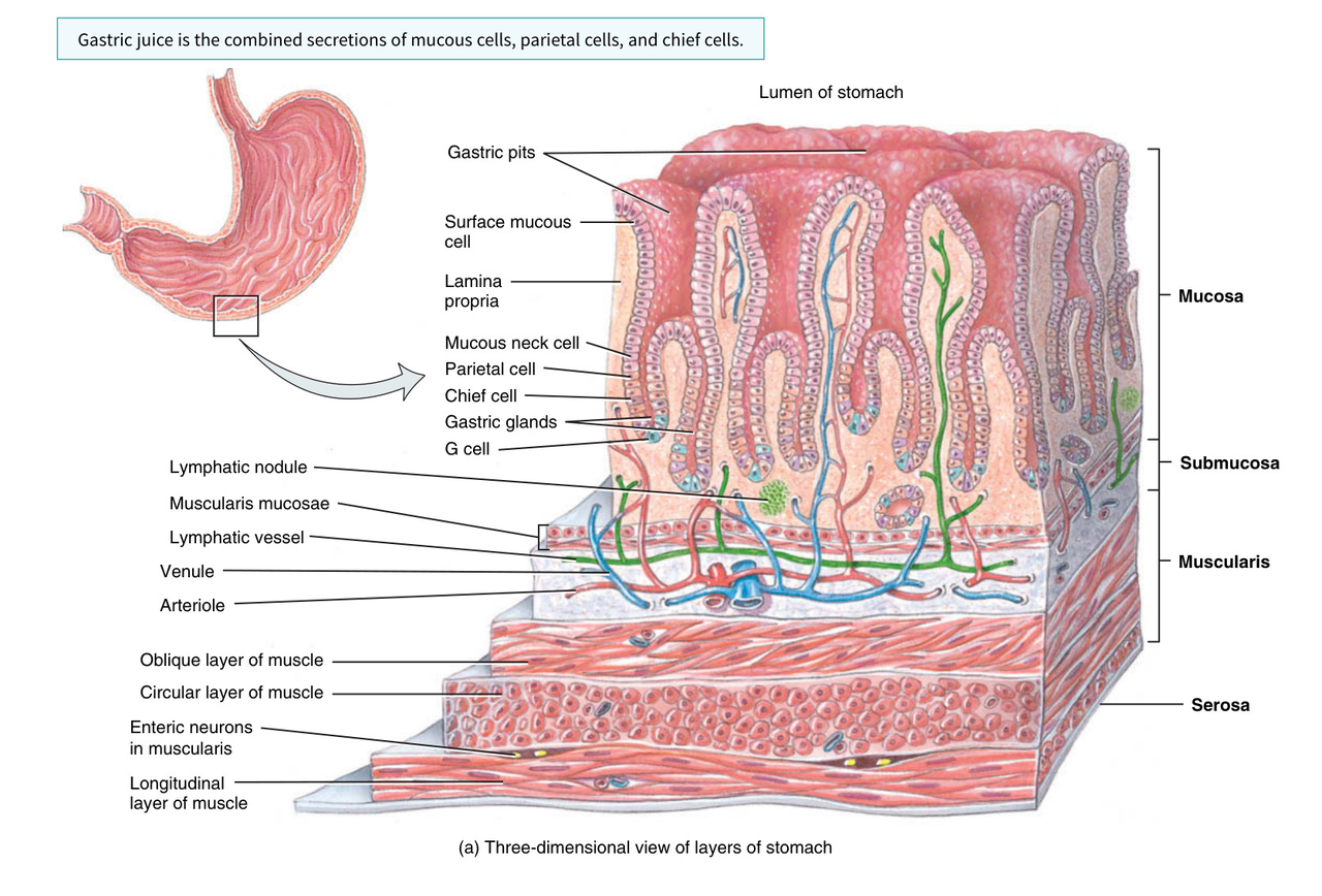
List the four types of cells found within the gastric glands.
Gastric juice is the combined secretion of _______, _______, and _______.
What is the function of surface mucous cells and mucous neck cells in the stomach?
Which substances are secreted by parietal cells in the stomach?
What is the primary function of intrinsic factor produced by parietal cells?
Which substances are secreted by the chief (zymogenic) cells?
What is the total daily volume of gastric juice produced?
The G cells, a type of enteroendocrine cell located mainly in the pyloric antrum, secrete the hormone _______ into the _______.
What is the main physiological role of gastrin?
Describe the composition of the stomach's submucosa layer.
How does the muscularis layer of the stomach differ from that of the esophagus, small intestine, and large intestine?
List the three layers of smooth muscle found in the stomach's muscularis.
The inner oblique layer of the stomach muscularis is limited primarily to the _______ of the stomach.
What tissues compose the stomach serosa?
What is the frequency of peristaltic waves in the stomach?
The process of _______ moves gastric contents from the body of the stomach toward the antrum, while _______ forces large food particles back into the body of the stomach to undergo further mechanical breakdown.
What is chyme?
What is gastric emptying, and how much chyme passes through the pyloric sphincter at a time?
The mixing of chyme with acidic gastric juice leads to the inactivation of _______ and the activation of _______, which digests triglycerides into fatty acids and diglycerides.
What is the primary role of the H+-K+ ATPase proton pump in parietal cells?
The enzyme _______ catalyzes the formation of _______ from water and carbon dioxide.
What is the 'alkaline tide' in relation to stomach digestion?
List the three main substances that stimulate parietal cells to secrete hydrochloric acid.
Histamine receptors on parietal cells are called _______ receptors, which differ in response from the _______ receptors involved in allergic reactions.
What is the function of pepsin, and how is it produced?
Why does pepsin not digest the stomach cells themselves?
Name the three substances that stimulate HCl secretion by parietal cells.
Surface mucous cells and mucous neck cells secrete _______, which forms a protective barrier preventing digestion of the stomach wall.
What is the primary function of intrinsic factor secreted by parietal cells?
What is the role of hydrochloric acid (HCl) in the stomach?
What substances are secreted by chief cells in the stomach?
G cells secrete _______, which stimulates parietal cells to secrete HCl and chief cells to secrete pepsinogen.
How does the muscularis layer contribute to digestion in the stomach?
The pyloric sphincter regulates the passage of chyme from the _______ to the _______ and prevents backflow.
What is the function of gastric lipase, and at what pH level does it operate best?
Pepsinogen is converted into active _______ only after it comes in contact with _______.
The stomach epithelium is protected from gastric juices by a layer of alkaline mucus that is _______ mm thick.
Which food group spends the least time in the stomach after a meal?
Vomiting, or _______, is the forcible expulsion of the contents of the upper GI tract through the mouth.
What are some common stimuli for vomiting?
What are the potential negative consequences of prolonged vomiting?
The pancreas is a _______ gland that is approximately _______ long and _______ thick.
What are the three distinct anatomical divisions of the pancreas?
Where is the head of the pancreas located in relation to the duodenum?
What is the alternative name for the pancreatic duct?
How is the hepatopancreatic ampulla (ampulla of Vater) formed?
The hepatopancreatic ampulla opens into the duodenum on an elevation known as the _______.
What is the secondary duct of the pancreas referred to as?
What is the approximate distance of the major duodenal papilla from the pyloric sphincter?
Which classes of nutrients are digested by pancreatic enzymes?
Where does the pancreatic duct empty its secretions?
The passage of pancreatic juice and bile into the duodenum is regulated through the _______.
What cells are responsible for secreting pancreatic juices into the small ducts?
Identify the relationship between the pancreas and the stomach based on their positions.
The _______ cells of the pancreas secrete pancreatic juices into small ducts that eventually form the pancreatic duct and the _______.
Flashcards in this deck (129)
-
What is the primary function of the cheeks and lips during mastication?
They keep food between the teeth to ensure foods are uniformly chewed.
anatomy digestive-system -
What are the two primary functions of saliva secreted by the salivary glands?
- It moistens and lubricates the lining of the mouth and pharynx.
- It softens, moistens, and dissolves food, while cleansing the mouth and teeth.
physiology digestion -
Salivary amylase is an enzyme that splits starch into smaller fragments, specifically maltose, maltotriose, and \(\alpha\)-dextrins.
enzymes digestion -
What is the role of extrinsic tongue muscles?
They move the tongue from side to side and in and out to maneuver food for mastication and shape it into a bolus.
anatomy muscular-system -
What is the function of intrinsic tongue muscles?
They alter the shape of the tongue to facilitate swallowing and speech.
anatomy muscular-system -
How is the secretion of saliva regulated by taste buds?
Taste buds act as receptors for gustation and presence of food, triggering nerve impulses that travel to salivary nuclei in the brain stem, which then stimulate the salivary glands.
physiology nervous-system -
Lingual glands secrete the enzyme lingual lipase, which breaks down triglycerides into fatty acids and diglycerides.
enzymes digestion -
Define a bolus.
A bolus is a soft, flexible, easily swallowed mass of food formed in the mouth through manipulation by the tongue, grinding by the teeth, and mixing with saliva.
definitions digestion -
Why must carbohydrates be broken down into monosaccharides?
Most carbohydrates ingested are starches or complex polysaccharides; only monosaccharides can be absorbed into the bloodstream.
physiology nutrition -
What happens to starch digestion after food is swallowed?
Salivary amylase continues to act on starches in the swallowed food for about another hour until stomach acids inactivate the enzyme.
physiology digestion -
When does lingual lipase become active?
It becomes activated in the acidic environment of the stomach.
physiology digestion -
Describe the composition of a diglyceride.
A diglyceride consists of a glycerol molecule that is attached to two fatty acids.
biochemistry lipids -
Describe the location and structure of the pharynx.
The pharynx is a funnel-shaped tube extending from the internal nares to the esophagus and larynx. It is composed of skeletal muscle and lined with mucous membrane.
anatomy respiratory-system -
What are the three parts of the pharynx?
- Nasopharynx
- Oropharynx
- Laryngopharynx
anatomy respiratory-system -
How do the functions of the nasopharynx differ from the oropharynx and laryngopharynx?
The nasopharynx functions only in respiration, whereas both the oropharynx and laryngopharynx have digestive as well as respiratory functions.
physiology respiratory-system -
What is the physical description and length of the esophagus?
The esophagus is a collapsible muscular tube that is approximately 25 cm (10 in.) long.
anatomy esophagus -
What is the anatomical pathway of the esophagus through the body?
- Begins at the inferior end of the laryngopharynx.
- Passes through the inferior aspect of the neck.
- Enters the mediastinum anterior to the vertebral column.
- Pierces the diaphragm through the esophageal hiatus.
- Ends in the superior portion of the stomach.
anatomy esophagus -
A hiatus hernia occurs when part of the stomach protrudes above the diaphragm through the esophageal hiatus.
pathology esophagus -
Name the three components that make up the mucosa of the esophagus.
- Nonkeratinized stratified squamous epithelium
- Lamina propria (areolar connective tissue)
- Muscularis mucosae (smooth muscle)
histology esophagus -
What tissues and structures are found in the submucosa of the esophagus?
- Areolar connective tissue
- Blood vessels
- Mucous glands
histology esophagus -
What is the functional significance of the nonkeratinized stratified squamous epithelium in the esophagus?
It provides protection against abrasion and wear and tear from food particles that are chewed, mixed with secretions, and swallowed.
histology function -
Describe the composition of muscle layers in the muscularis of the esophagus from superior to inferior.
- Superior third: Skeletal muscle
- Intermediate third: Skeletal and smooth muscle
- Inferior third: Smooth muscle
histology muscularis -
What is the function of the upper esophageal sphincter (UES) and what is it made of?
The UES consists of skeletal muscle and regulates the movement of food from the pharynx into the esophagus.
anatomy sphincter -
What is the function of the lower esophageal (cardiac) sphincter (LES) and what is it made of?
The LES consists of smooth muscle, is located near the heart, and regulates the movement of food from the esophagus into the stomach.
anatomy sphincter -
Why is the superficial layer of the esophagus called the adventitia rather than a serosa?
The adventitia consists of areolar connective tissue that is not covered by mesothelium, and it merges with the connective tissue of surrounding structures rather than being free-floating.
histology adventitia -
What are the primary physiological functions of the esophagus?
- Secretes mucus
- Transports food to the stomach
physiology esophagus -
What digestive processes does the esophagus lack?
The esophagus does not produce digestive enzymes and does not perform absorption.
physiology esophagus -
What is the primary purpose of the process known as deglutition?
Deglutition, or swallowing, is the process of moving food from the mouth into the stomach.
physiology digestive -
What substances facilitate the process of deglutition?
The movement of food is facilitated by the secretion of saliva and mucus.
physiology digestive -
Name the three stages of deglutition.
- Voluntary stage
- Pharyngeal stage
- Esophageal stage
physiology digestive -
During the voluntary stage of swallowing, the bolus is passed into the oropharynx by the movement of the tongue.
physiology digestive -
What anatomical movements occur to prevent food from entering the nasopharynx during swallowing?
The soft palate and uvula move upward to close off the nasopharynx.
physiology digestive -
What prevents food from entering the larynx during the pharyngeal stage of swallowing?
The epiglottis closes off the opening to the larynx.
physiology digestive -
Where is the deglutition center located in the brain?
It is located in the medulla oblongata and the lower pons of the brain stem.
neurology physiology -
What is the definition of peristalsis?
Peristalsis is a progression of coordinated contractions and relaxations of the circular and longitudinal layers of the muscularis.
physiology digestive -
Besides the esophagus, in what structures does peristalsis occur?
- Other parts of the GI tract (to the anus)
- Ureters
- Bile ducts
- Uterine tubes
physiology anatomy -
How does the esophagus move the bolus toward the stomach?
Circular muscle fibers superior to the bolus contract, constricting the esophageal wall and squeezing the bolus downward.
physiology digestive -
The esophageal stage of swallowing is controlled by the medulla oblongata.
physiology digestive -
The involuntary stage where the bolus is passed through the pharynx into the esophagus is known as the pharyngeal stage.
physiology digestive -
What is the primary function of circular muscles in the esophagus during swallowing?
Circular muscles contract to push the food bolus forward through the esophagus.
biology digestive swallowing -
What role do longitudinal fibers play inferior to the bolus during swallowing?
They contract to shorten the inferior section of the esophagus and push its walls outward, preparing the region to receive the bolus.
biology digestive swallowing -
Secretions from esophageal glands provide lubrication to the bolus, which significantly reduces friction during its transit.
biology digestive swallowing -
How long does it typically take for solid or semisolid food to travel from the mouth to the stomach?
It takes approximately 4 to 8 seconds.
biology digestive swallowing -
What is the medical definition of gastroesophageal reflux disease (GERD)?
GERD is a condition where the lower esophageal sphincter fails to close adequately after food enters the stomach, allowing stomach contents to reflux into the inferior portion of the esophagus.
pathology digestive gerd -
The burning sensation known as heartburn is caused by hydrochloric acid (HCl) from the stomach irritating the esophageal wall.
pathology digestive gerd -
Which specific food types are identified as triggers that can stimulate stomach acid and potentially worsen GERD symptoms?
- Coffee
- Chocolate
- Tomatoes
- Fatty foods
- Orange juice
- Peppermint
- Spearmint
- Onions
pathology digestive gerd -
How do histamine-2 (H2) blockers function as a management strategy for GERD?
They block acid secretion and are typically taken 30 to 60 minutes before eating.
pathology digestive gerd -
Antacids like Tums or Maalox manage GERD symptoms by neutralizing acid that has already been secreted into the stomach.
pathology digestive gerd -
What is the anatomical description and location of the stomach?
The stomach is a J-shaped enlargement of the GI tract situated directly inferior to the diaphragm in the abdomen, connecting the esophagus to the duodenum.
anatomy stomach -
What are the primary digestive functions of the stomach?
The stomach serves as a mixing chamber and a holding reservoir for food.
biology stomach -
With each inhalation, the diaphragm pushes the stomach inferiorly, and with each exhalation, it pulls the stomach superiorly.
anatomy stomach -
What is the pharyngeal stage of deglutition's effect on the airway?
It closes the air passageways.
physiology deglutition -
List the four specific activities performed by the esophagus during the digestive process as summarized in Table 24.2.
- Relaxation of upper esophageal sphincter
- Esophageal stage of deglutition (peristalsis)
- Relaxation of lower esophageal sphincter
- Secretion of mucus
physiology esophagus -
What are the four primary anatomical regions of the stomach?
- Cardia
- Fundus
- Body
- Pyloric part
stomach anatomy -
The stomach functions to mix saliva, food, and gastric juice to form chyme.
stomach physiology -
The stomach serves as a reservoir for food before its release into the small intestine.
stomach physiology -
What are the components of gastric juice and their specific physiological roles?
- HCl: Kills bacteria and denatures proteins
- Pepsin: Begins protein digestion
- Intrinsic factor: Aids in vitamin \(B_{12}\) absorption
- Gastric lipase: Aids in triglyceride digestion
stomach gastric-juice -
The stomach secretes the hormone gastrin into the blood.
stomach endocrine -
What are the three distinct layers of the muscularis externa in the stomach?
- Longitudinal layer
- Circular layer
- Oblique layer
stomach histology -
The internal folds of the stomach mucosa that allow for expansion are called rugae.
stomach anatomy -
Which specific anatomical feature regulates the passage of contents from the stomach into the duodenum?
The pyloric sphincter
stomach anatomy -
The pyloric antrum is the wider, proximal portion of the pyloric part of the stomach.
stomach anatomy -
The pyloric canal is the narrowed distal portion of the pyloric part that leads into the duodenum.
stomach anatomy -
What is the function of the serosa in the context of the stomach wall?
It serves as the outermost layer of the stomach wall, often referred to as the visceral peritoneum.
stomach histology -
What is the medical specialty that deals with the structure, function, diagnosis, and treatment of diseases of the stomach and intestines?
Gastroenterology
medicine stomach -
The stomach has four main regions: the cardia, the fundus, the body, and the pyloric part.
anatomy stomach -
What are the three specific regions that divide the pyloric part of the stomach?
- Pyloric antrum
- Pyloric canal
- Pylorus
anatomy stomach -
When the stomach is empty, the mucosa lies in large folds called rugae.
anatomy stomach -
Describe the difference between the lesser and greater curvatures of the stomach.
- Lesser curvature: The concave medial border.
- Greater curvature: The convex lateral border.
anatomy stomach -
What is the hallmark symptom and primary treatment for pyloric stenosis?
- Symptom: Projectile vomiting.
- Treatment: Surgical correction.
clinical stomach -
Pylorospasm occurs when smooth muscle fibers of the pyloric sphincter fail to relax normally, causing the stomach to become overly full.
clinical stomach -
Name the four basic layers of the stomach wall.
- Mucosa
- Submucosa
- Muscularis
- Serosa
histology stomach -
What specific cell type forms the surface of the stomach mucosa?
Surface mucous cells
histology stomach -
Identify the three distinct layers of muscle found in the muscularis of the stomach.
- Oblique layer
- Circular layer
- Longitudinal layer
histology stomach -
List the four types of cells found within the gastric glands.
- Mucous neck cell
- Parietal cell
- Chief cell
- G cell
histology stomach -
Gastric juice is the combined secretion of mucous cells, parietal cells, and chief cells.
physiology stomach -
What is the function of surface mucous cells and mucous neck cells in the stomach?
They secrete mucus.
stomach histology cells -
Which substances are secreted by parietal cells in the stomach?
- Hydrochloric acid
- Intrinsic factor
stomach histology cells -
What is the primary function of intrinsic factor produced by parietal cells?
It is needed for the absorption of vitamin B12.
stomach histology physiology -
Which substances are secreted by the chief (zymogenic) cells?
- Pepsinogen
- Gastric lipase
stomach histology cells -
What is the total daily volume of gastric juice produced?
2000–3000 mL (roughly 2–3 qt) per day.
stomach physiology -
The G cells, a type of enteroendocrine cell located mainly in the pyloric antrum, secrete the hormone gastrin into the bloodstream.
stomach histology hormones -
What is the main physiological role of gastrin?
It stimulates several aspects of gastric activity.
stomach physiology hormones -
Describe the composition of the stomach's submucosa layer.
It is composed of areolar connective tissue.
stomach anatomy -
How does the muscularis layer of the stomach differ from that of the esophagus, small intestine, and large intestine?
The stomach has three layers of smooth muscle, whereas the esophagus and intestines only have two.
stomach anatomy -
List the three layers of smooth muscle found in the stomach's muscularis.
- Outer longitudinal layer
- Middle circular layer
- Inner oblique layer
stomach anatomy -
The inner oblique layer of the stomach muscularis is limited primarily to the body of the stomach.
stomach anatomy -
What tissues compose the stomach serosa?
- Simple squamous epithelium (mesothelium)
- Areolar connective tissue
stomach anatomy -
What is the frequency of peristaltic waves in the stomach?
Peristaltic waves occur every 15 to 25 seconds.
stomach digestion peristalsis -
The process of propulsion moves gastric contents from the body of the stomach toward the antrum, while retropulsion forces large food particles back into the body of the stomach to undergo further mechanical breakdown.
stomach digestion propulsion retropulsion -
What is chyme?
Chyme is a soupy liquid formed when gastric contents are mixed with gastric juice.
stomach digestion chyme -
What is gastric emptying, and how much chyme passes through the pyloric sphincter at a time?
Gastric emptying is the process where chyme enters the small intestine; only about 3 mL of chyme moves through the pyloric sphincter at a time.
stomach digestion gastric-emptying -
The mixing of chyme with acidic gastric juice leads to the inactivation of salivary amylase and the activation of lingual lipase, which digests triglycerides into fatty acids and diglycerides.
stomach digestion enzymes -
What is the primary role of the H+-K+ ATPase proton pump in parietal cells?
It actively transports \(H^+\) into the stomach lumen while bringing \(K^+\) into the parietal cell.
stomach parietal-cells physiology -
The enzyme carbonic anhydrase catalyzes the formation of carbonic acid (\(H_2CO_3\)) from water and carbon dioxide.
stomach enzymes biochemistry -
What is the 'alkaline tide' in relation to stomach digestion?
It is the surge of bicarbonate ions (\(HCO_3^-\)) entering the bloodstream in exchange for chloride ions, which may be enough to elevate blood pH and make urine more alkaline after a meal.
stomach physiology alkaline-tide -
List the three main substances that stimulate parietal cells to secrete hydrochloric acid.
- Acetylcholine (ACh) (released by parasympathetic neurons)
- Gastrin (secreted by G cells)
- Histamine (a paracrine substance released by nearby mast cells)
stomach secretion physiology -
Histamine receptors on parietal cells are called H_2 receptors, which differ in response from the H_1 receptors involved in allergic reactions.
stomach histamine receptors -
What is the function of pepsin, and how is it produced?
Pepsin is the only proteolytic (protein-digesting) enzyme in the stomach, secreted by chief cells. It severs peptide bonds between amino acids to break proteins into smaller fragments.
stomach enzymes pepsin -
Why does pepsin not digest the stomach cells themselves?
Pepsin is secreted in an inactive form called pepsinogen, which cannot digest the proteins in the chief cells.
stomach digestion pepsinogen -
Name the three substances that stimulate HCl secretion by parietal cells.
- Acetylcholine (ACh)
- Gastrin
- Histamine
stomach physiology digestion -
Surface mucous cells and mucous neck cells secrete mucus, which forms a protective barrier preventing digestion of the stomach wall.
stomach digestion histology -
What is the primary function of intrinsic factor secreted by parietal cells?
It is needed for the absorption of vitamin \(B_{12}\), which is used in red blood cell formation (erythropoiesis).
stomach physiology nutrition -
What is the role of hydrochloric acid (HCl) in the stomach?
- Kills microbes in food
- Denatures proteins
- Converts pepsinogen into pepsin
stomach physiology digestion -
What substances are secreted by chief cells in the stomach?
- Pepsinogen
- Gastric lipase
stomach physiology digestion -
G cells secrete gastrin, which stimulates parietal cells to secrete HCl and chief cells to secrete pepsinogen.
stomach physiology digestion -
How does the muscularis layer contribute to digestion in the stomach?
It performs mixing waves (gentle peristaltic movements) that churn and physically break down food, mixing it with gastric juice to form chyme.
stomach physiology motility -
The pyloric sphincter regulates the passage of chyme from the stomach to the duodenum and prevents backflow.
stomach digestion anatomy -
What is the function of gastric lipase, and at what pH level does it operate best?
Gastric lipase splits triglycerides into fatty acids and monoglycerides; it operates best at a pH of 5–6.
stomach enzymes digestion -
Pepsinogen is converted into active pepsin only after it comes in contact with hydrochloric acid.
stomach digestion enzymes -
The stomach epithelium is protected from gastric juices by a layer of alkaline mucus that is 1–3 mm thick.
stomach histology protection -
Which food group spends the least time in the stomach after a meal?
Foods rich in carbohydrates.
stomach digestion nutrition -
Vomiting, or emesis, is the forcible expulsion of the contents of the upper GI tract through the mouth.
clinical pathology gi -
What are some common stimuli for vomiting?
- Irritation and distension of the stomach
- Unpleasant sights
- General anesthesia
- Dizziness
- Certain drugs (e.g., morphine and derivatives of digitalis)
clinical pathology gi -
What are the potential negative consequences of prolonged vomiting?
- Alkalosis (higher than normal blood pH)
- Dehydration
- Damage to the esophagus and teeth
clinical pathology gi -
The pancreas is a retroperitoneal gland that is approximately 12–15 cm long and 2.5 cm thick.
anatomy pancreas -
What are the three distinct anatomical divisions of the pancreas?
- Head
- Body
- Tail
anatomy pancreas -
Where is the head of the pancreas located in relation to the duodenum?
The head is the expanded portion of the organ located near the curve of the duodenum.
anatomy pancreas -
What is the alternative name for the pancreatic duct?
The duct of Wirsung.
anatomy ducts -
How is the hepatopancreatic ampulla (ampulla of Vater) formed?
It is formed by the union of the common bile duct and the pancreatic duct.
anatomy ducts -
The hepatopancreatic ampulla opens into the duodenum on an elevation known as the major duodenal papilla.
anatomy ducts -
What is the secondary duct of the pancreas referred to as?
The accessory duct (or duct of Santorini).
anatomy ducts -
What is the approximate distance of the major duodenal papilla from the pyloric sphincter?
It is approximately 10 cm (4 in.) inferior to the pyloric sphincter.
anatomy digestive -
Which classes of nutrients are digested by pancreatic enzymes?
- Starches (polysaccharides)
- Proteins
- Triglycerides
- Nucleic acids
physiology enzymes -
Where does the pancreatic duct empty its secretions?
It empties into the small intestine.
physiology pancreas -
The passage of pancreatic juice and bile into the duodenum is regulated through the hepatopancreatic ampulla.
anatomy digestive -
What cells are responsible for secreting pancreatic juices into the small ducts?
Exocrine cells.
physiology histology -
Identify the relationship between the pancreas and the stomach based on their positions.
The pancreas lies posterior to the greater curvature of the stomach.
anatomy pancreas -
The exocrine cells of the pancreas secrete pancreatic juices into small ducts that eventually form the pancreatic duct and the accessory duct.
physiology pancreas