Sign up to unlock more features
- Save this deck to your account
- Study flashcards with spaced repetition
- Export to Anki (.apkg) or PDF
- Process documents up to 100 pages
- Images extracted from PDFs and documents
- Better text extraction from your PDFs and documents
- Better flashcards with our more advanced AI model
What are 'aphasias' in the study of language?
What early hypothesis did Bouillaud propose about speech in 1825?
What key claim did Broca make in 1861 about language localization?
Describe the main speech characteristics of Broca's aphasia.
Which brain areas and structures are listed as commonly damaged in Broca's aphasia?
What is the effect of damage restricted to Broca's area alone?
How does Wernicke's aphasia differ from Broca's in speech fluency and comprehension?
Where did Wernicke report lesions that disrupted normal speech?
What features in the Mr. Ford transcript illustrate Broca's aphasia?
Why is it important to monitor one's own speech and the speech of others?
What general property of spontaneous speech is stated in the text?
List notable features of the example labeled 'A Wernicke's aphasic' in the text.
How did the interviewer describe their ability to interrupt the Wernicke's aphasic speaker?
Which supplementary material illustrates the Mr. Gorgan speech sample?
What brain region is damaged in the posterior sector associated with language deficits?
What additional damage occurs in severe, persistent language cases?
How do Broca's aphasics perform on sentence comprehension dependent on content words versus complex grammar?
Give an example sentence type Broca's aphasics can understand and one they typically cannot.
What specific processing difficulty contributes to Broca's aphasics' trouble with complex sentences?
In Wernicke's original model, which fibre tract linked Broca's and Wernicke's areas?
According to Wernicke's model, what deficit results from damage to the arcuate fasciculus?
What did early studies of aphasics show about hemispheric dominance for language?
What was a key split-brain finding about objects placed in the right versus left hand?
What language functions are associated with the right hemisphere?
How do anterior versus posterior right hemisphere lesions differently affect prosody?
What is the WADA procedure's basic method for testing hemispheric function?
Which illustration can be used as an explanatory aid for language-related brain areas (Broca, Wernicke, auditory cortex, motor cortex, arcuate fasciculus)?
How is a patient's ability to speak commonly assessed in language studies?
In right‑handers, what percentages show speech representation by hemisphere?
In left‑handers, what percentages show speech representation by hemisphere?
Which language‑related brain areas are labeled in the provided illustration?
What extension did Lichtheim add to Wernicke's model in 1885 regarding aphasias?
What is the defining repetition ability of transcortical motor aphasia (TMA)?
What are the comprehension and repetition features of transcortical sensory aphasia (TSA)?
List the key characteristics of Broca's aphasia from the table.
List the key characteristics of Wernicke's aphasia from the table.
What site and features define conduction aphasia in the table?
How does the table describe global aphasia?
What site and speech features characterize transcortical motor aphasia in the table?
What site and features characterize transcortical sensory aphasia in the table?
What are the features of anomic aphasia according to the table?
According to the text, how does the Wernicke–Geschwind model route spoken words?
According to the text, how does the Wernicke–Geschwind model route written words?
What illustration can help visualize Geschwind's model of language neurology?
What does the Lichtheim schematic illustrate about disconnection syndromes?
In the study comparing five conditions, which cortical regions were activated by passively viewing words (minus rest)?
Which regions were activated by listening to words (minus rest)?
Were the specific visual and auditory regions activated by non-word stimuli in the study?
After subtracting passive viewing (or listening) from repeating written (or spoken) words, which areas remained active?
What brain regions remained active when generating new words compared with repeating words?
What key criticism does the text give about the Wernicke-Geschwind model regarding reading?
What does the study reveal about overlap between speech production and comprehension?
What evidence is cited that argues against a single, unitary semantic/comprehension system?
What important brain regions does the Wernicke-Geschwind model omit that the text highlights?
What surprising finding about cortico-cortical connections and the arcuate fasciculus is mentioned?
Summarize the main processing steps for written and spoken words in the model (use image as illustration).
What image illustrates activation patterns during different language tasks, and what tasks are shown?
Flashcards in this deck (57)
-
What are 'aphasias' in the study of language?
Disorders of language often occurring without other cognitive impairment or inability to move speech muscles.
neuroscience language -
What early hypothesis did Bouillaud propose about speech in 1825?
That speech was controlled by the frontal lobes.
history localization -
What key claim did Broca make in 1861 about language localization?
Broca identified a region in the frontal lobes essential for language production and suggested language depended on the left hemisphere.
history localization -
Describe the main speech characteristics of Broca's aphasia.
Speech is laboured, slow, with impaired articulation; word selection for nouns often correct, but verbs and grammatical conjunctions are poorly selected or missing; comprehension is relatively preserved.
broca aphasia -
Which brain areas and structures are listed as commonly damaged in Broca's aphasia?
- Broca's area (Brodmann's areas 44/45)
- Surrounding frontal fields
- Underlying white matter
- Insula and basal ganglia
broca lesion -
What is the effect of damage restricted to Broca's area alone?
Damage restricted to Broca's area produces a far less severe and transient aphasia.
broca lesion -
How does Wernicke's aphasia differ from Broca's in speech fluency and comprehension?
Wernicke's aphasia is effortless, melodic and produced at a normal rate (fluent) but the content is unintelligible and comprehension of sentences is impaired.
wernicke aphasia -
Where did Wernicke report lesions that disrupted normal speech?
Lesions on the superior surface of the left temporal lobe.
wernicke lesion -
What features in the Mr. Ford transcript illustrate Broca's aphasia?
Slow, effortful speech with halting syllables, poor articulation, preserved comprehension (he completes or understands phrases).
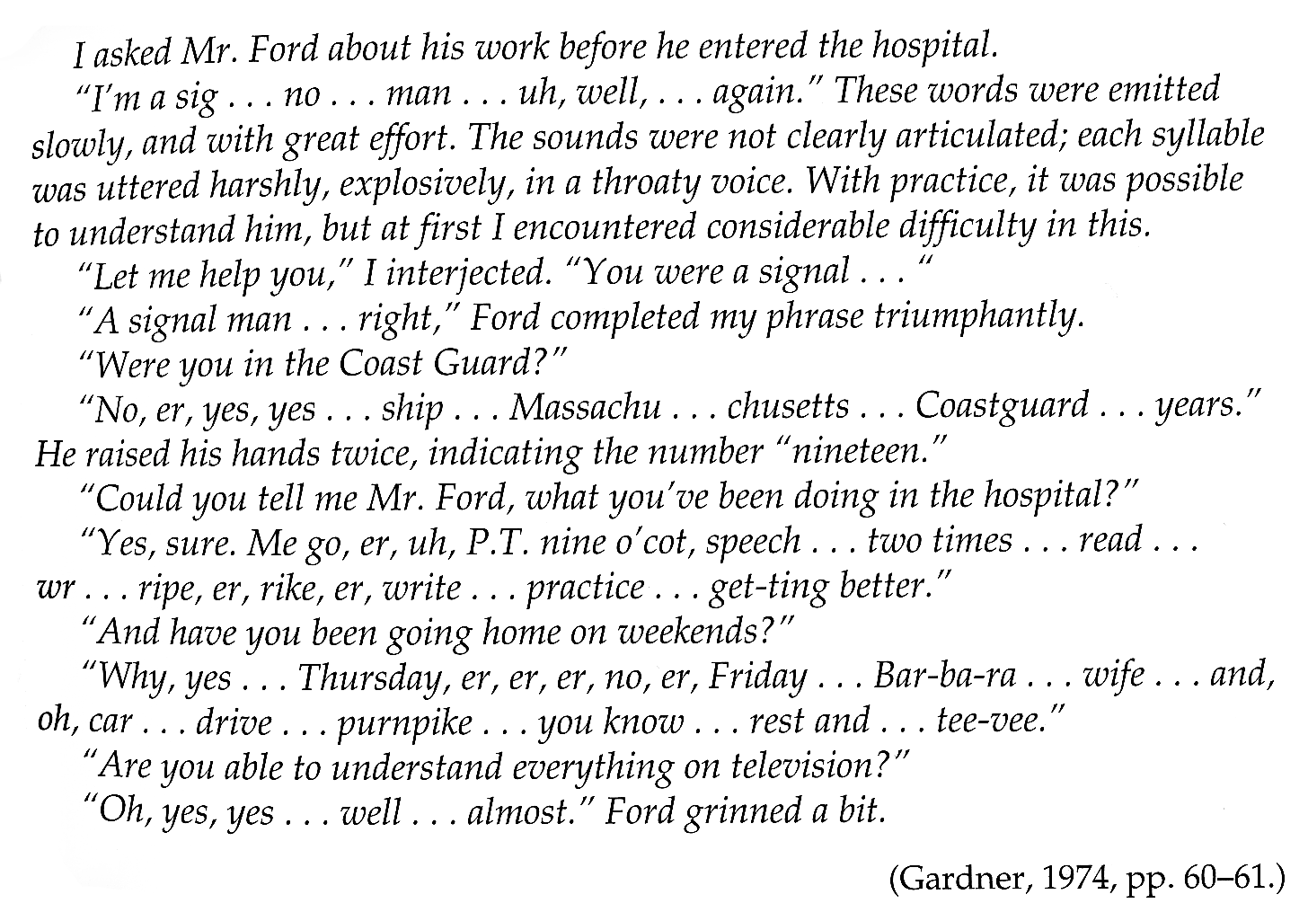 broca example
 -
Why is it important to monitor one's own speech and the speech of others?
- Because spontaneous speech is full of errors and requires monitoring to understand and correct them.
speech monitoring -
What general property of spontaneous speech is stated in the text?
- Spontaneous speech is full of errors.
speech spontaneous -
List notable features of the example labeled 'A Wernicke's aphasic' in the text.
- Rapid, relentlessly steady outflow of speech
- Frequent paraphasic/neologistic words (e.g., 'tarripoi', 'trebbin')
- Difficulty for an interlocutor to break in
aphasia wernicke -
How did the interviewer describe their ability to interrupt the Wernicke's aphasic speaker?
- The interviewer attempted several times but was unable to break in against the speaker's relentlessly steady and rapid outflow.
interview aphasia -
Which supplementary material illustrates the Mr. Gorgan speech sample?
- See image:
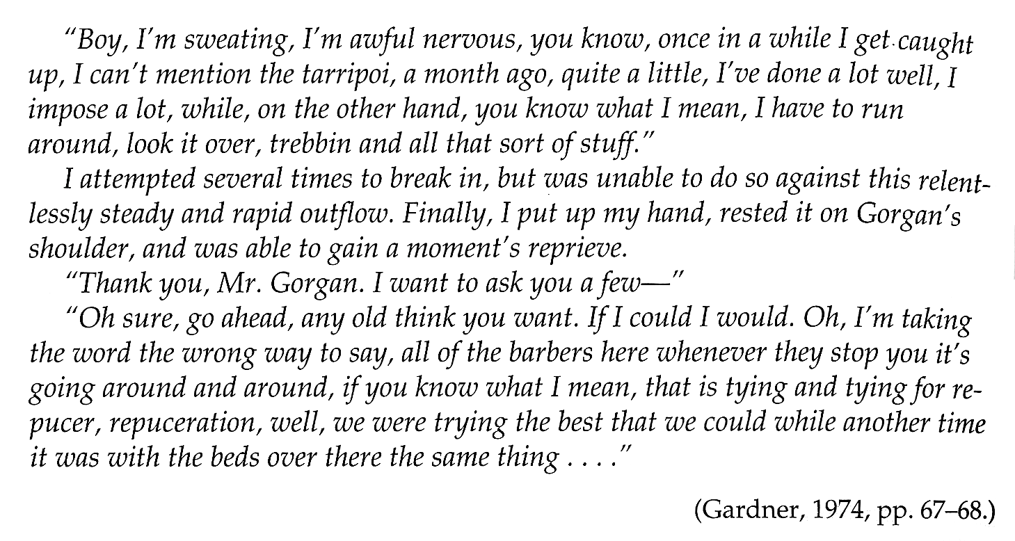 (contains transcript of the quoted fluent, error-filled speech).
media example - See image:
-
What brain region is damaged in the posterior sector associated with language deficits?
- Posterior sector of the left auditory association cortex (Brodmann's area 22)
neuroanatomy language -
What additional damage occurs in severe, persistent language cases?
- Middle temporal gyrus and underlying white matter
neuroanatomy pathology -
How do Broca's aphasics perform on sentence comprehension dependent on content words versus complex grammar?
- Can understand sentences when meaning is pieced from content words/world knowledge but are impaired when comprehension depends on complex grammar
aphasia broca -
Give an example sentence type Broca's aphasics can understand and one they typically cannot.
- Can: 'The apple that the boy is eating is red'
- Cannot: 'The boy that the girl chased is tall'
aphasia syntax -
What specific processing difficulty contributes to Broca's aphasics' trouble with complex sentences?
- Difficulty using grammar when elements to be linked are in different parts of the sentence, requiring short-term memory
aphasia memory -
In Wernicke's original model, which fibre tract linked Broca's and Wernicke's areas?
- Arcuate fasciculus
models anatomy -
According to Wernicke's model, what deficit results from damage to the arcuate fasciculus?
- Impairment in repetition of the spoken word without spontaneous speech or word comprehension deficits (conduction aphasia)
aphasia conduction -
What did early studies of aphasics show about hemispheric dominance for language?
- Language deficits were associated primarily with damage to the left hemisphere
laterality language -
What was a key split-brain finding about objects placed in the right versus left hand?
- Object in right hand could be named; object in left hand could not be named and got only rudimentary description
splitbrain laterality -
What language functions are associated with the right hemisphere?
- Emotional and tonal colouring of language (prosody); interpreting emotion, narrative coherence, social language use, and jokes
righthemisphere prosody -
How do anterior versus posterior right hemisphere lesions differently affect prosody?
- Anterior right hemisphere damage: produce inappropriate intonation
- Posterior right hemisphere damage: difficulty interpreting emotion in others' speech
prosody lesions -
What is the WADA procedure's basic method for testing hemispheric function?
- Inject a short-acting anaesthetic (e.g., sodium amytal) into either left or right carotid artery to anaesthetise the ipsilateral hemisphere
wada methods -
Which illustration can be used as an explanatory aid for language-related brain areas (Broca, Wernicke, auditory cortex, motor cortex, arcuate fasciculus)?
- Illustration of brain areas related to language:
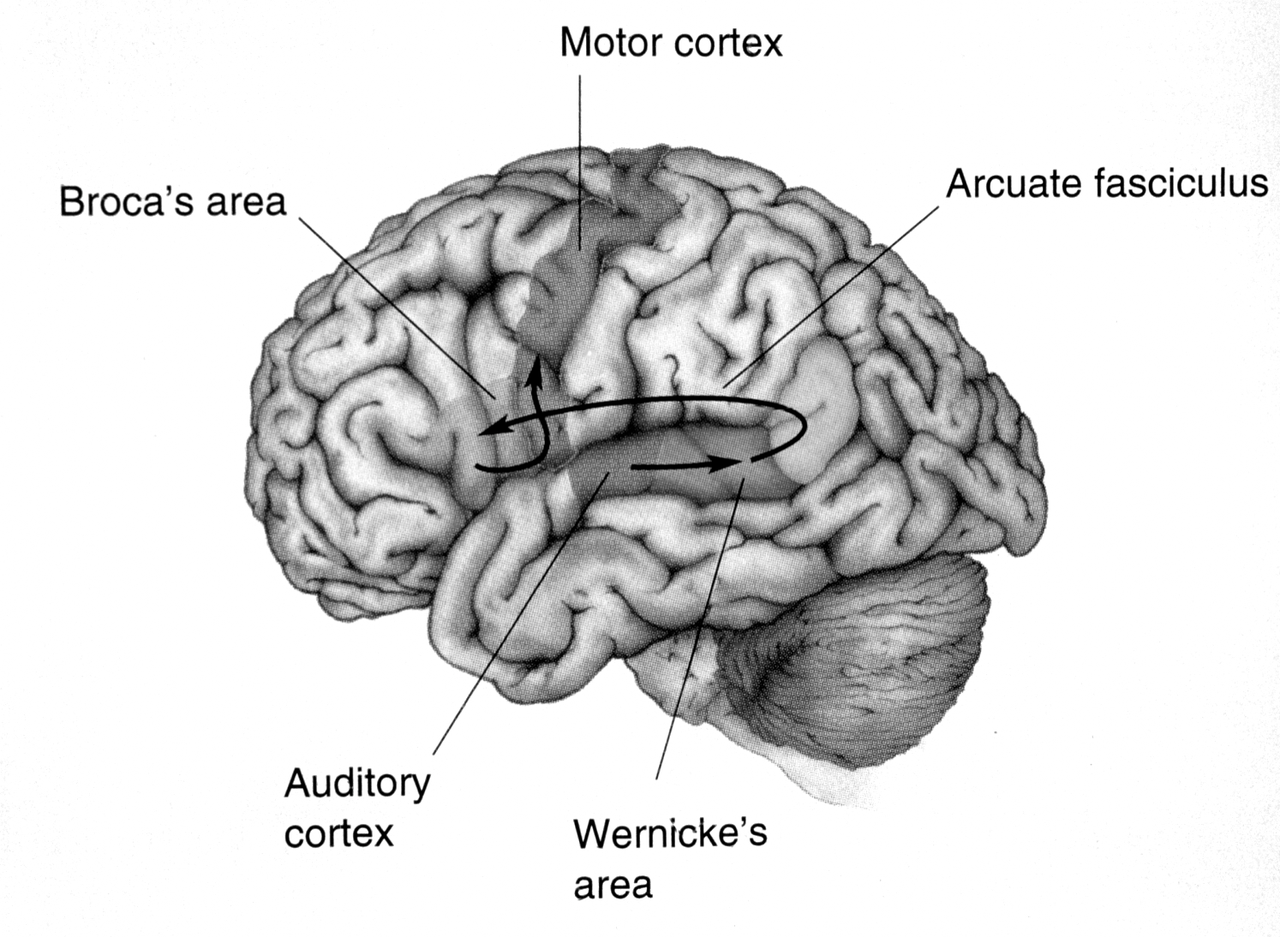
visual anatomy - Illustration of brain areas related to language:
-
How is a patient's ability to speak commonly assessed in language studies?
- The patient is asked questions to assess their ability to speak.
assessment speech -
In right‑handers, what percentages show speech representation by hemisphere?
- Left hemisphere: 96%
- Right hemisphere: 4%
- Bilateral: 0%
laterality handedness -
In left‑handers, what percentages show speech representation by hemisphere?
- Left hemisphere: 70%
- Right hemisphere: 15%
- Bilateral: 15%
laterality handedness -
Which language‑related brain areas are labeled in the provided illustration?
- Motor cortex
- Broca's area
- Arcuate fasciculus
- Auditory cortex
- Wernicke's area
neuroanatomy image -
What extension did Lichtheim add to Wernicke's model in 1885 regarding aphasias?
Lichtheim described two additional aphasias: transcortical motor aphasia (TMA) and transcortical sensory aphasia (TSA), which are complements of conduction aphasia.
lichtheim aphasia -
What is the defining repetition ability of transcortical motor aphasia (TMA)?
Patients with TMA speak nonfluent but can repeat even very long sentences.
tma repetition -
What are the comprehension and repetition features of transcortical sensory aphasia (TSA)?
Patients with TSA show poor comprehension but can repeat sentences and can make grammatical corrections to sentences they don't understand.
tsa comprehension -
List the key characteristics of Broca's aphasia from the table.
- Site: Motor association cortex of frontal lobe
- Comprehension: Good
- Speech: Nonfluent, agrammatical
- Impaired repetition: Yes
- Paraphasic errors: Yes
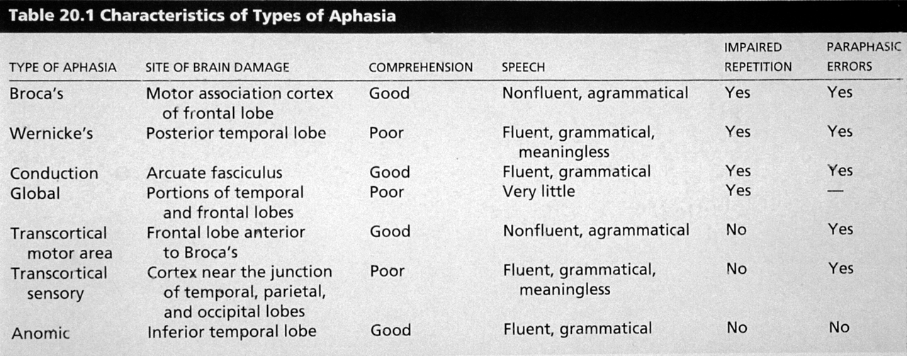 broca aphasia table
 -
List the key characteristics of Wernicke's aphasia from the table.
- Site: Posterior temporal lobe
- Comprehension: Poor
- Speech: Fluent, grammatical, meaningless
- Impaired repetition: Yes
- Paraphasic errors: Yes
wernicke aphasia table -
What site and features define conduction aphasia in the table?
- Site: Arcuate fasciculus
- Comprehension: Good
- Speech: Fluent, grammatical
- Impaired repetition: Yes
- Paraphasic errors: Yes
conduction aphasia arcuate -
How does the table describe global aphasia?
- Site: Portions of temporal and frontal lobes
- Comprehension: Poor
- Speech: Very little
- Impaired repetition: Yes
global aphasia -
What site and speech features characterize transcortical motor aphasia in the table?
- Site: Frontal lobe anterior to Broca's
- Comprehension: Good
- Speech: Nonfluent, agrammatical
- Impaired repetition: No
- Paraphasic errors: Yes
tma aphasia table -
What site and features characterize transcortical sensory aphasia in the table?
- Site: Cortex near junction of temporal, parietal, and occipital lobes
- Comprehension: Poor
- Speech: Fluent, grammatical, meaningless
- Impaired repetition: No
- Paraphasic errors: Yes
tsa aphasia table -
What are the features of anomic aphasia according to the table?
- Site: Inferior temporal lobe
- Comprehension: Good
- Speech: Fluent, grammatical
- Impaired repetition: No
- Paraphasic errors: No
anomic aphasia -
According to the text, how does the Wernicke–Geschwind model route spoken words?
Spoken words arrive via auditory cortex (areas 41, 42) and are recognized after processing in Wernicke's area.
geschwind spoken -
According to the text, how does the Wernicke–Geschwind model route written words?
Written words arrive via visual cortex (area 17, 18, 19), are processed in the angular gyrus (area 39), then in Wernicke's area to be converted into an auditory format.
geschwind written angular -
What illustration can help visualize Geschwind's model of language neurology?
A diagram shows cortical regions and arrows for routes of language processing (spoken word via areas 41,42 → Wernicke; written word via areas 17,18,19 → area 39 → Wernicke).
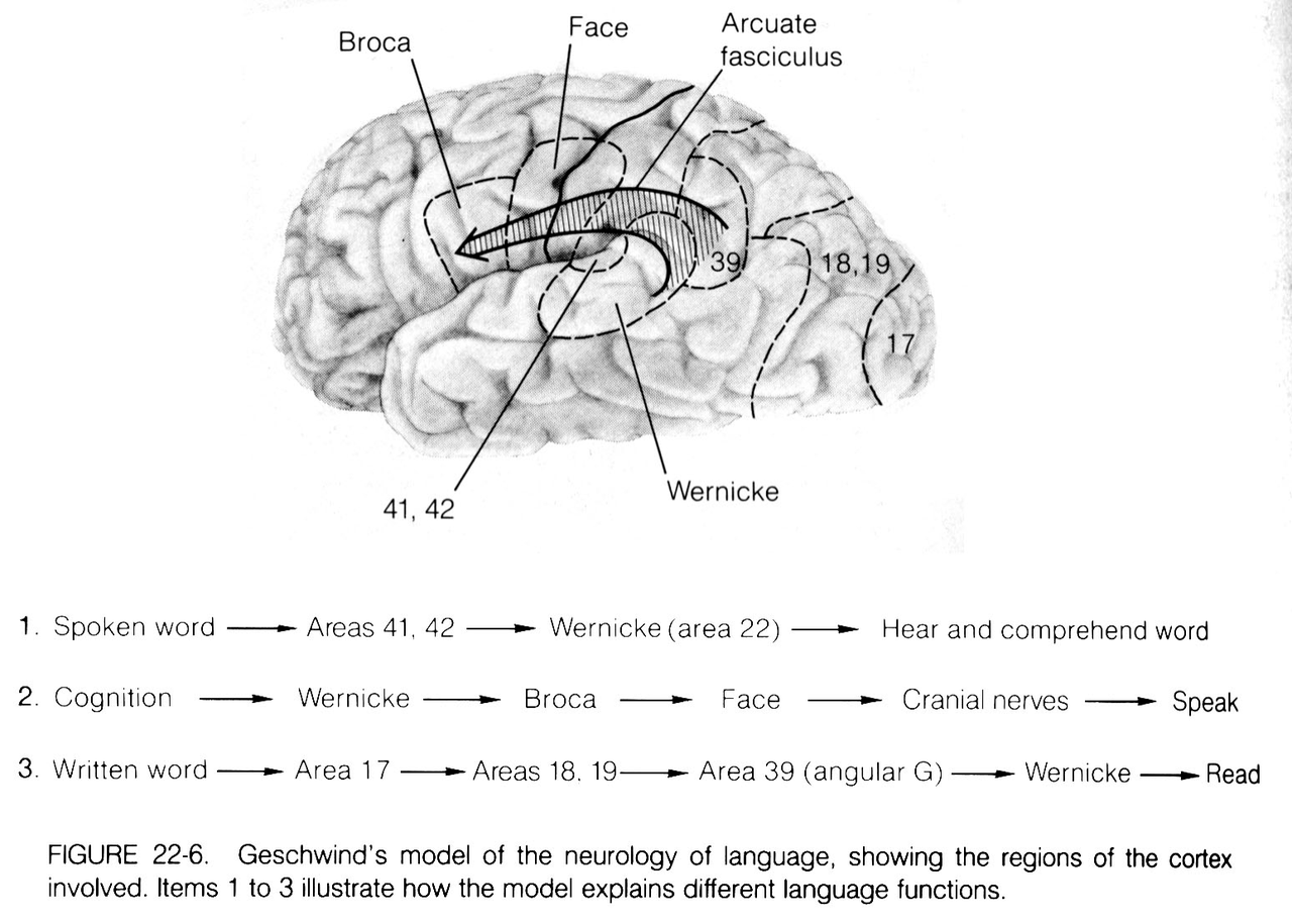 geschwind diagram model
 -
What does the Lichtheim schematic illustrate about disconnection syndromes?
The schematic represents hypothetical disconnection syndromes linking motor images of words and auditory images of words, labeling C.A. (conduction aphasia), T.M.A., and T.S.A..
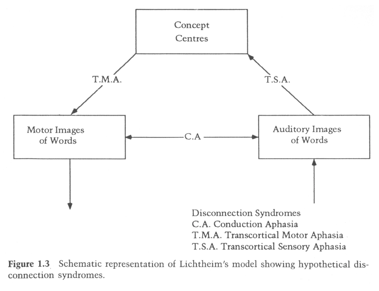 lichtheim disconnection
 -
In the study comparing five conditions, which cortical regions were activated by passively viewing words (minus rest)?
- Striate cortex
- Extrastriate cortex
visual neuroimaging -
Which regions were activated by listening to words (minus rest)?
- Primary auditory cortex
- Secondary auditory cortex
- Wernicke's area
auditory neuroimaging -
Were the specific visual and auditory regions activated by non-word stimuli in the study?
- No — these specific visual and auditory regions were not activated by non-word stimuli
stimuli neuroimaging -
After subtracting passive viewing (or listening) from repeating written (or spoken) words, which areas remained active?
- Primary motor cortex
- Supplementary motor cortex
- Broca's area
motor language -
What brain regions remained active when generating new words compared with repeating words?
- Frontal cortex
- Temporal cortex
(Thought to relate to the word association task.)
generation language -
What key criticism does the text give about the Wernicke-Geschwind model regarding reading?
- Visual input during reading does not go via the angular gyrus or Wernicke's area; there is an alternative route to anterior speech areas
models critique -
What does the study reveal about overlap between speech production and comprehension?
- There is considerable overlap between speech production and word comprehension; these abilities are not completely independent
overlap language -
What evidence is cited that argues against a single, unitary semantic/comprehension system?
- Category-specific naming deficits (anomias) and modality-specific deficits (e.g., naming animals worse than tools; naming words worse than picture comprehension)
semantics aphasia -
What important brain regions does the Wernicke-Geschwind model omit that the text highlights?
- Subcortical structures, including the basal ganglia
anatomy omission -
What surprising finding about cortico-cortical connections and the arcuate fasciculus is mentioned?
- These connections and the arcuate fasciculus can apparently be removed without incurring aphasia
connectivity aphasia -
Summarize the main processing steps for written and spoken words in the model (use image as illustration).
- Written word: Early visual processing: Striate cortex → Visual word recognition: Extrastriate cortex
- Spoken word: Early auditory processing: Primary auditory cortex → Auditory word recognition: Temporoparietal cortex (angular gyrus) / Anterior superior temporal cortex
- Semantic association: Inferior frontal cortex
- Premotor coding: Supplementary motor area
- Motor control of speech: Primary motor cortex
Illustration:
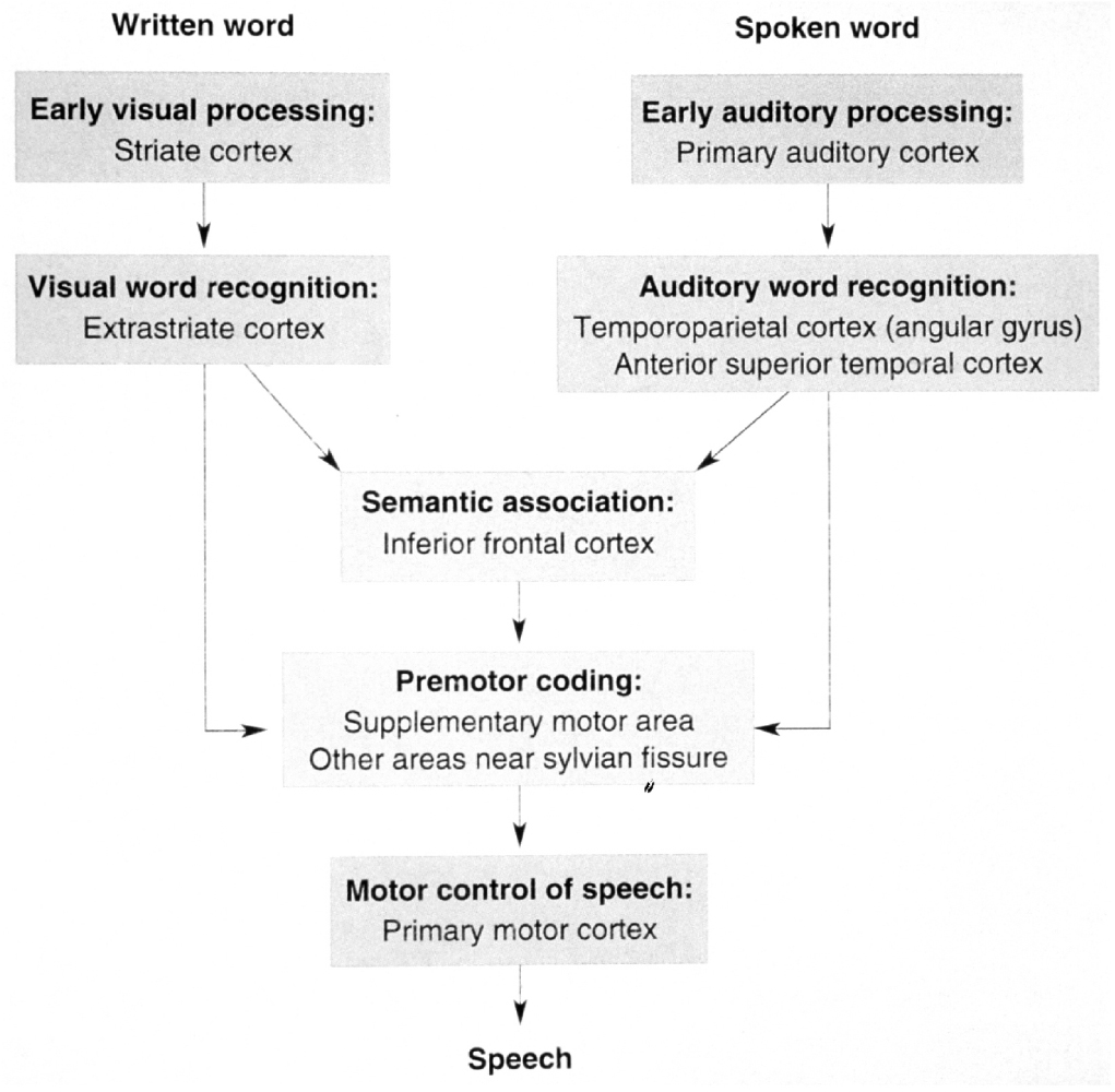 processing flowchart
 -
What image illustrates activation patterns during different language tasks, and what tasks are shown?
- Brain activation maps image showing: passively viewing words, listening to words, speaking words, generating verbs
Image:
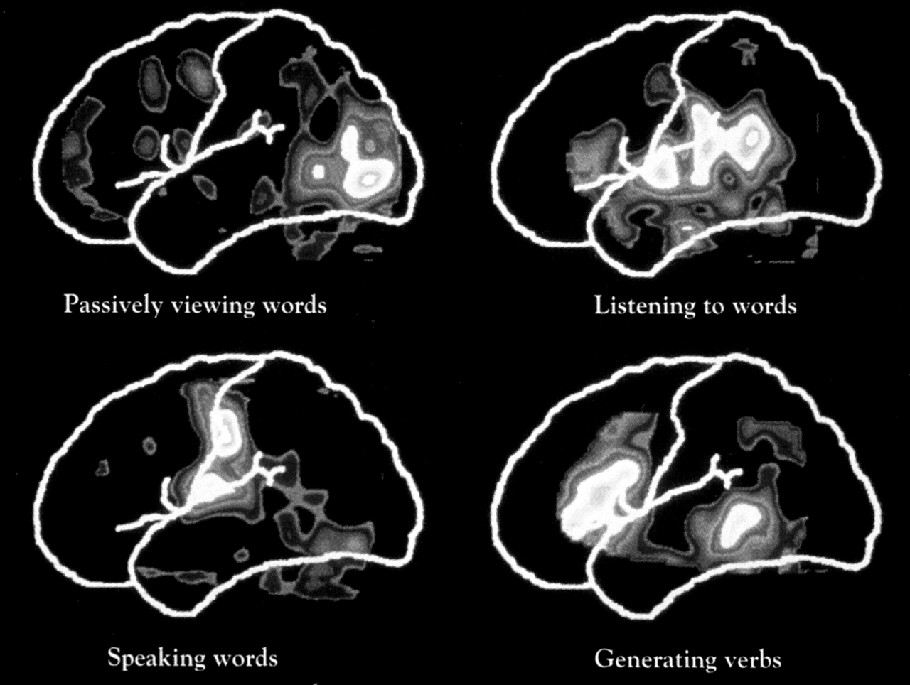 image neuroimaging
Neural basis of language — concise study notes
Overview
- Focus: how brain regions support language, classical aphasia syndromes, models of language organization, hemispheric specialisation, and neuroimaging evidence.
- Approach: historical lesion studies (Broca, Wernicke, Lichtheim), extended by modern imaging and clinical observations.
Historical background
- Early localisation: Bouillaud (1825) suggested frontal lobes; Broca (1861) identified a left inferior frontal region critical for speech production and left‑hemisphere dominance.
- Wernicke (1874) described a distinct posterior temporal lesion causing fluent but meaningless speech and impaired comprehension.
Broca's aphasia (expressive / motor aphasia)
- Key features:
- Nonfluent, effortful speech with poor articulation and short utterances.
- Comprehension relatively preserved for simple sentences and individual words.
- Impaired repetition, especially complex sentences.
- Common paraphasias (word-finding difficulty) and grammatical omissions.
- Typical lesion sites: Broca's area (Brodmann 44/45), adjacent frontal cortex, underlying white matter, insula, basal ganglia.
- Lesions limited strictly to Broca's area usually cause milder, transient deficits.
Example (patient Mr. Ford)
- Illustration: slow, effortful syllable-by-syllable speech with preserved ability to select content words (nouns) but difficulty assembling fluent phrases.
Wernicke's aphasia (receptive / sensory aphasia)
- Key features:
- Fluent, melodic, normal-rate speech that is often semantically empty or unintelligible.
- Frequent paraphasic errors (wrong words, phoneme substitutions) and jargon.
- Marked comprehension impairment for spoken language.
- Impaired repetition.
- Typical lesion sites: posterior superior temporal lobe (Brodmann area 22) and, in severe cases, middle temporal gyrus and underlying white matter.
Example (patient Mr. Gorgan)
- Illustration: rapid, fluent output with many incorrect or invented words and poor ability to answer coherently.
Other aphasia types — key distinctions
- Conduction aphasia: fluent speech and good comprehension but poor repetition; often linked to damage of the arcuate fasciculus.
- Transcortical motor aphasia (TMA): nonfluent like Broca's but preserved repetition (lesion anterior to Broca's).
- Transcortical sensory aphasia (TSA): fluent, poor comprehension but preserved repetition (lesion near temporo‑parieto‑occipital junction).
- Global aphasia: large lesions of frontal and temporal regions; severe impairment across modalities.
- Anomic aphasia: primary deficit is word‑finding (naming); comprehension relatively spared.
- Use the table to compare comprehension, fluency, repetition, and lesion locations at a glance.
Classical models of language organization
- Wernicke–Lichtheim model: separate centres for "motor images" (Broca) and "auditory images" (Wernicke) connected by pathways; used to explain conduction and transcortical aphasias via disconnection.
- Wernicke–Geschwind model (1960s): extended Lichtheim by specifying cortical routes for spoken and written words (visual → angular gyrus → Wernicke → Broca → motor cortex). Useful as a teaching model but oversimplified.
Hemispheric specialisation
- Classical lesion studies and later techniques show left hemisphere dominance for core language in most people.
- Split‑brain studies: left hemisphere names objects and follows written commands; right hemisphere handles nonverbal, pictorial information and struggles to name objects when disconnected.
- Right hemisphere functions: important for prosody, emotional tone, discourse cohesion, social language use, and understanding nonliteral language (jokes, inferences).
WADA test (sodium amytal)
- Temporarily anaesthetizes one hemisphere to test lateralisation of language.
- Typical findings:
- Right‑handers: ~96% left‑dominant for language.
- Left‑handers: more variability (≈70% left, 15% right, 15% bilateral).
Contributions from functional neuroimaging
- PET/fMRI tasks compare conditions like: (i) passive viewing, (ii) listening, (iii) repeating, (iv) generating words, (v) rest.
- Key findings:
- Visual word viewing activates striate/extrastriate visual cortex; auditory words activate primary/secondary auditory cortex including Wernicke's region.
- Subtracting sensory input from production reveals Broca's area, premotor/supplementary motor areas, and primary motor cortex tied to speech production.
- Word generation (semantic retrieval) engages inferior frontal and temporal regions beyond simple repetition.
Limitations and modern revisions of classical models
- The Wernicke–Geschwind model is too linear and modular: visual input often reaches anterior speech areas without strict routing via the angular gyrus.
- There is considerable overlap between comprehension and production networks; they are not fully separable.
- Category‑specific and modality‑specific deficits (e.g., naming living vs nonliving items) argue against a single, unitary semantic store.
- Classical models underemphasize subcortical structures (basal ganglia) and the distributed, bilateral contribution to some language functions.
- Some cortico‑cortical connections can be lesioned without producing classic aphasia, showing redundancy and plasticity.
Clinical and cognitive implications — study tips
- Distinguish fluency (production rate) from comprehension and repetition when classifying aphasia.
- Map symptoms to likely lesion locations (e.g., Broca: left inferior frontal; Wernicke: posterior superior temporal; conduction: arcuate fasciculus).
- Remember exceptions: transcortical syndromes preserve repetition despite impaired fluency/comprehension.
- Use imaging + behavioural testing (including WADA when needed) to assess lateralisation and guide rehabilitation.
Quick summary — what to remember
- Broca: nonfluent, good comprehension, impaired repetition; left inferior frontal lesions.
- Wernicke: fluent but meaningless, poor comprehension, impaired repetition; posterior left temporal lesions.
- Conduction: poor repetition with preserved comprehension and fluent speech; arcuate fasciculus.
- Transcortical syndromes: like Broca/Wernicke but repetition spared.
- Left hemisphere dominant for propositional language; right hemisphere supports prosody and discourse.
- Modern imaging shows distributed, overlapping networks rather than strict serial routes.